
Heart CVS
Contents of this page
- Thrombus in the left ventricle
- Chordae tendinae calcification
- Ebstein anomaly
- Rhabdomyoma of heart (cardiac Rhabdomyoma) in left ventricle
- Artificial cardiac Pacemaker
- Dissecting aneurysm of abdominal aorta
- apical-aneurysm-left-ventricle
- mitral-stenosis-with-MR
- right-atrial-thrombus
- non-compaction-cardiomyopathy
- left-atrial-myxoma
Thrombus in the left ventricle
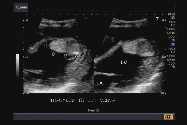
Large clot seen in ventricle
This young adult male patient underwent echocardiography to rule out cardiac pathology for pyrexia. Ultrasound image shows a large thrombus adherent to the wall of the left ventricle. The thrombus is seen as an echogenic, well defined mass in the ventricular lumen. Cardiac thrombi are seen following ventricular dysfunction, cardiomyopathy, myocardial infarction and ventricular aneurysm. Ultrasound image courtesy of Dr. Prasenjeet Singh, Delhi, India.
Reference: http://pediatrics.aappublications.org/cgi/content/full/107/2/421 (free article)
Chordae tendinae calcification
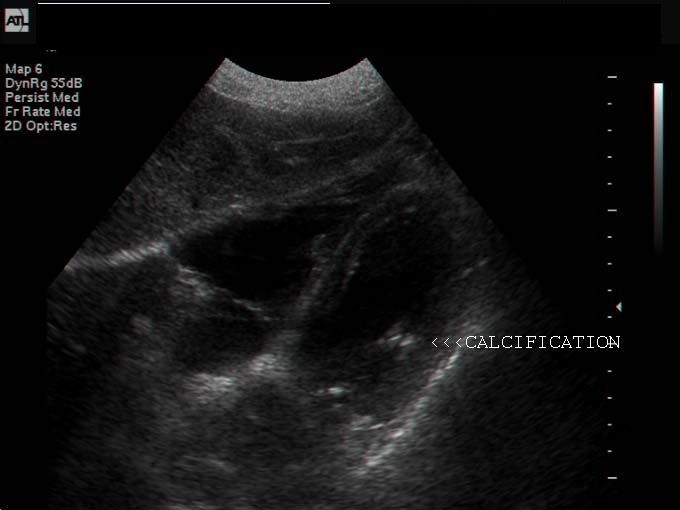
Echocardiography of the heart in this patient shows calcification of the chordae tendinae in the right ventricle. The causes of such calcific lesions of the chordae tendinae include acute or sub-acute bacterial endocarditis. These lesions may cause weakening and even rupture of the chordae. The chordae tendinae are string like fibrous chords that connect the papillary muscles to the borders of the tricuspid and mitral valves. Ultrasound image courtesy of Shlomo Gobi, Israel.
Reference:http://archinte.highwire.org/cgi/content/summary/127/4/574
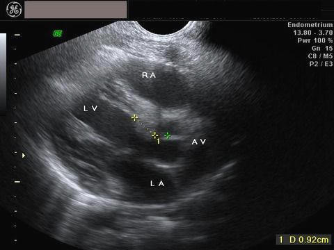
Ebstein anomaly
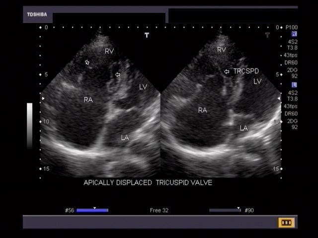
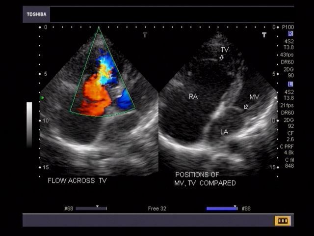
This adult patient presented with dyspnoea and palpitation. Echocardiography and Color Doppler imaging of the heart revealed apically displaced tricuspid valve with resultant atrialization of a major part of the right ventricle. There was also evidence of tricuspid regurgitation (image on right- Color Doppler). The right ventricle and the right atrium appear enlarged. These ultrasound images suggest Ebstein anomaly with tricuspid regurgitation. Both images courtesy of Dr. Vikas Arora, MD, India.
References:http://emedicine.medscape.com/article/154447-diagnosis
Rhabdomyoma of heart (cardiac Rhabdomyoma) in left ventricle
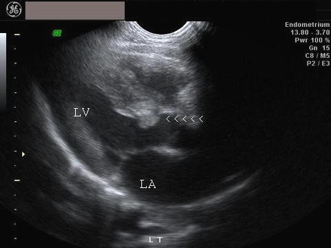
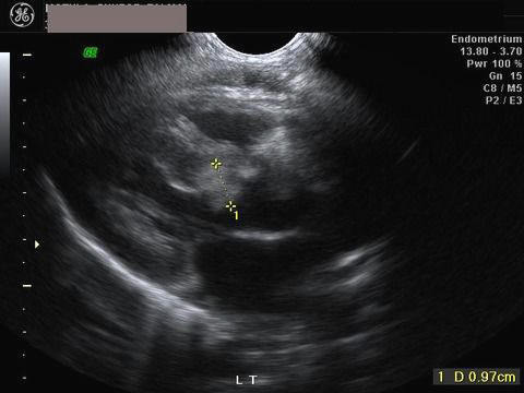
Sonography of cardiac masses
These ultrasound images of the heart show Rhabdomyoma of the left ventricle, measuring 10 mm. in size. The mass is seen as an echogenic (hyperechoic), well-defined mass of 10 mm., in the upper part of the interventricular septum. The patient is a neonate with proven diagnosis of the disease Tuberous sclerosis. Earlier sonography (not shown here) showed considerable obstruction to the left ventricular outflow tract. Rhabdomyoma is one of few rare tumours of the heart, seen in 1 in 10,000 births and are usually benign. Ultrasound image courtesy of Mr. Shlomo Gobi, Israel.
Reference: http://www.thefetus.net/page.php?id=506
Artificial cardiac Pacemaker

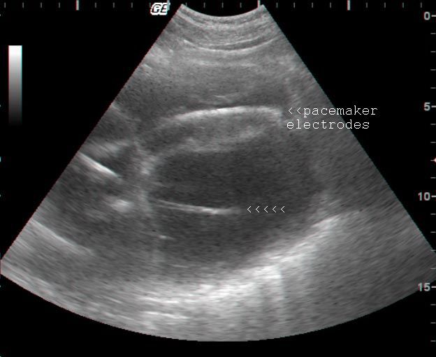
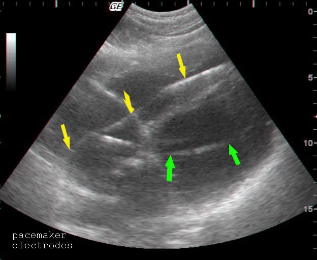
Sonography of the heart (echocardiography) was done in this patient of congestive cardiac failure (CCF) or congestive heart failure (CHF). This patient had undergone implantation of dual chamber pacemakers into the heart. The pacemaker electrodes are seen as hyperechoic linear echoes within the heart. Ultrasound images are courtesy of Mr. Shlomo Gobi, Israel.
Reference: http://en.wikipedia.org/wiki/Artificial_pacemaker
Dissecting aneurysm of abdominal aorta
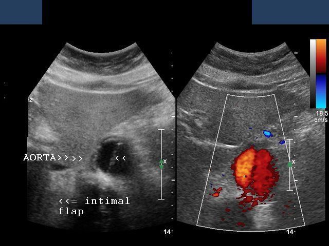
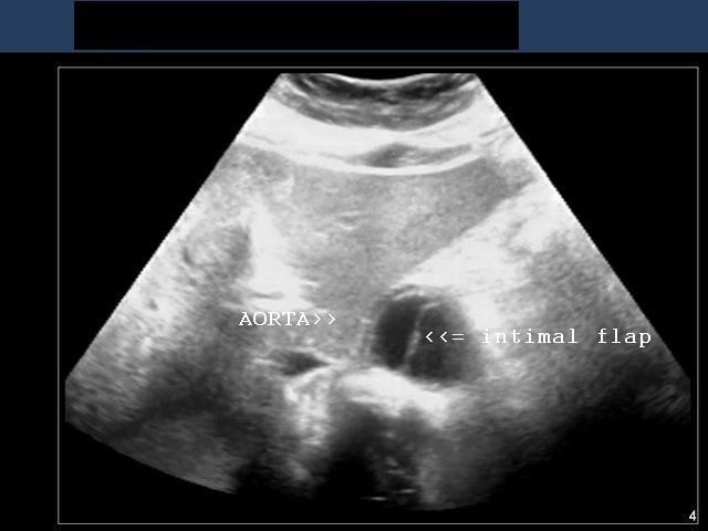
This color Doppler image (left) and ultrasound images show a cross section of the abdominal aorta with the separated intimal flap seen as an echogenic linear structure in the lumen of the aorta. Color Doppler image shows flow on both sides of the intimal flap suggesting a dissecting aortic aneurysm/ aortic dissection. Images are courtesy of Ravi Kadasne, MD, UAE.
Reference: /article/756835-overview (free article and images)
apical-aneurysm-left-ventricle
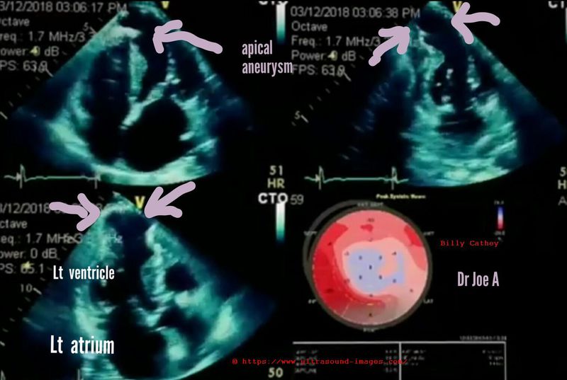

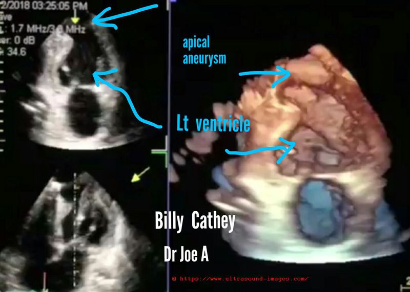
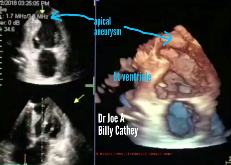

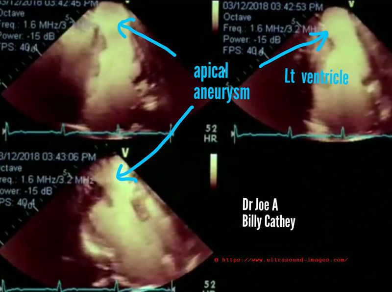
B-mode and contrast enhanced B-mode echocardiography (cardiac ultrasound) along with 3-D/ 4-D echo/ ultrasound show an aneurysm of the apical part of left ventricle (blue arrows). This type of aneurysm is the result of myocardial infarction involving the left ventricle resulting in weakening of the myocardium causing out-pouching or aneurysm formation in the apical region with each contraction or systole of the left ventricle.
Apical aneurysm of the left ventricle is the commonest site of aneurysm formation as a sequelae to myocardial infarction. (Images are courtesy of Billy Cathey).
D/d: pseudo-aneurysm of the heart--> pseudo-aneurysm results from rupture of the myocardium following a myocardial infarction with formation of a sac lined by pericardium. This type of lesion has no myocardial tissue lining the sac.
True aneurysms (like the case here), have lining of myocardium which can be traced to the adjacent part of the ventricle.
References:
1) Aneurysm of the left ventricle
2) What is an aneursym of the ventricle
3) pseudo-aneurysm of the Left ventricle
mitral-stenosis-with-MR
Above echocardiography (echo) video shows MS (mitral stenosis) with MR (mitral regurgitation) with mild AR or aortic regurgitation.
= severe rheumatic mitral stenosis evident with mitral valve orifice markedly reduced in size (see the MS jet on color Doppler study)
= markedly enlarged left atrium due to severe mitral stenosis
= also evident is an eccentric mitral regurgitation (MR jet on color Doppler)
= a mild AR is also present (AR jet on color Doppler ultrasound)
This patient was due for surgery for mitral valvular disease.
Ultrasound video (echo) taken using Toshiba Nemio XG ultrasound system by Joe Antony, MD
right-atrial-thrombus
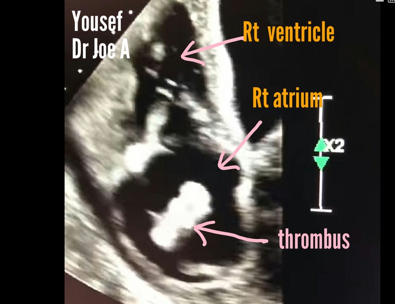
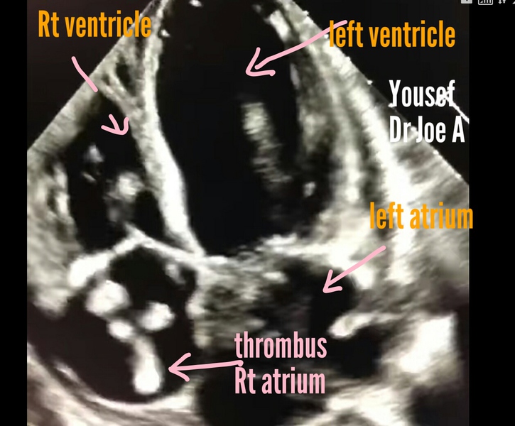

Echocardiography or echo study of adult heart showing
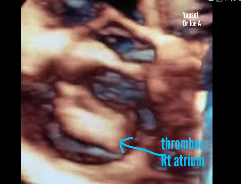
= huge thrombus in Right atrium (see 2D and 3D echo ultrasound images above)
= such freely floating thrombus or thrombi are the result of transit from the inferior vena cava or the sequel to thrombus in deep veins of the lower limb (DVT or deep vein thrombosis)
= can result in pulmonary embolism if not managed in time especially surgically removed
See the echocardiography or 3D echo ultrasound video above which shows the thrombus freely floating in the right atrium and causing intermittent obstruction at the Rt. atrio-ventricular valves
Video of Rt. atrial thrombus is courtesy of Yousef (cardiac sonographer)
Edits: by Dr. Joe Antony (me)
References: echo study of adult heart for Right atrial thrombus
non-compaction-cardiomyopathy
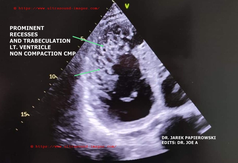

Non compaction cardiomyopathy is characterized by (see adult echo/ echocardiography images and video above) :
= prominent trabeculation in left ventricle
= non compaction usually affects the left ventricle but may affect the right ventricle or both also
= (the echocardiography video and images are courtesy of Dr. Jarek Papierowski. Edits by Dr. Joe A)
= it is a congenital anomaly of the heart and may be associated with other cardiac anomalies
= there are multiple prominent recesses within the endocardium and myocardium of the left ventricle resulting in a spongy appearance (spongy myocardium)
= D/d: left ventricular thrombus and apical hypertrophy
= it is important to visualize the apical region of the heart well during echo to diagnose this condition
Prognosis: can cause ventricular arrythmias, heart failure and thromus formation
References: non compaction cardiomyopathy echo
left-atrial-myxoma
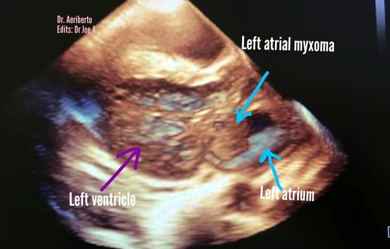

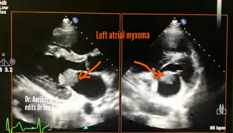
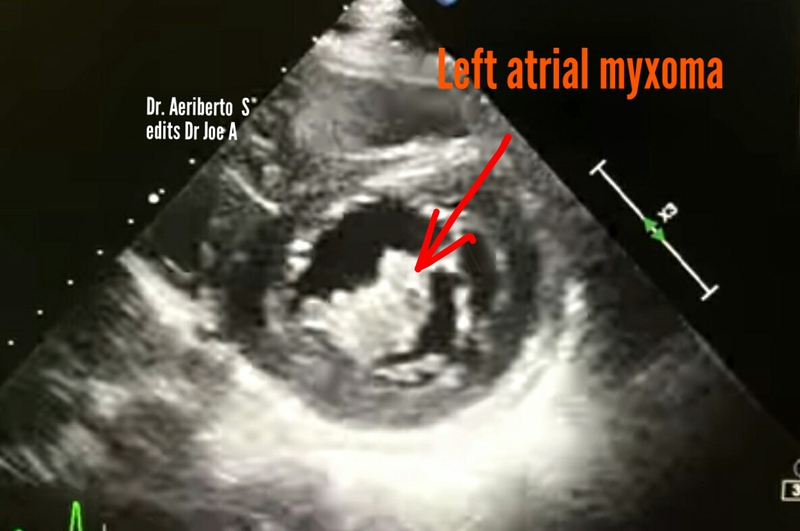
2D B mode as well as 3D and 4D ultrasound echo images and video of left atrial myxoma:
= benign solid cardiac mass usually seen in left atrium
= can be attached to the inter atrial septum or Atrio-ventricular (mitral) valve
(as in this case)
= the 3D/ 4D ultrasound and echo video above show the left atrial myxoma moves within the mitral valve and oscillates between the left atrium and left ventricle
= in such locations, left atrial myxoma usually produces mitral incompetence
= Ultrasound and echo video of left atrial myxoma is courtesy of Dr. Aeriberto Souza;
edits by Dr. Joe A
= left atrial myxoma is the commonest cardiac mass accounting for more than 50 % of all benign cardiac masses
= chief differential diagnosis is cardiac thrombus. Left atrial myxoma is usually oval in shape and has a pedicle. Thrombi within the heart occur mostly in patients with h/o of myocardial infarction or coronary artery disease.
= some authors feel that left atrial myxoma is the result of long standing organized intra cardiac thrombus
© 2019, Joe Antony, MD All images and content in this site are copyrighted. www.ultrasound-images.com When visiting this site, we may use cookies to improve the performance of this site. Use of this site means you accept the use of cookies. Read our privacy policy at this page: https://www.ultrasound-images.com/privacy-policy/