
Ultrasound images of ovarian tumours
Contents of this page
- Benign complex cystic masses of ovary
- Hemorrhagic cyst of ovary
- A typical serous cystadenoma of the ovary: (case-2)
- Serous cystadenoma- case-3
- Mucinous cystadenoma of ovary
- Mucinous cystadenoma- Case-2
- Cystadenocarcinoma
- Calcific masses of the ovary
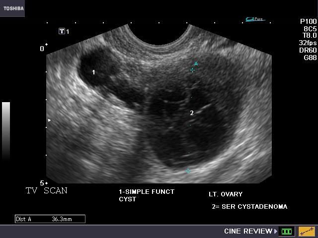
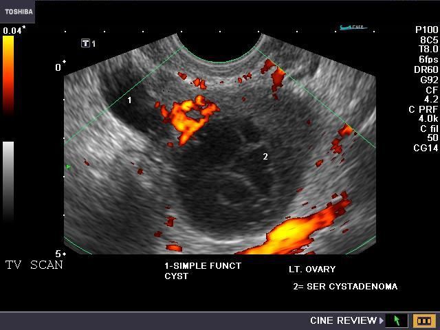
Benign complex cystic masses of ovary
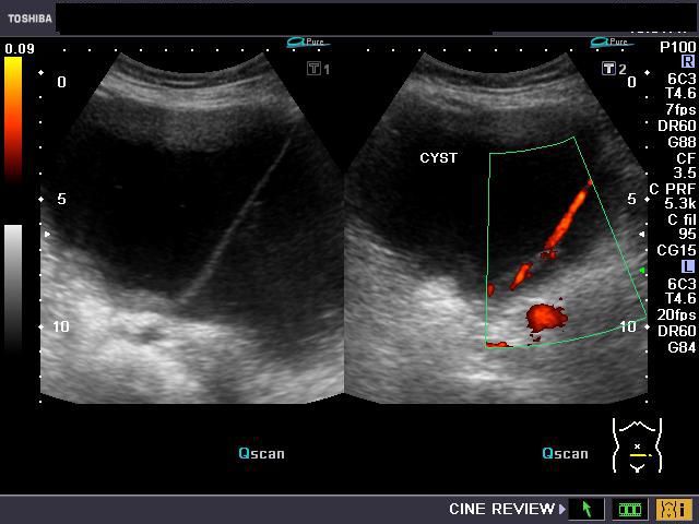
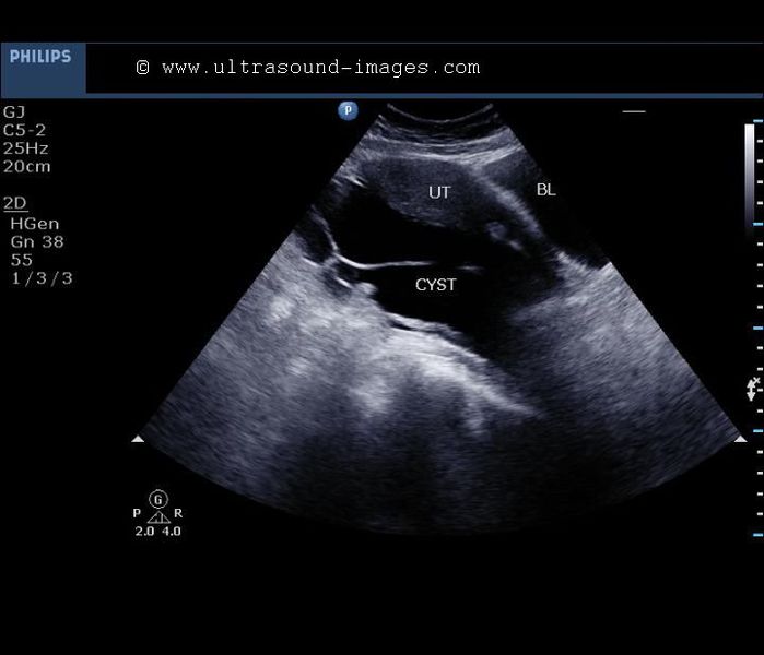
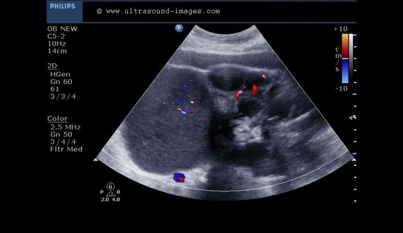
Hemorrhagic cyst of ovary
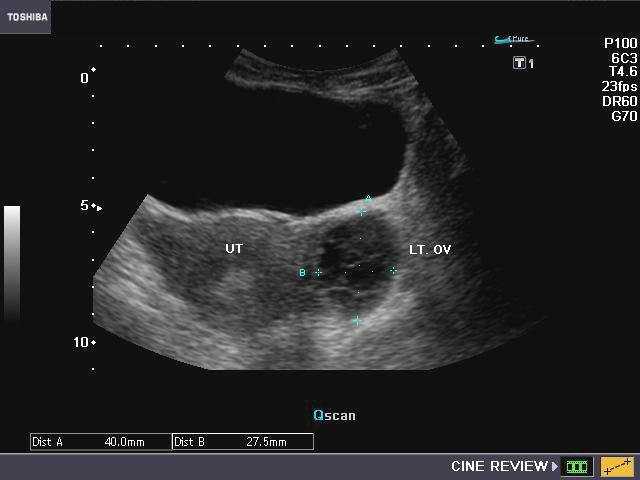

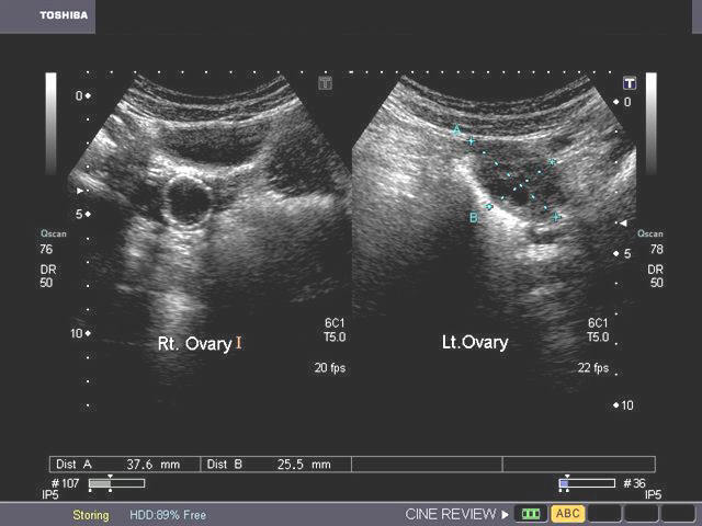
This young adult female patient underwent sonography of the ovaries. Upper Left image shows a cystic mass with multiple thin septae, in the left ovary. Power Doppler image (upper-right) of the left ovary shows no vascularity within the septae. Transvaginal ultrasound imaging of the ovary shows (bottom -Left) thin septae with clear fluid within the cyst. Smaller cystic lesion, appears to be a coexisting functional cyst of the ovary. Power Doppler image (lower right) confirms the findings described earlier. These ultrasound images suggest hemorrhagic cyst of the left ovary. Note the absence of solid tissue within the cystic lesion.
Reference:
2)http://emedicine.medscape.com/article/255865-diagnosis
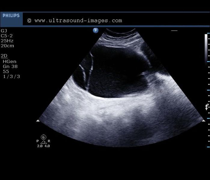
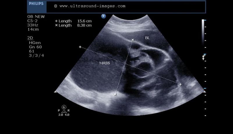
A typical serous cystadenoma of the ovary: (case-2)

The above transabdominal ultrasound images show a huge serous cystadenoma of the ovary. Note the cyst is separate from the urinary bladder, which is compressed upon by the cystic mass. With huge cystic lesions of this size (15 x 9 cms.), it is often difficult to determine the source of the mass or the side from which it originates. Here the cyst apparently arises from the left ovary (but I cant be sure). This patient was an elderly female. There is a prominent septum passing through the cyst, which shows considerable vascularity (see Power Doppler images in bottom row). This vascular septum is an ominous sign and hence malignancy cannot be ruled out (cystadenocarcinoma). However, no solid nodules are seen in this cystic tumor, suggesting a more benign nature. A differential diagnosis in this case would be a urinary bladder diverticulum. However this is unlikely, as both the cyst and bladder are clearly identified separately. The clear fluid contents suggest a diagnosis of serous cystadenoma of the ovary. Mesenteric cyst is another possibility in this case. In either case, this cyst has to be surgically removed considering a possible malignancy, and also due to the large size of this cystic mass.
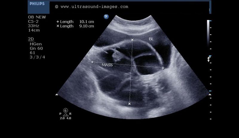
Serous cystadenoma- case-3
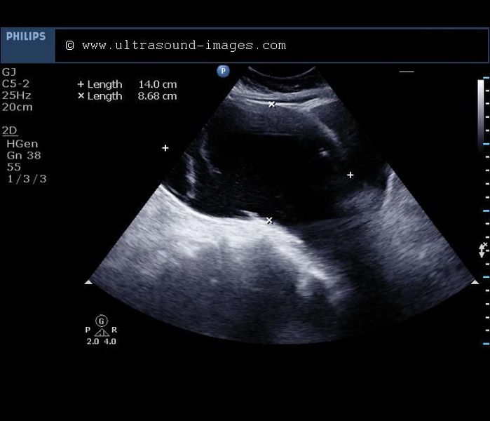
This is yet another case of serous cystadenoma of the ovary (again it is difficult to determine the side of origin of this huge ovarian cyst from the ultrasound images above. There is clear fluid within this multiseptate cyst, with minimal particulate matter. The septae are relatively thin walled again suggesting a benign nature. No mural nodules are present in this serous cystadenoma of the ovary. This sonography was performed using a Philips HD 15 ultrasound system.
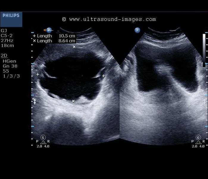
Mucinous cystadenoma of ovary

The right ovary in this middle aged female patient (see ultrasound images above), shows a large, primarily cystic mass with multiple septae. The cystic neoplasmmeasures 14 x 7 cms. in size and part of it shows coarse particulate matter producing fine echoes within the fluid. These appearances are typical of mucinous cystadenoma of the right ovary. Mucinous cystadenomas are ovarian epithelial tumors and rank only second in incidence to the commonest such cystic tumor, namely serous cystadenoma. The characteristic features of mucinous cystadenomas include the large size (15 to 30 cms. in size) and presence of low level echoes within the fluid (mucinous matter). The septae in this type of neoplasm are comparatively thicker than in the serous type of cystadenomas. Ultrasound images of mucinous cystadenoma are courtesy of Gunjan Puri, MD, India.
References:
http://en.wikipedia.org/wiki/Mucinous_cystadenoma(free article)
Mucinous cystadenoma- Case-2
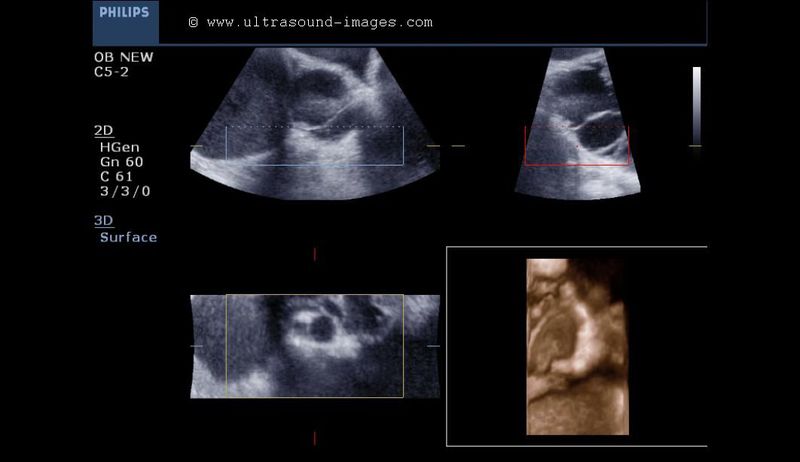
B- mode transabdominal ultrasound images showing huge, multiseptate cyst with thick, viscid fluid and thick walled septae posterior to the urinary bladder.Color Doppler image showing thick septae within the huge cyst, with multiple vessels within the septae.3D ultrasound image (right- orange image)- showing internal details of the cyst. Note the thick septae. Final diagnosis based on these images-huge mucinous cystadenoma or mucinous cystadenocarcinoma of the ovary (this cystic ovarian mass measures more than 15 x 10 cms.). Again the large size of the lesion making it difficult to determine the side of origin of this cyst.
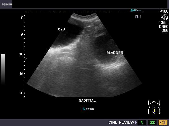
Cystadenocarcinoma
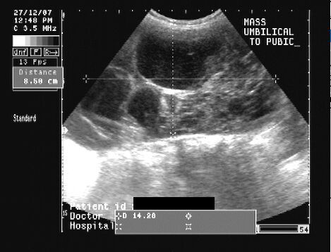
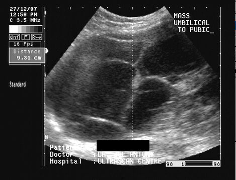

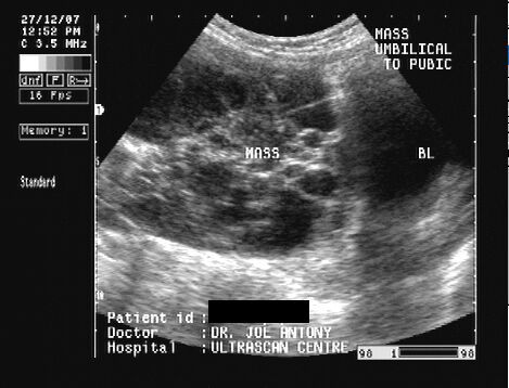
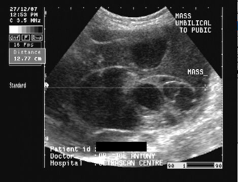
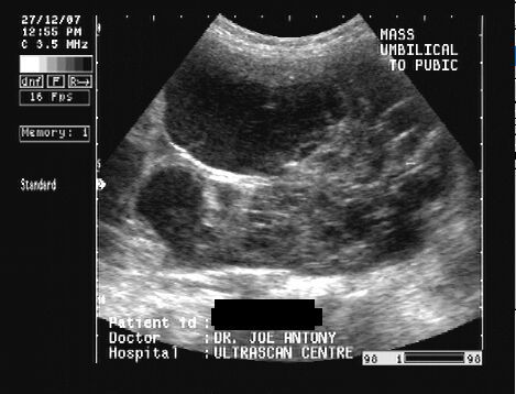
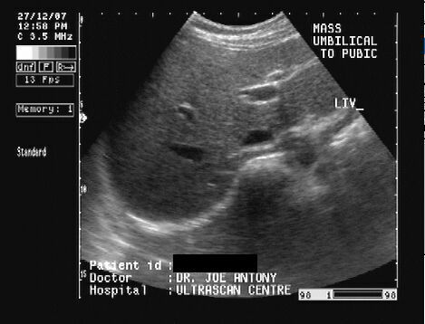


This middle aged female patient presented with abdominal mass extending from pelvis to above the umbilical region. On sonography there was a complex mass having both solid and cystic components extending upwards from the pelvis. The mass measured about 14 x 9 x 10 cms. in size. There was no evidence of ascites. The liver, spleen and kidneys appeared normal. Multiple loculations with walled septae were seen in the tumour. The ovaries were not separately visualized. These ultrasound images suggest cystadenocarcinoma of the ovary; either mucinous or serous. Images taken using a Pie Scanner 100 Falco, by Dr. Joe Antony, MD, Cochin, India.
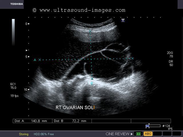
Calcific masses of the ovary
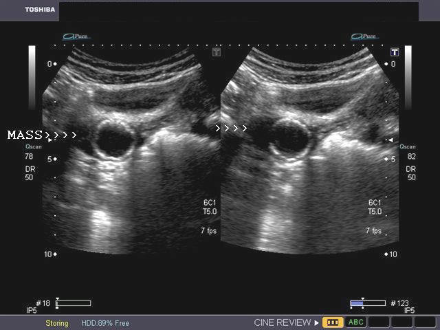

The right ovary shows a calcified mass (rim calcification) measuring 2.2 cms. The contents of the lesion appear anechoic suggesting a cystic nature. There is no acoustic shadowing posterior to the mass. These ultrasound findings suggest calcified dermoid cyst of the right ovary. The other diagnostic possibility is that of ovarian fibroma with rim calcification. The left ovary appears normal. Ultrasound images courtesy of Dr. Gunjan Puri, MD, India.
References:
http://radiographics.rsna.org/content/20/5/1445.full.pdf+html(free article and images)
http://radiographics.rsna.org/content/9/5/959.full.pdf+html (free article and images)
© 2019, Joe Antony, MD All images and content in this site are copyrighted. www.ultrasound-images.com When visiting this site, we may use cookies to improve the performance of this site. Use of this site means you accept the use of cookies. Read our privacy policy at this page: https://www.ultrasound-images.com/privacy-policy/