
Ultrasound images of thyroid disease-2
Contents of this page
- Hemorrhagic cyst of Left lobe thyroid
- A typical case of parathyroid adenoma
- densely calcific colloid nodule
- multinodular goitre with embedded carotid artery
- normal thyroid variant
- lateral spread of multinodular goiter
- hashimoto-thyroiditis-pretracheal-nodes
- De-Quervains-thyroiditis-or-subacute-thyroiditis
- Papillary-carcinoma-in-thyroglossal-duct-cyst
- metastases-to-lymph-nodes
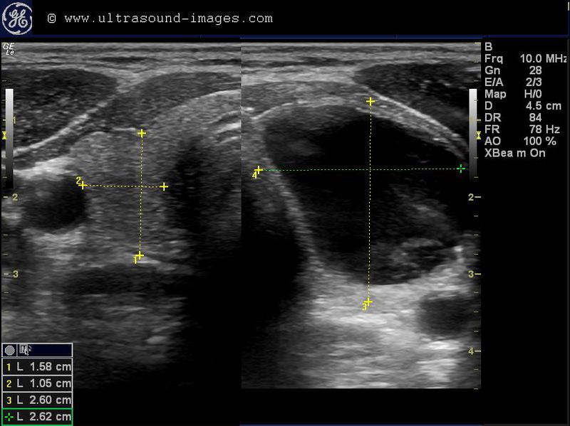
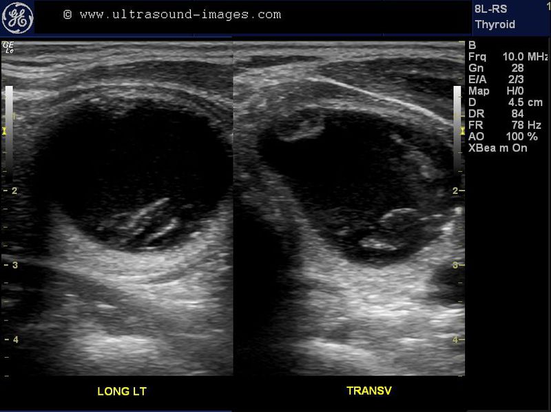

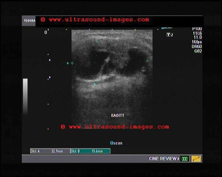
This is a classic case of hemorrhage in the left lobe of the thyroid resulting in a fluid and debris filled hemorrhagic cyst of the left lobe.
The ultrasound images of the thyroid cyst show the lesion filled with clear fluid with some septation towards the posterior half of the hrmorrhagic cyst. Fine debris is present in parts of the cystic mass which is seen to occupy most of the left lobe of thyroid.
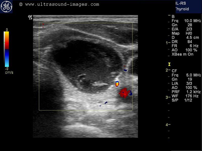
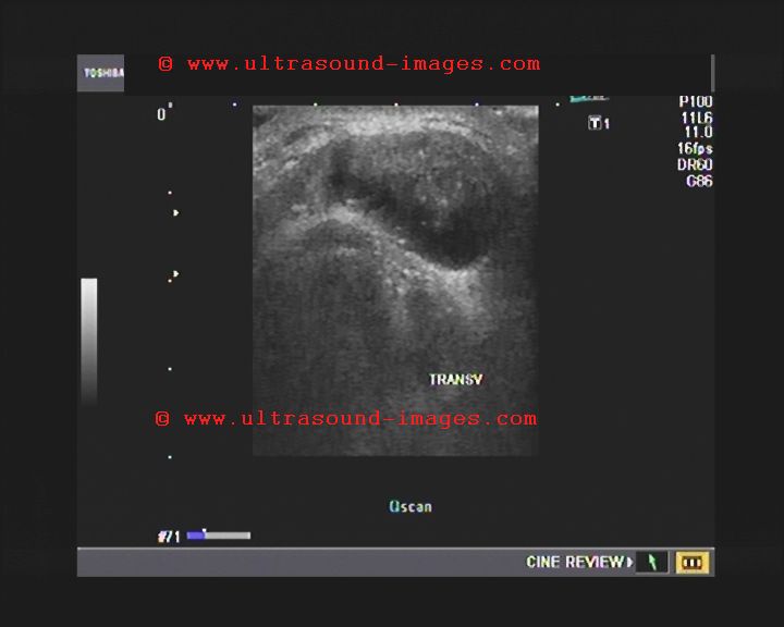
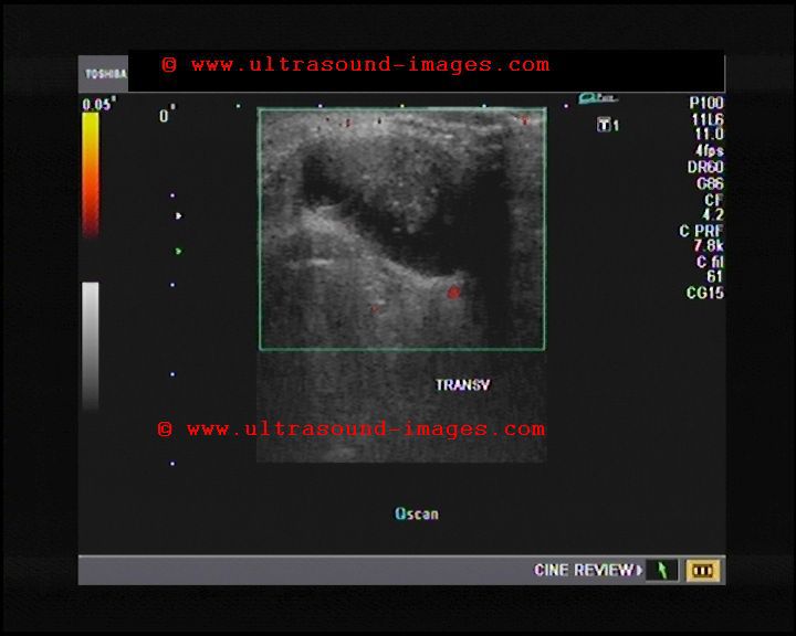
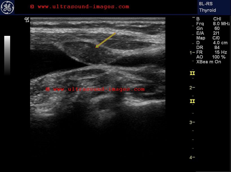
A typical case of parathyroid adenoma
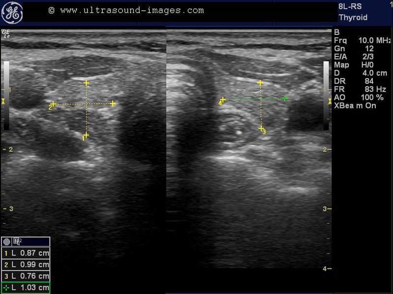
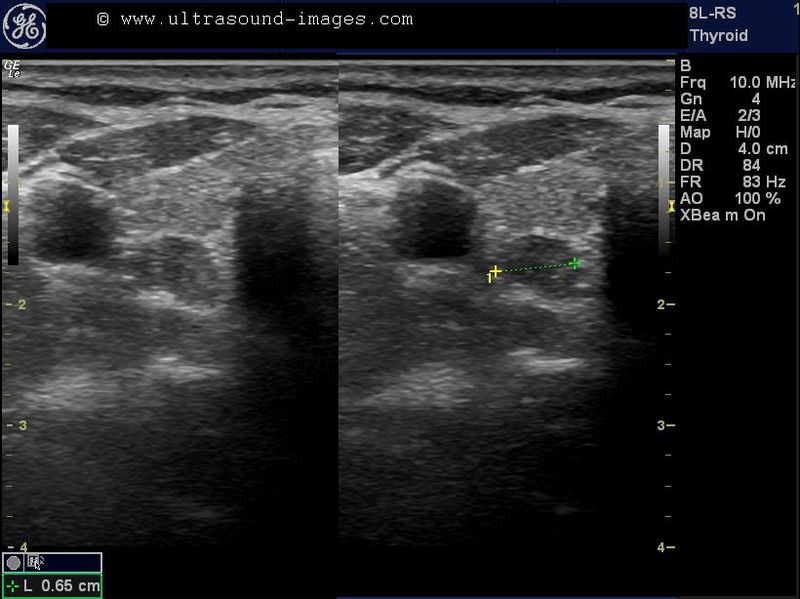
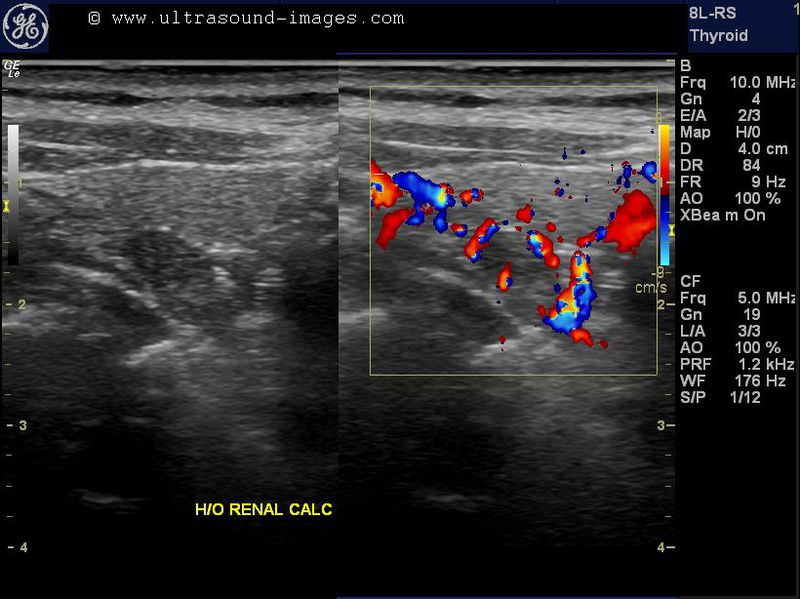

This is the case of a 57-year-old lady with history of multiple renal calculi. On ultrasound imaging of the thyroid, we found this hypoechoic nodular lesion posterior tothe right lobe of the thyroid towards the lower pole. This typical location, the well encapsulated and distinct nature of the nodule, with moderate internal and external vascularity of the lesion all point to the possibility of this mass being a parathyroid adenoma.
please see: http://www.ultrasound-images.com/thyroid.htm#Parathyroid%20adenoma
where we have discussed and presented for similar cases with stunning ultrasound images of parathyroid adenoma.
densely calcific colloid nodule


This young adult male patient complained of discomfort in the left side of the neck. Sonography detected this colloid nodule in the upper half of the left lobe of the thyroid. Ultrasound images show a densely calcific colloid nodule of 2 x 1 cms. in the left lobe thyroid. Color Doppler ultrasound shows typical twinkling artefacts in this calcific colloid nodule of the thyroid. FNAC was advised and would show a classic gritty feel as the needle traverses this densely calcific lesion. Such thyroid lesions are highly likely to be benign due to the presence of macro-calcification and other characterisitic such as well defined margins of this calcific thyroid lesion.
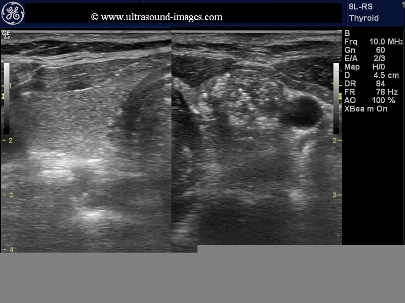
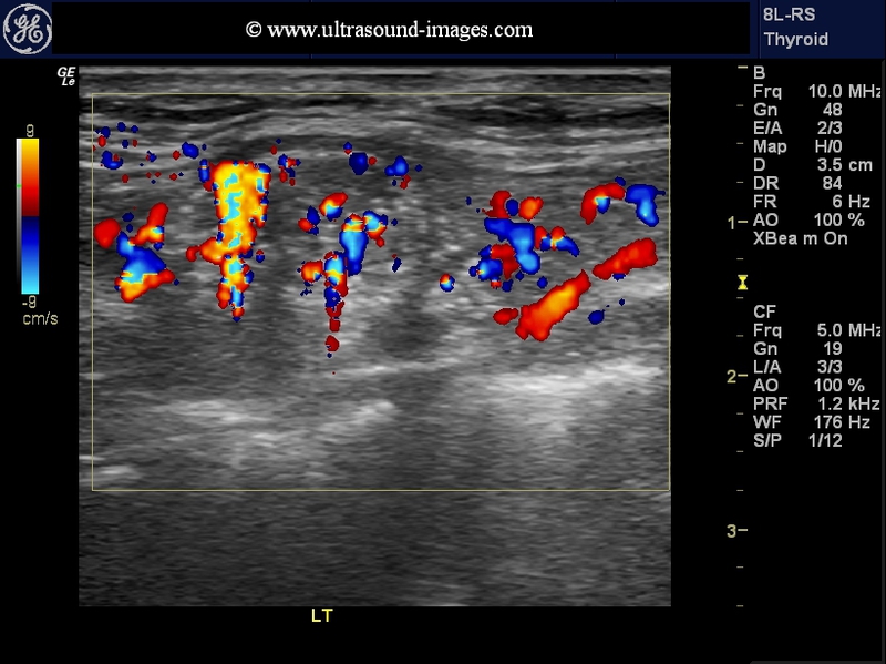
multinodular goitre with embedded carotid artery
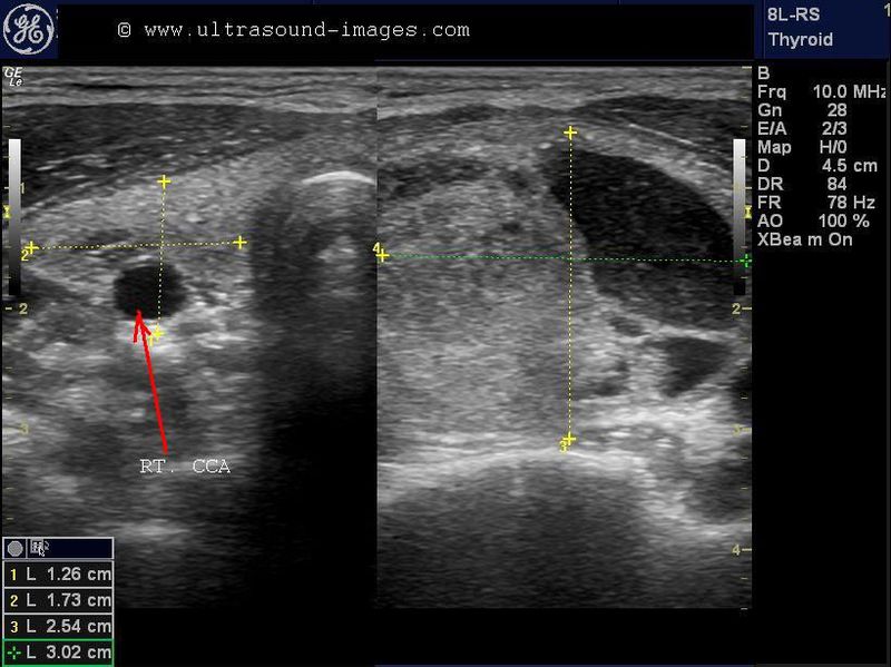
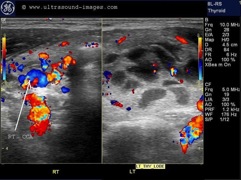
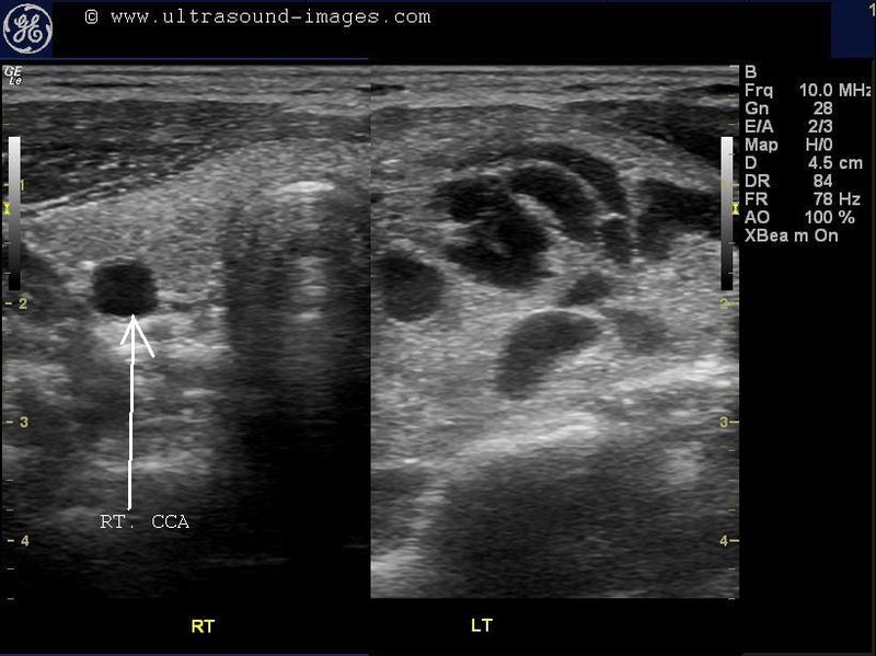
In this middle aged female patient, there is a large, primarily cystic goitre involving the left lobe. Multiple septae are present in the cystic lesion of the left lobe of thyroid. However, the interesting feature is the right lobe of thyroid, which actually covers the right common carotid artery,which is partially embedded within the right lobe of thyroid. This is a rare discovery and the first time I have actually seen this variant of this vessel. This finding itself of the right common carotid artery embedded in the right lobe of the thyroid may by itself be just an incidental finding with little significance.
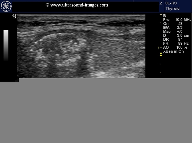
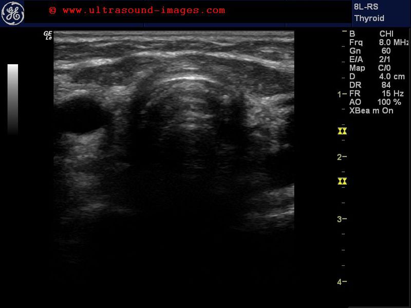
normal thyroid variant

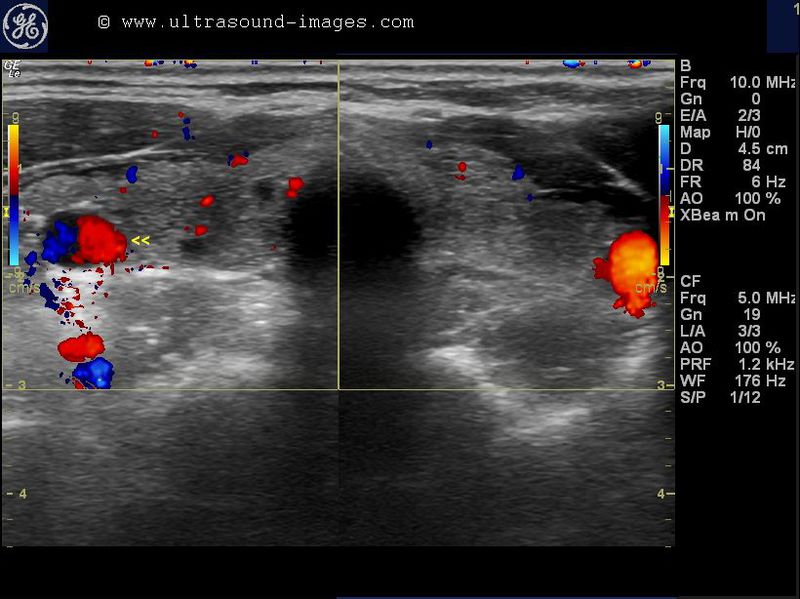
this thyroid shows a small aberration, or normal variant, wherein the right lobe of thyroid surrounds the right common carotid artery. The result is the right common carotid artery is embedded within the right lobe of thyroid. In addition, the left lobe shows a small colloid nodule.
lateral spread of multinodular goiter
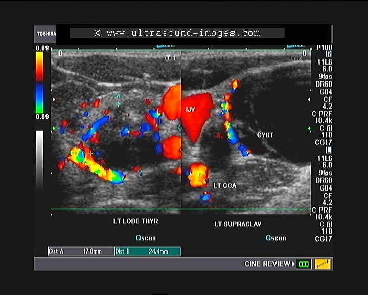

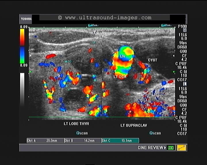
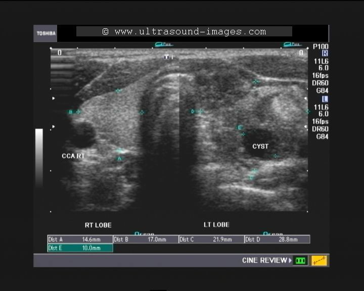
this male patient had a large supraclavicular mass on the left side. Ultrasound images show a multinodular goitre involving the left lobe of thyroid. The multinodular goitre appears to extend laterally into the left supraclavicular region (see ultrasound images above). Thus this appears to be a case of multinodular goitre with lateral extension of the goitre into the left supraclavicular region- a form of ectopic thyroid tissue with goitrous transformation. The left supraclavicular mass is composed of a large cystic lesion along with multiple solid nodues also present. the left common carotid artery and internal jugular vein appear to be trapped between the left lobe of thyroid and the ectopic thyroid tissue in the left supraclavicular region. The ectopic thyroid goitre appears to be very large and size and almost entirely cystic in nature. The cyst shows plenty of particulate matter, suggesting possible colloid material.
References:ectopic thyroid tissue in the left supraclavicular region
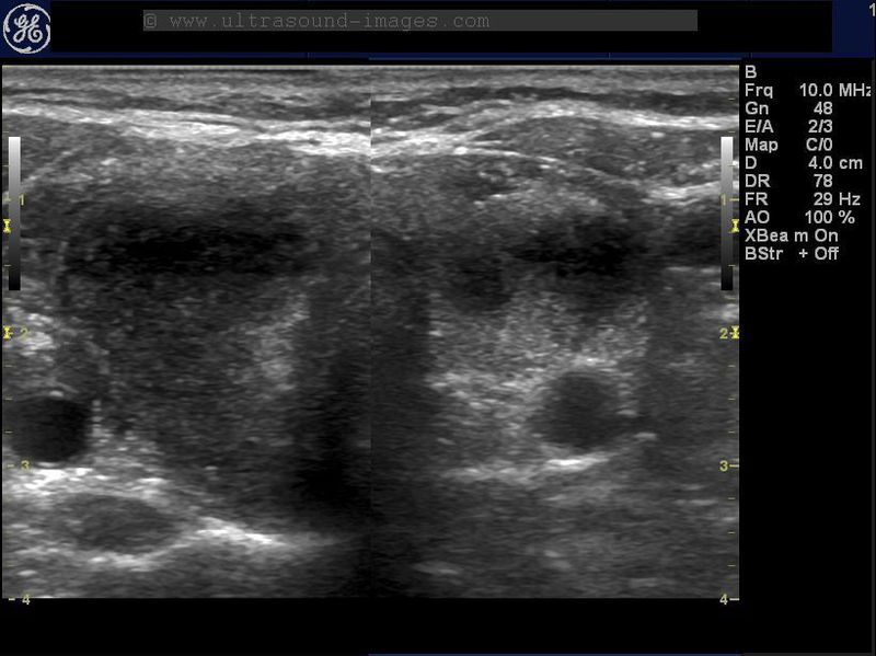
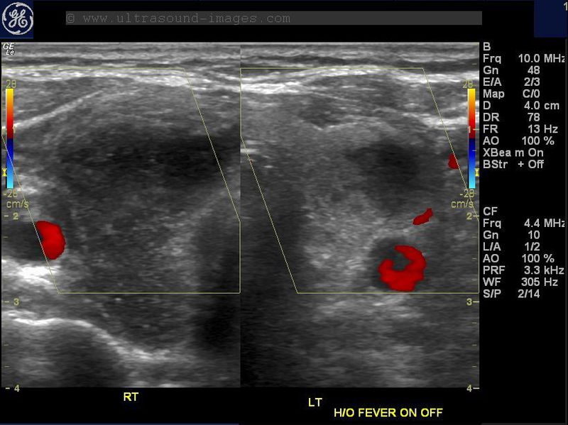
hashimoto-thyroiditis-pretracheal-node
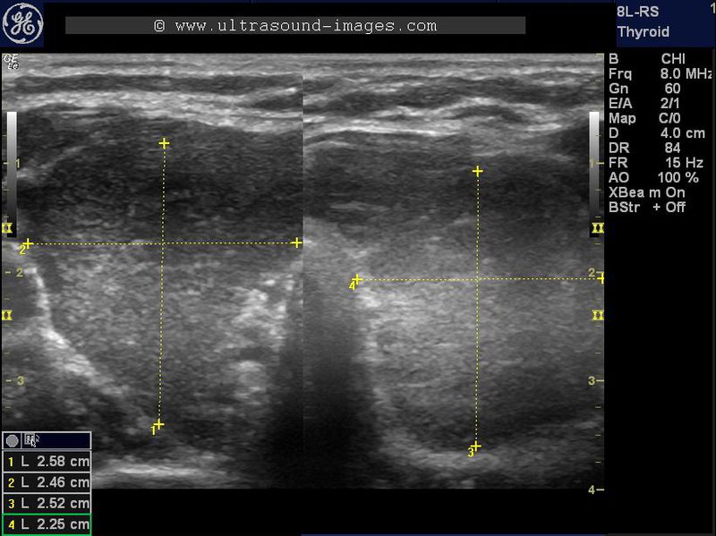
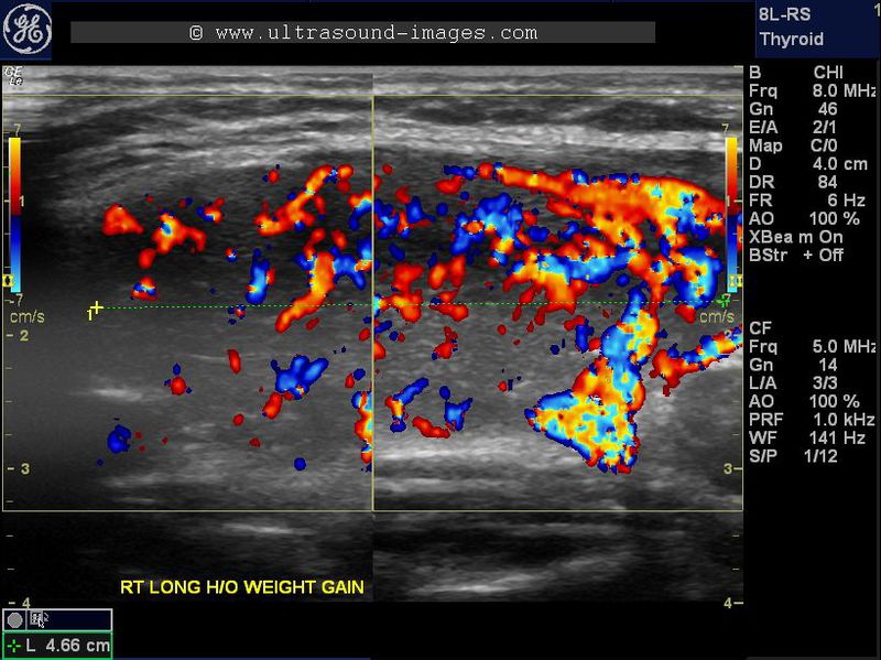
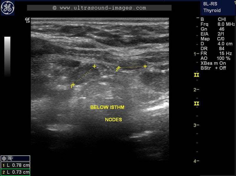
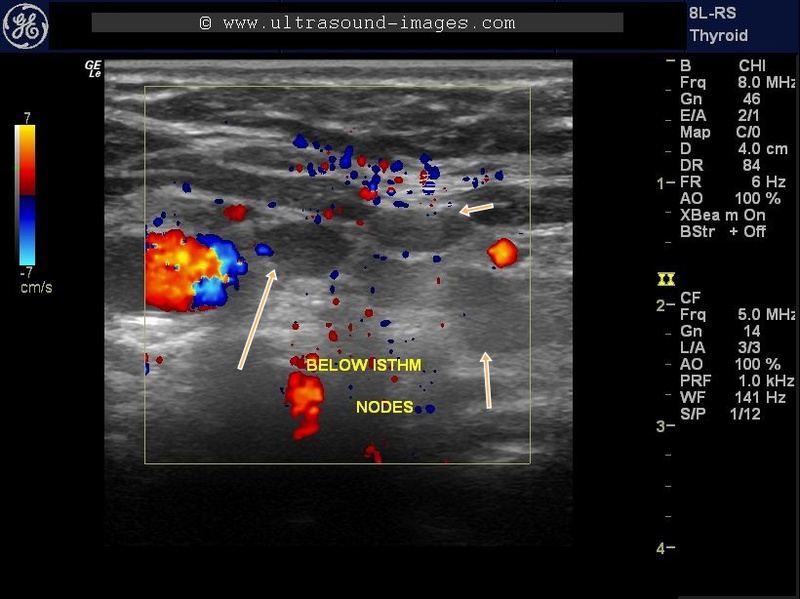
This female patient shows an enlarged, inhomogenous hypoechoic thyroid with 3 enlarged pretracheal lymph nodes suggesting Hashimoto's thyroiditis. The presence of enlarged pretracheal lymph nodes is a common finding in patients with Hashimoto's thyroiditis and further confirms this diagnosis and hence does not merit any alarm or the need for further investigation.
De-Quervains-thyroiditis-or-subacute-thyroiditis

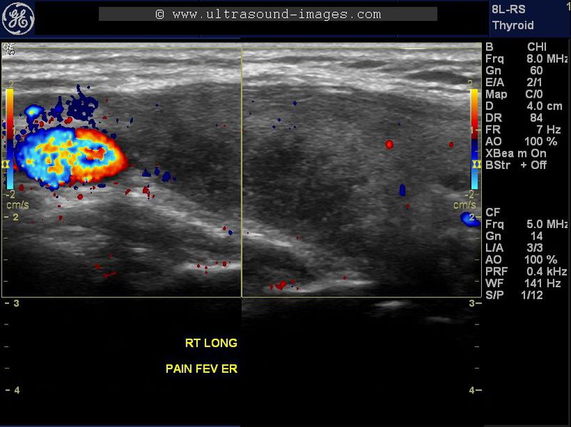
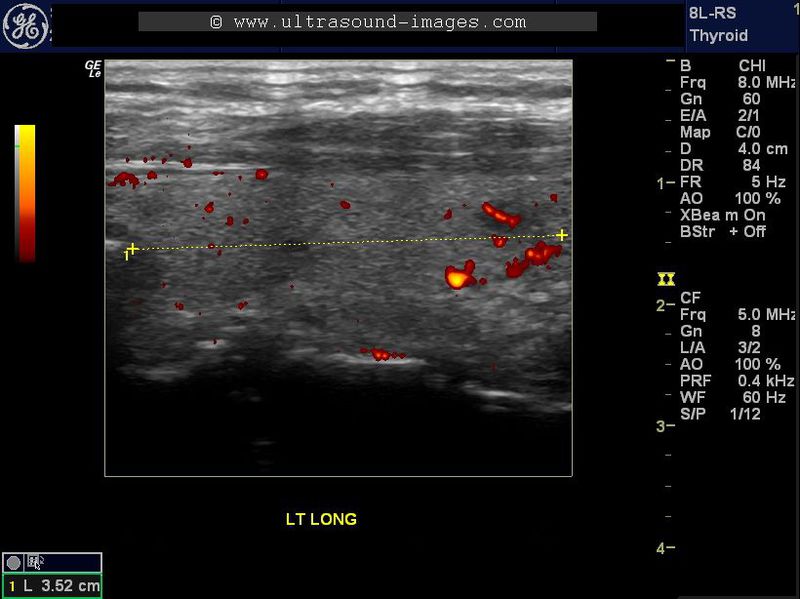
This elderly female patient presented with pain in the neck and intermittent fever. Ultrasound imaging of the thyroid gland shows markedly hypoechoic lesions in the right lobe. Color and Power Doppler ultrasound failed to show significant vascularity within the affected area (lesion) in the right lobe. The hypoechoic thyroid lesion shows irregular borders and is seen to infiltrate along the long axis of the affected lobe. Follow up ultrasound images show that the lesion has increased in size and also shows patchy involvement of the left lobe also. Again there is no vascularity in the affected hypoechoic lesions and marked tenderness is presented in these lesions on probe pressure. These sonographic findings are suggestive of De Quervain's thyroiditis, also known as subacute thyroiditis.
Papillary-carcinoma-in-thyroglossal-duct-cyst

A 40 yr old male patient presented with midline swelling just above the isthmus of thyroid. Ultrasound imaging showed a normal orthotopic thyroid with normal sized lobes and isthmus. However, just above then isthmus, just to the left of midline, a complex mass was seen on sonography. The larger cystic area of 3 cms. was seen to contain a papilliform growth bulging into the cystic area. This mural nodule showed irregular margins with multiple microcalcifications. No significant wascularity was see on color Doppler imaging. FNAC was repeated and confirmed papillary carcinoma within the thyrglossal duct cyst. Papillary carcinoma within a thyroglossal duct cyst is an extremely rare entity, but not unknown. Papillary carcinoma is the commonest form of malignancy within a thyroglossal duct cyst.
Reference: Ultrasound imaging of papillary carcinoma in thyroglossal duct cyst
metastases-to-lymph-nodes
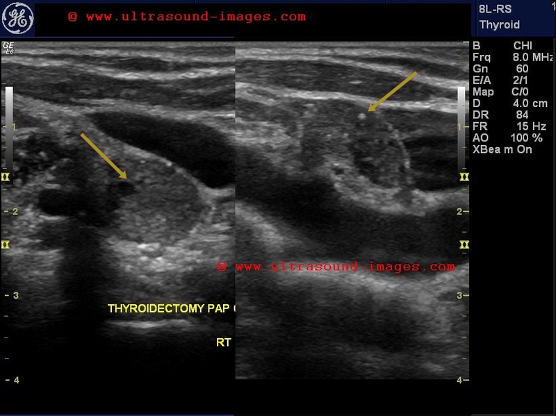
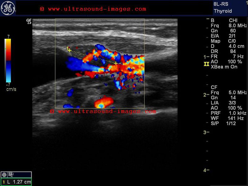
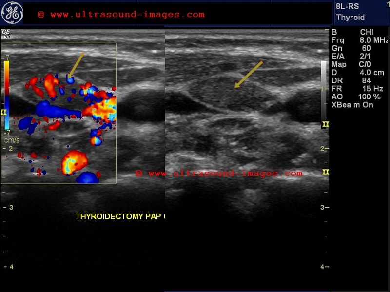
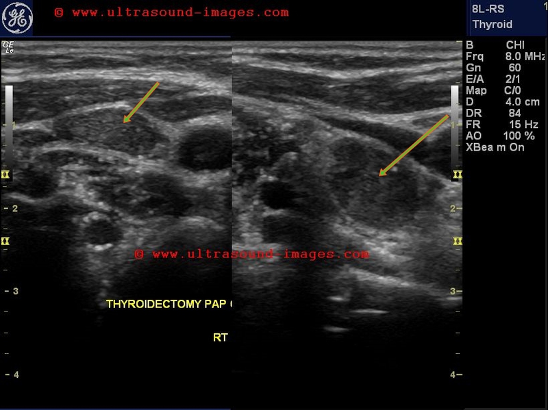
This patient underwent thyroidectomy for papillary carcinoma of the thyroid. Ultrasound and color Doppler images show multiple lymph nodes - Rt. cervical and supraclavicular which are- rounded in shape, show microcalcifications and marked peripheral and internal vascularity.
These are the halllmarks of metastases of the papillary carcinoma to the affected lymph nodes.
References: sonography of lymph node metastases
© 2019, Joe Antony, MD All images and content in this site are copyrighted. www.ultrasound-images.com When visiting this site, we may use cookies to improve the performance of this site. Use of this site means you accept the use of cookies. Read our privacy policy at this page: https://www.ultrasound-images.com/privacy-policy/