
acute scrotum
Contents of this page
- Idiopathic scrotal edema
- Torsion of testes
- Case-2: partial torsion of testis (synonym- incomplete torsion)
- Testicular trauma (rupture of testes or fracture testes)
- Case- 2: Ruptured left testicle
- Case-3: Rupture left testis with hematoma
- Ischemic orchitis
- Torsion of appendix testes with hydrocele
- Case-2 : Torsion of testicular appendage/ appendix testis
- Epididymal abscess (bilateral abscess of epididymis)
- Inguinal hernia
- Strangulated inguinal hernia
- Funiculitis or corditis
- Ruptured-testis-case-4
Idiopathic scrotal edema
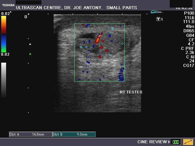
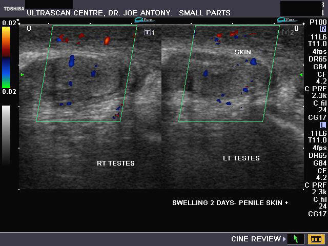
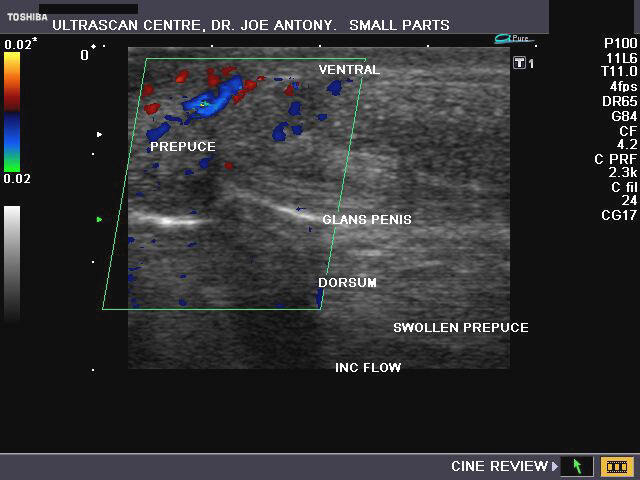
Sonography of the scrotum was done in this child who had acute swelling of the scrotum and penis. Ultrasound images reveal 1) thickening (11mm.) of the scrotal and penile skin and subcutaneous tissue and 2) marked hyperemia of the scrotum as well as the prepuce. The testes show normal appearance on sonography. These ultrasound and color doppler images are diagnostic of idiopathic scrotal edema. Images taken using a Nemio XG color doppler machine, by Joe Antony, MD.
Reference: http://www.duj.com/Article/Thomas.html
Torsion of testes
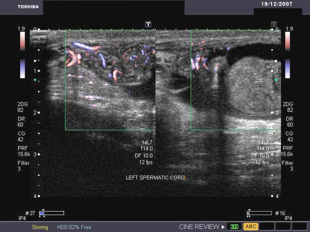
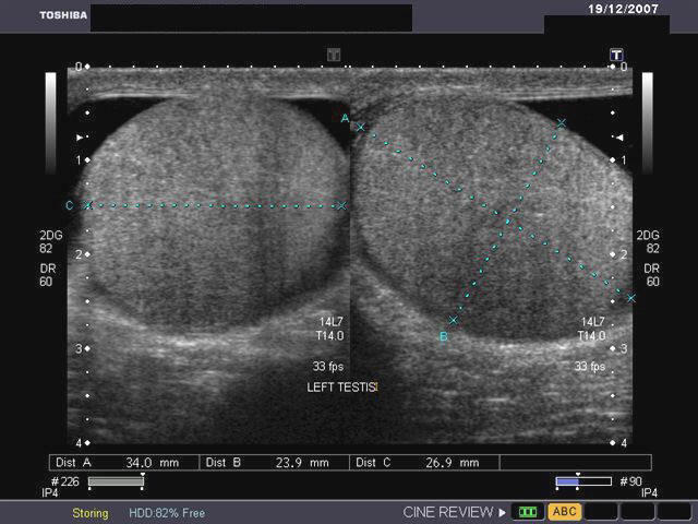

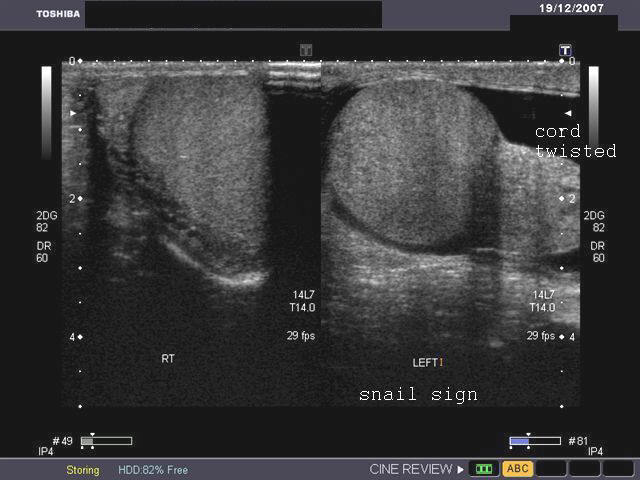

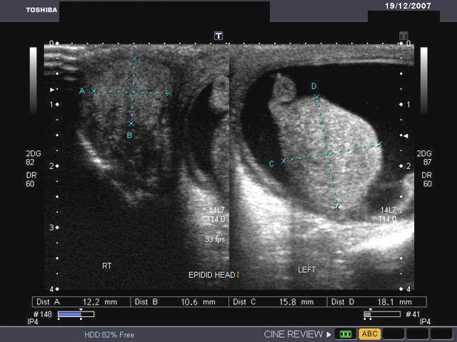

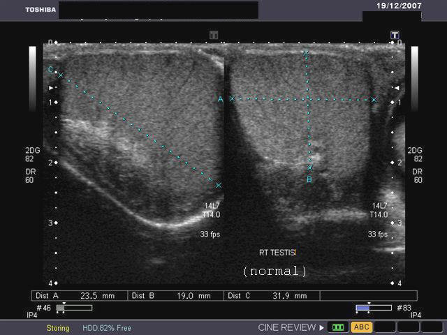
Ultrasound images showing torsed left testes. Testicular torsion occurs most commonly due to long spermatic cord. There are 2 types of torsion: extravaginal (usually in newborns) and intravaginal torsion (usually at puberty). In the ultrasound images above, the left testes appears swollen and mildly hypoechoic. A small left hydrocele is also present. On color doppler imaging, the left testes shows absence of blood flow. The "snail sign" is also seen in the left testes, caused by swollen testes (shell of snail) and twisted, bulky spermatic cord (body and head of snail). These ultrasound images suggest torsion of the left testes. The affected testes was removed and found to be gangrenous. Images taken using a Toshiba Xario color doppler and ultrasound machine, courtesy of Dr. Gunjan Puri, Surat, India.
Reference:
Case-2: partial torsion of testis (synonym- incomplete torsion)

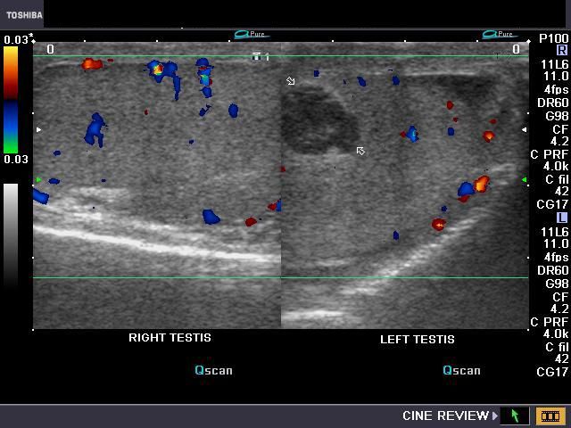
This young adult male patient had a history of left testicular pain of acute onset. Color Doppler ultrasound images of teh scrotum.It is obvious that the Color Doppler image of the left testis shows diminished vascularity as compared to the normal right testis. The most common cause of such an appearance is a partial torsion of the left testis. In this case the twisting of the left testis on the spermatic cord is less than 360 degrees (partially torsed testis). Current medical literature and studies have it that torsion or twisting of the testis is not an all or none phenomenon, with torsion being classified as both complete and incomplete or partial. The color Doppler / ultrasound appearances of such a case vary depending on the duration and degree of torsion. In this case (incomplete torsion), the arterial supply to the left testis is only partially cut off. Above case and images are courtesy of Dr. Vikas Shukla, MD, India).
References:http://www.jultrasoundmed.org/cgi/content/full/27/11/1629 (excellent article and images)- free.
Also: http://emedicine.medscape.com/article/778086-overview (free article and images).
Testicular trauma (rupture of testes or fracture testes)


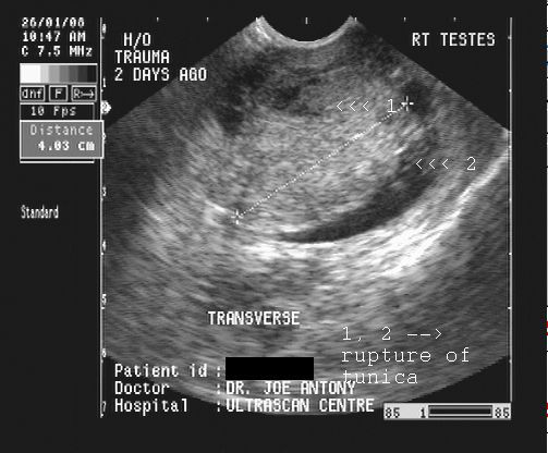
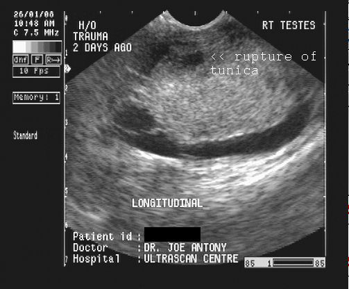
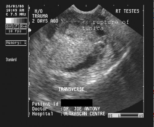

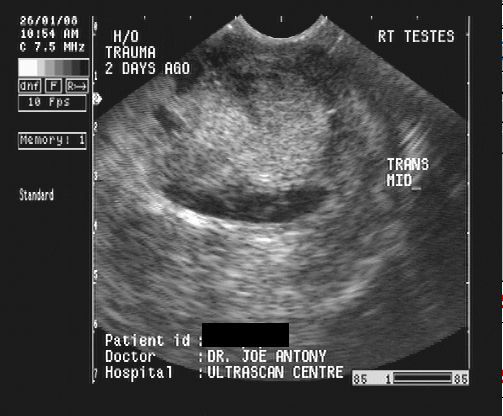
This 34 yr. old male patient had a severe blunt injury to the right scrotum 2 days prior to sonography. Ultrasound images of the scrotum reveal
1) a small hypoechoic fluid collection (hematocele)l in the right tunica vaginalis sac. .
2) multiple septations within the Rt. tunica vaginalis sac
3) rupture of the tunica albuginea of the right testes
4) hypoechoic areas within the right testes s/o contusions
5) thickening of the scrotal wall (edema).
6) edematous and swollen right testes
The left testes appears normal. These ultrasound images suggest contusion and rutpture of the right testes with hematocele. These sonographic images were taken using a Pie scanner 100 Falco ultrasound machine, by Dr. Joe Antony, MD, India.
Reference:
1) http://www.emedicine.com/MED/topic2859.htm (free article and images)
2) http://www.emedicine.com/radio/topic682.htm
Case- 2: Ruptured left testicle

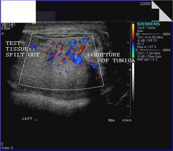
The above ultrasound images show traumatic rupture of the left testicle with herniation of the testicular tissue through the defect in the tunica albuginea. There is also an organized hematoma surrounding the left testis. Color Doppler image (bottom row- left) shows evidence of vascularity in the ruptured/ herniated tissue, suggesting viable testicular parenchyma. Ultrasound images are courtesy of Shlomo Gobi, Israel.
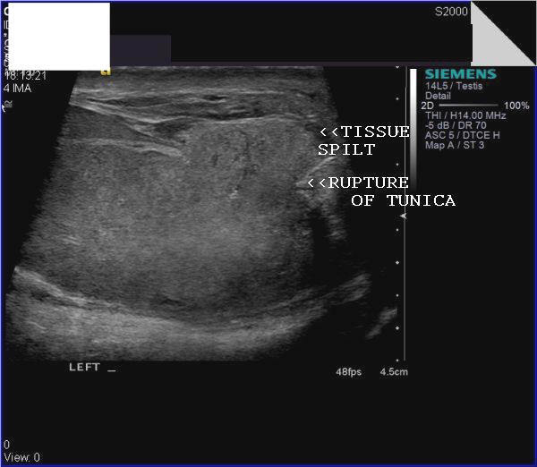
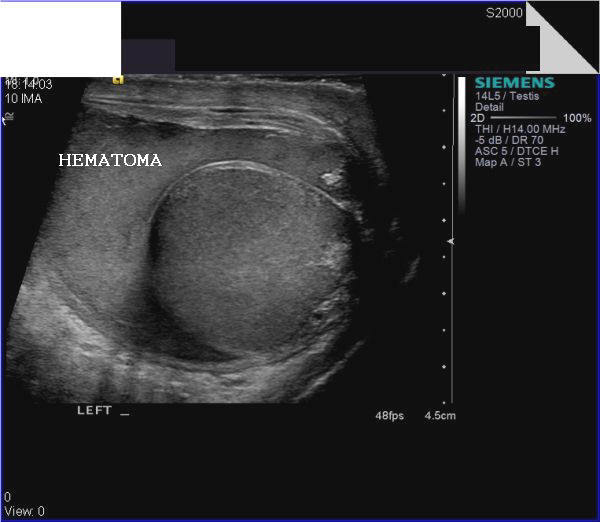
Case-3: Rupture left testis with hematoma
This adult male patient has a history of blunt trauma to the left scrotum. The result (see ultrasound images above)- left hematocele with intratesticular hematoma (large hypoechoic lesion (arrows) inside the the left testis) with rupture of tunica albuginea and herniation of testicular parenchyma (tissue).
For ultrasound and color Doppler video clips of this case visit:
http://ultrasound-videos.blogspot.com/2010/12/blunt-trauma-to-testes-with-rupture.html
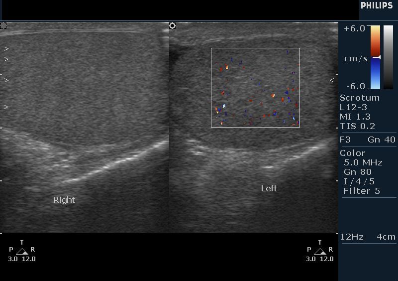
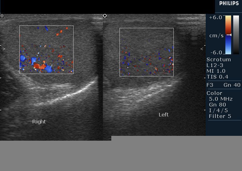
Ischemic orchitis
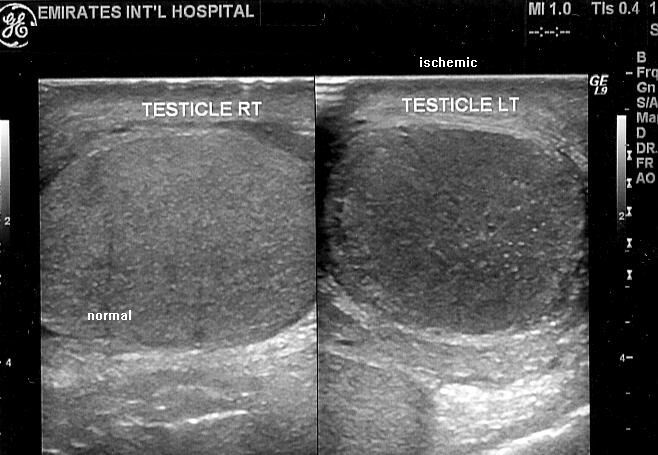
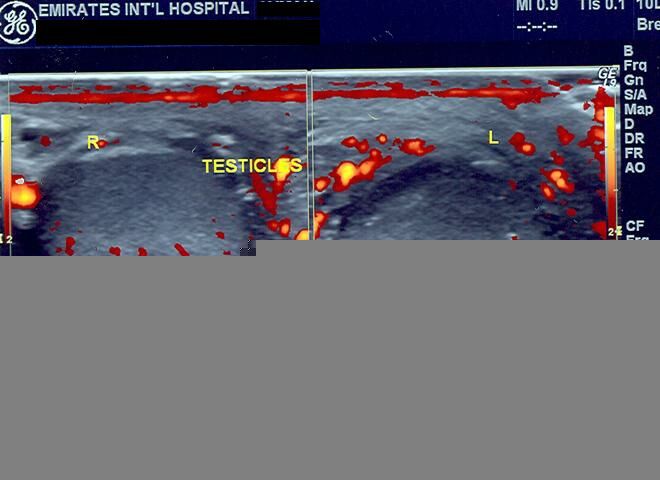
This young male patient underwent hernia repair, following which he complained of pain and swelling of left scrotum. Sonography of the scrotum shows: 1) hypoechoic left testes 2) swelling of the left testes 3) absence of vascularity in left testes. These ultrasound and color doppler images suggest left ischemic orchitis. Ischemic orchitis is a known complication of herniorraphy. Ultrasound images courtesy of Dr. Ravi Kadasne, UAE.
Reference:
1)http://www.tabexperts.com/TesticularAtrophy.htm
2)http://en.wikipedia.org/wiki/Orchitis
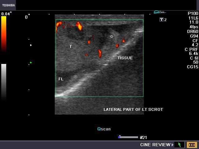
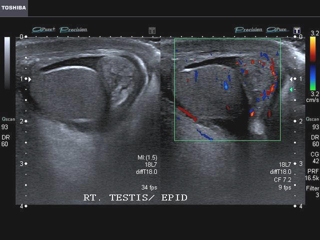
Torsion of appendix testes with hydrocele
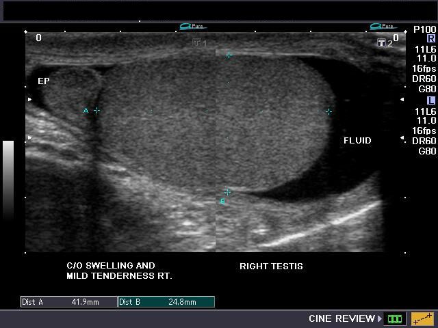
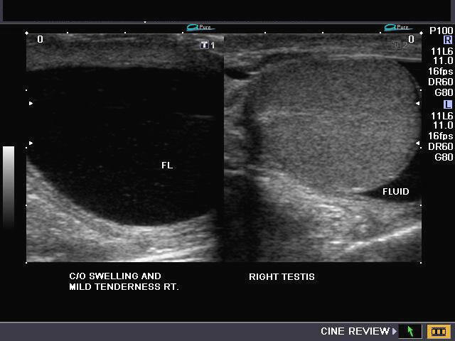
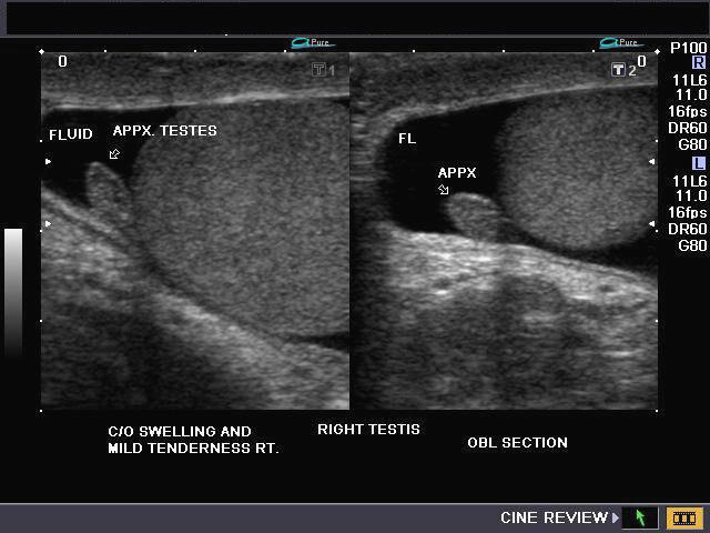


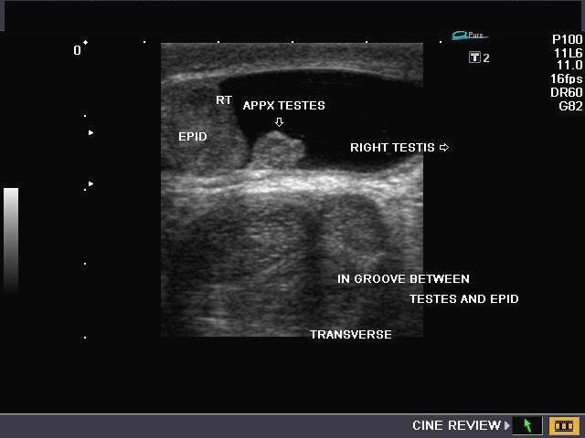
This patient, a young adolescent male had selling, pain and mild tenderness of the right scrotum. High frequency ultrasound probe shows a large right hydrocele with clear fluid content surrounding the the right testes. The right testes and epididymis are well outlined by the anechoic fluid. Ultrasound images also show the appendix testes occupying the groove between the testes and epididymis on the upper pole. The testicular appendix shows echogenic rim with hypoechoic central part and no vascularity on color Doppler imaging (topmost image). These ultrasound findings suggest torsion of the right testes with hydrocele.
Reference:
1)http://emedicine.medscape.com/article/778170-overview
2) http://www.jultrasoundmed.org/cgi/reprint/24/1/87.pdf
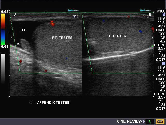
Case-2 : Torsion of testicular appendage/ appendix testis


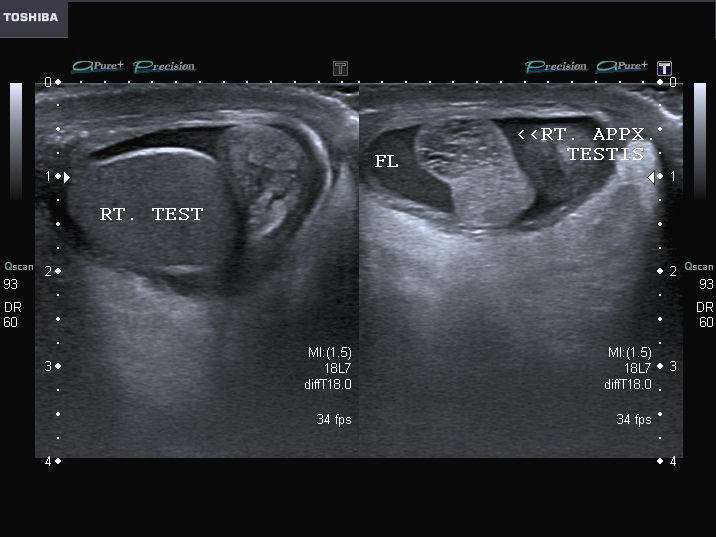
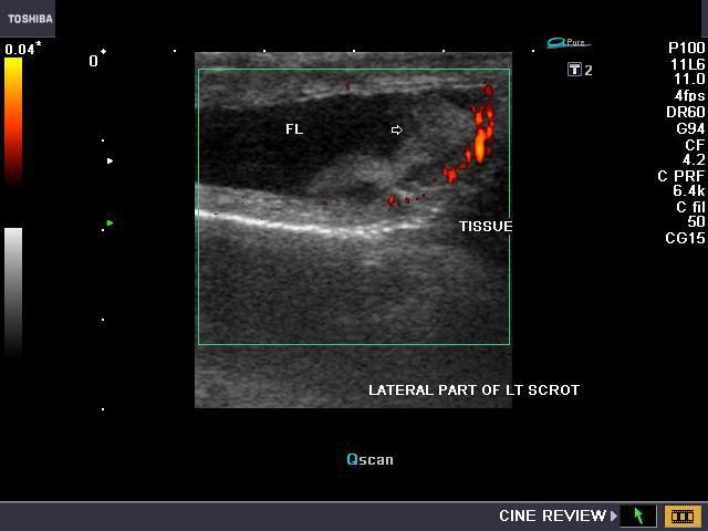
The above images show the typical features of torsion of the appendix testis in a young child. The boy had severe pain in the right scrotum with mild swelling. The ultrasound and color Doppler study of the right scrotum show- large, swollen testicular appendage (appendix testis- Right) with small amount of fluid in the tunica vaginalis sac (hydrocele). Color Doppler images show absent flow/ vascularity in the appendix testis with marked increase of flow in the surrounding region, especially the head of the right epididymis. Note also the globular/ round shape of the appendix testis which is so typical of torsed appendix testis. Above ultrasound images are courtesy of Ravi Kadasne, MD, UAE. (FL= fluid; APPX. = appendix testis; EPI= epididymis). (The main differential diagnosis in this case is possible inguinal hernia. Clinical examination would help rule out this pathology).
Epididymal abscess (bilateral abscess of epididymis)
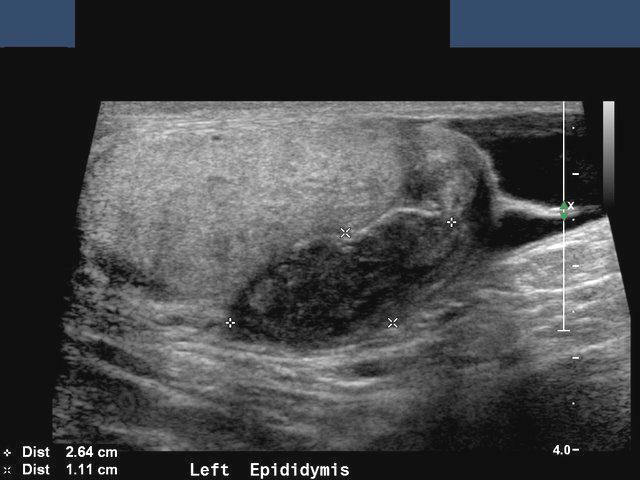

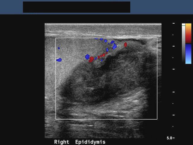
Hypoechoic, swollen, tender epididymis.
This male patient complained of pain and swelling of the scrotum. Sonography of the scrotum showed bilateral swollen epididymis. Ultrasound images of the epididymis showed them to be markedly hypoechoic (hyperemia with edema) and had increased vascularity (on Color Doppler imaging). The affected regions are mainly the body and tail of these structures. The testes also showed multiple small hypoechoic lesions (image on bottom right- arrowed), possibly due to obstructed seminiferous tubules (sperm granuloma formation). These ultrasound images suggest bilateral epididymal inflammation (epididymitis) with abscess formation. All images are courtesy of Dr. Ravi Kadasne, UAE. Images were taken using a Philips IU-22 ultrasound system.
References:
http://www.ajronline.org/cgi/reprint/164/2/376.pdf(free article and images)
http://emedicine.medscape.com/article/777181-overview(free article and images).
Inguinal hernia
The above ultrasound images show herniated bowel showing peristalsis within the right scrotal sac. (T= right testis; H= herniated bowel). This is the typical finding in inguinal hernia. Inguinal hernias may be direct or indirect depending on the relation of the hernial sac with the inferior epigastric artery. Those that are medial to the inferior epigastric artery are direct while those lateral to the artery are said to be indirect. Ultrasound images are courtesy of Dr. Ravi Kadasne, UAE.
Reference:http://www.medscape.com/viewarticle/527887_8
Strangulated inguinal hernia
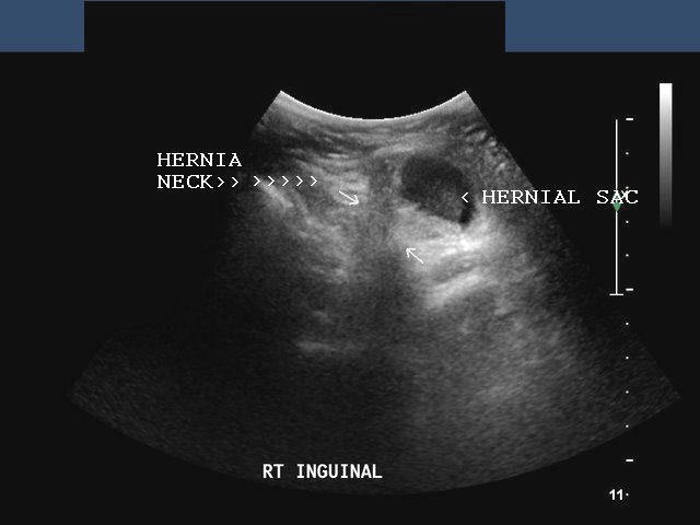
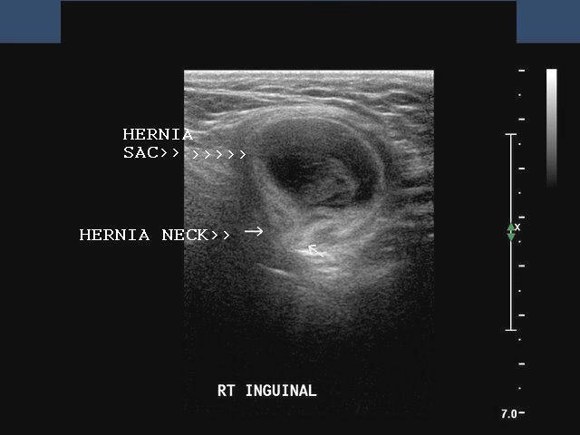
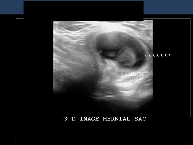
This male patient presented with severe pain and tenderness in the right inguinal region. On examination, there was a tender mass in the right inguinal area. Sonography of the inguinal mass showed a hypoechoic collection (the hernia sac with echogenic tissue- bowel loop) with a very neck. Color Doppler imaging of the mass showed absent flow in the structures within the sac (not shown here). These ultrasound images suggest strangulated right inguinal hernia. This was confirmed on surgery. The strangulated inguinal hernia contained an ischemic and edematous bowel loop. The image in bottom row shows 3-D visualization of the hernia. Images courtesy of Dr. Ravi Kadasne, UAE.
Funiculitis or corditis
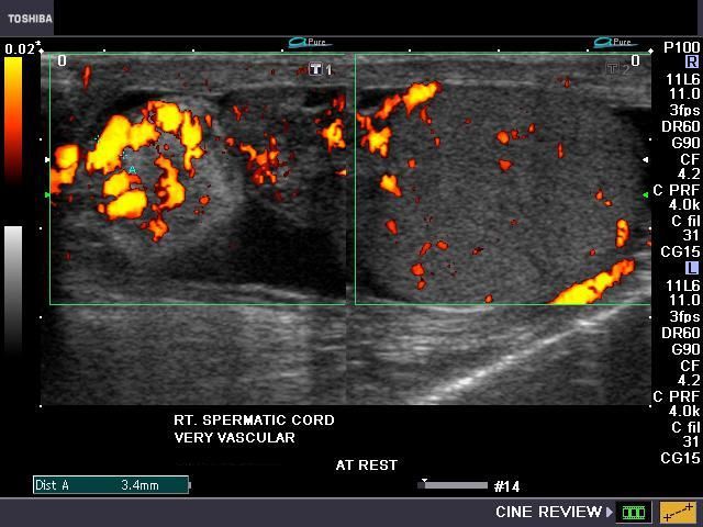

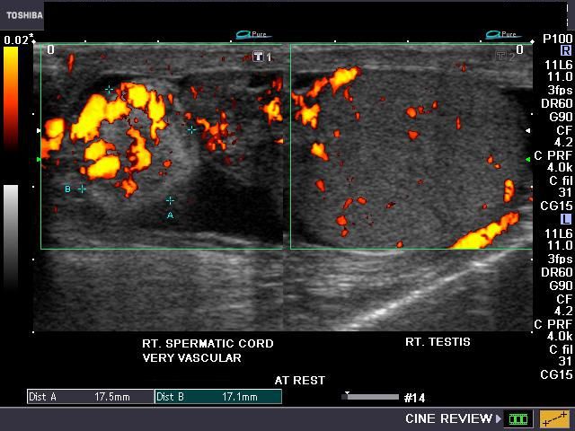
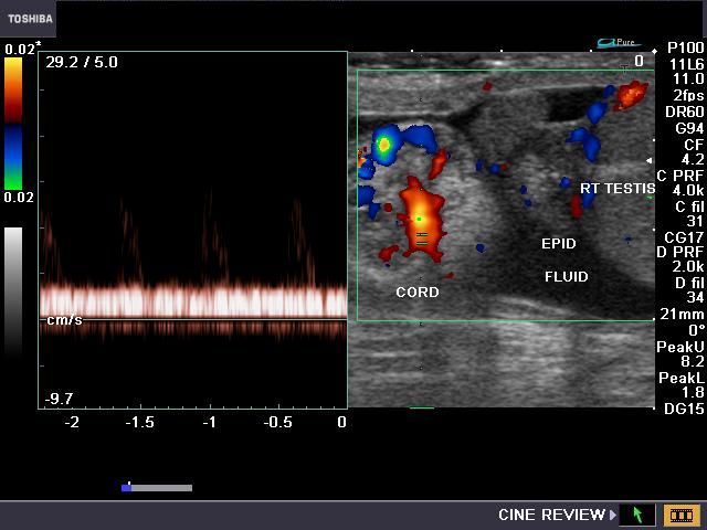
This middle aged male patient presented with scrotal pain and nodular mass on the right side. Grey scale B-mode ultrasound image showed a mildly hyperechoic oval, inhomogenous mass in the region of the right spermatic cord. The mass was located just above the right epididymis and showed marked vascularity on Power and Color Doppler imaging. To rule out a varicocele, I made the patient perform a Valsalva maneuver. However there was no difference on breath holding. The right epididymis also showed mild hypervascularity. Spectral Doppler trace showed a typical venous flow pattern in this mass. There is also evidence of a small right hydrocele. The final diagnosis based on these ultrasound and color Doppler images was funiculitis (inflammation of the spermatic cord) or corditis.
Reference:http://www.aafp.org/afp/980215ap/junnila.html
torsion appendix of epididymis
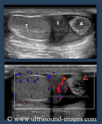
This 6 year old male child complained of severe right scrotal pain and tenderness. Ultrasound images show normal rights testis and epididymis. However, an oval structure attached to the right epididymis (the apppendix of the right epididymis) appears swollen, and shows increased echogenicity around its rim with central hypoechoic area. In addition, the colour Doppler ultrasound image shows lack of vascularity of the structure- namely the appendix of the epididymis. a small right hydrocele is also present These ultrasound findings suggest a diagnosis of torsion of the apppendix of the right epididymis. These images of torsion of apppendix of epididymis are courtesy of Ravi Kadasne, MD, UAE.
Key: A= appendix epididymis
E= epididymis
T= testis
Ruptured-testis-case-4
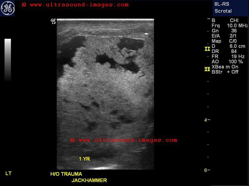
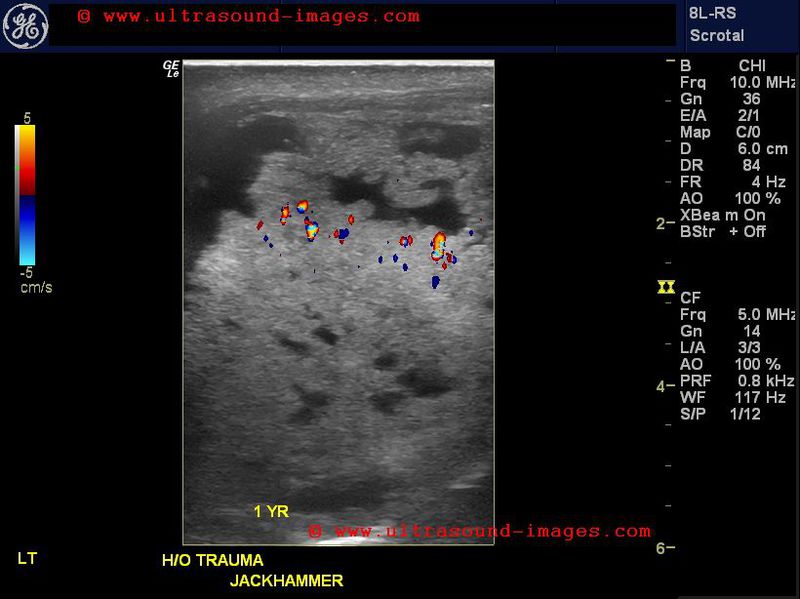
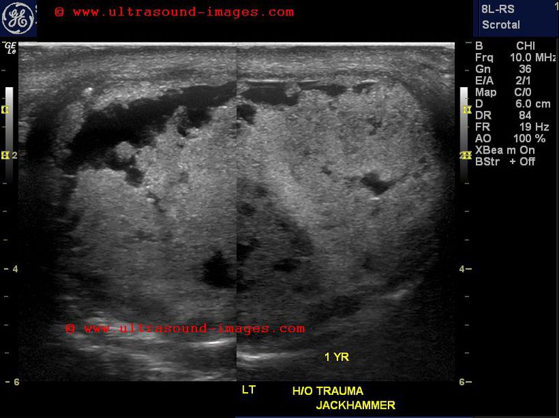
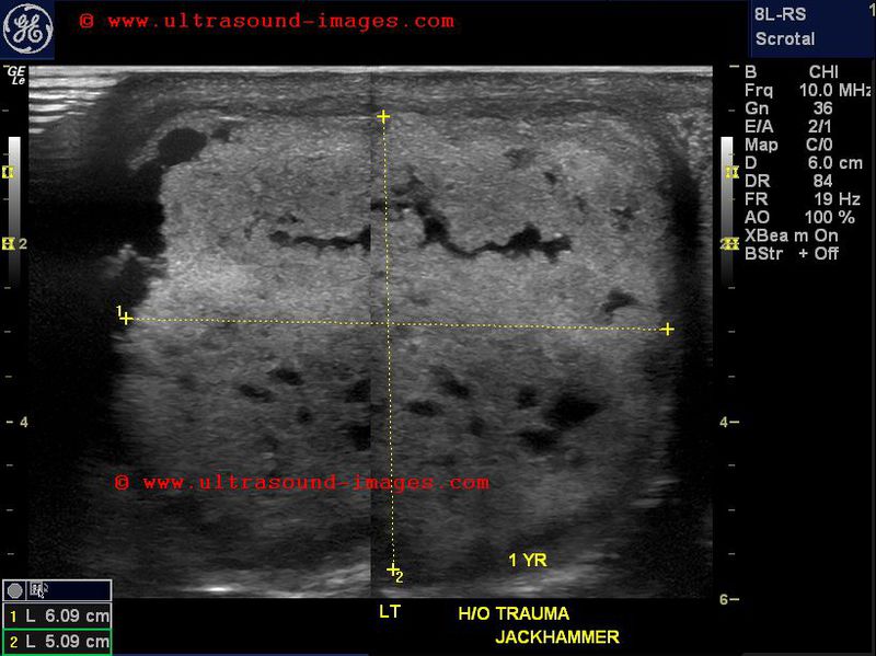
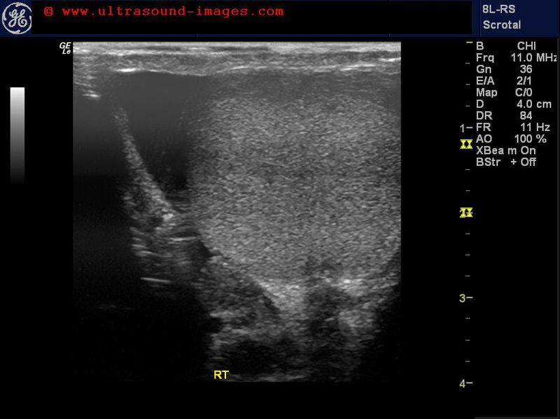
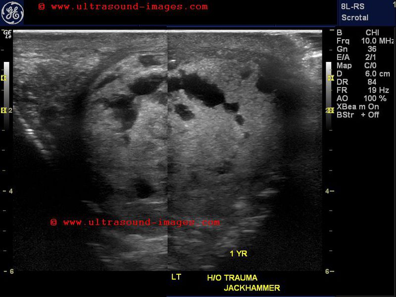
This middle aged male had traumatic injury to the left testis from a jackhammer more than a year ago. This old severe injury to the testis is seen as multiple areas of rupture of the tunica albuginea of the left testis with multiple areas of necrosis within the testis. The normal right testis is shown for comparison. Color Doppler ultrasound reveals lack of flow in the injured testis suggesting that this left testis is non viable and cannot be saved. The large size of the left testis further confirms the severity of the injury with resultant fluid collection and edema of the affected testis.
© 2019, Joe Antony, MD All images and content in this site are copyrighted. www.ultrasound-images.com When visiting this site, we may use cookies to improve the performance of this site. Use of this site means you accept the use of cookies. Read our privacy policy at this page: https://www.ultrasound-images.com/privacy-policy/