
Fetal Face and neck
Contents of this page
- Normal fetal lens
- cleft-lip
- Multiple facial anomalies
- Hypotelorism, proboscis, with Holoprosencephaly and overriding of aorta in fetus
- Cystic hygroma
- Fetus sucking thumb- 3D ultrasound images
- cleft lip- 3-D ultrasound images
- fetal cataract
- multiple cystic hygromas in neck and chest
- micrognathia with club foot
- fetal eyelashes
- 3-D alobar holoprosencephaly
- saddle-nose-with-cleft-lip-in-holoprosencephaly
- fetal-thyroid-goiter
Normal fetal lens

Download my E book for Amazon Kindle (download free Kindle reader for i Phone or Android)
Assorted ultrasound cases- by Joe Antony, MD
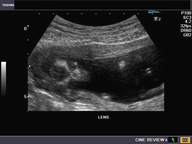
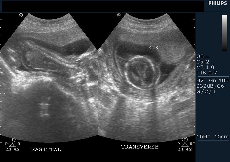
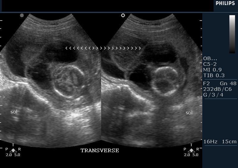
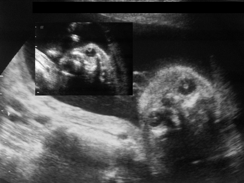
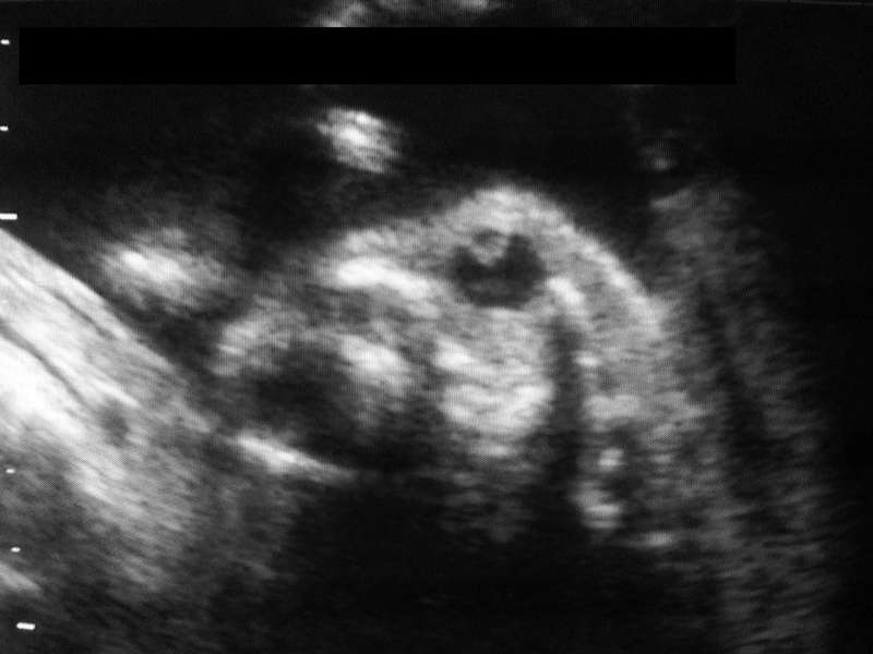
Ultrasound images of fetal eye
The above ultrasound images show a coronal section of the normal fetal face (at 14 weeks gestation), with the fetal eyeballs visualized. The fetal lens of the eyes are visualized as minute echogenic rings in the orbit. Real time sonography would show the fetal lens as moving with fetal eyeball movements. Observing the fetal lens is a sure way of confirming the presence of the eyeballs.
Another set of ultrasound images of the normal fetal lenses
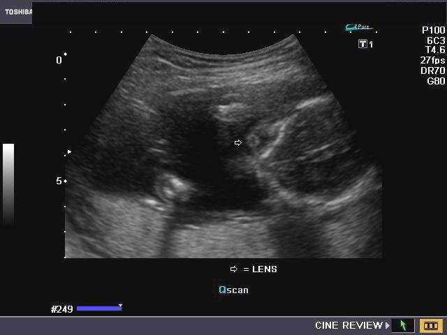
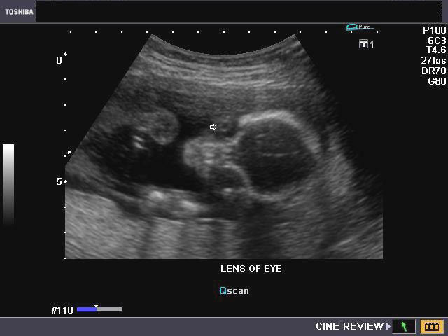
This fetus is 21 weeks gestational age. The presence of the lens rules out anopthalmia (absence of the eyeballs).
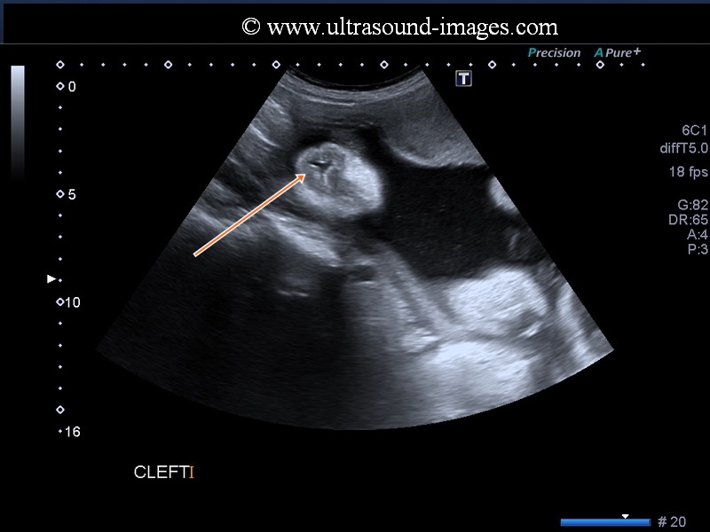
cleft-lip
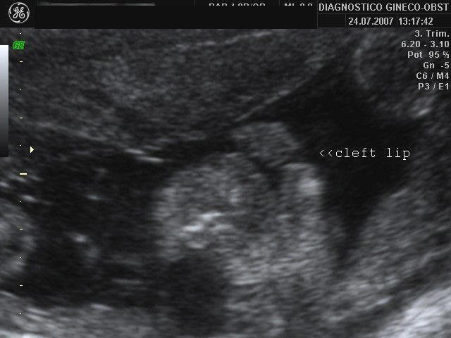
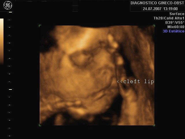
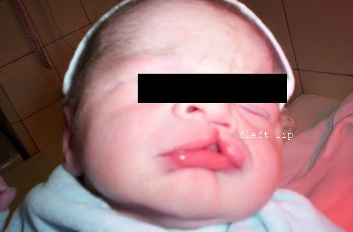
These images reveal isolated cleft lip anomaly. The first ultrasound image is a B-mode coronal section through the face. The cleft is seen well. The image on the right uses 3-D sonography reveals the cleft by surface rendering. The follow up (post-natal) snap of the baby confirms these findings. Images courtesy of Dr. Martin Horenstein, Argentina.
Multiple facial anomalies
Hypotelorism, proboscis, with Holoprosencephaly and overriding of aorta in fetus

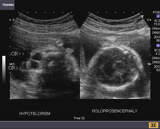
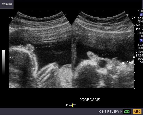
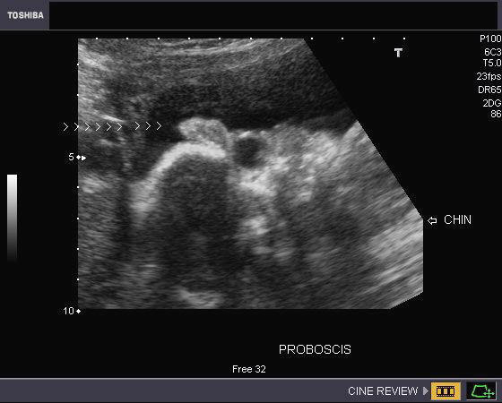

This late 2nd trimester fetus underwent routine obstetric sonography. Ultrasound imaging showed hypotelorism (both orbits are very close to each other), a small fleshy projection above the eyes in the region of the forehead (proboscis), holoprosencephaly (single fused monoventricle) and cardiac anomaly (over riding of aorta). The presence of hypotelorism and proboscis is called ethmocephaly. Holoprosencephaly, especially the severe form, (alobar) is often associated with multiple facial anomalies of this nature. The prognosis, in this fetus is very bleak. Trisomy-13 and Trisomy-18 are the common chromosomal disorders found in such cases. Ultrasound images are courtesy of Dr. Durr-e- Sabih, Pakistan. These images were taken using a Toshiba Nemio-30 Ultrasound system. (OR= Orbits)
Reference: http://emedicine.medscape.com/article/409265-overview (free article and images).
Google book reference on hypotelorism
Cystic hygroma
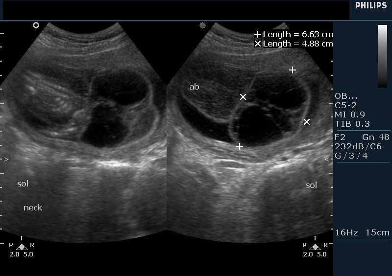
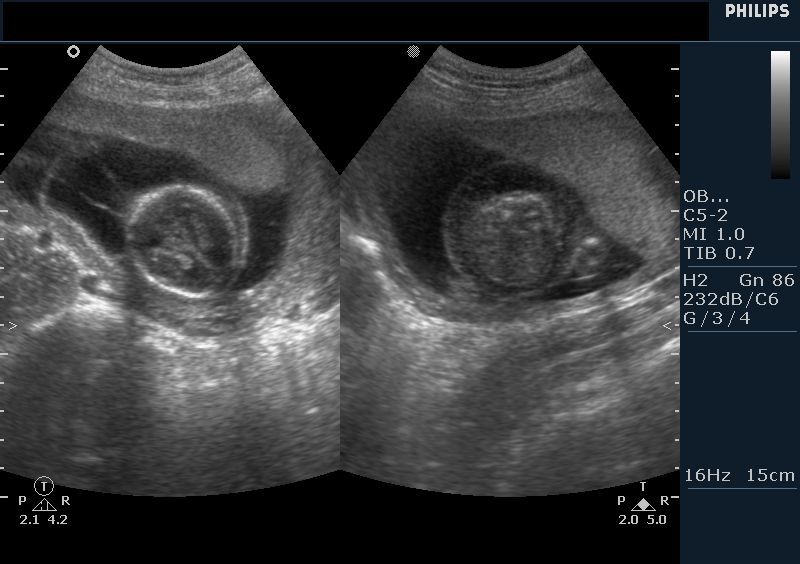
The above ultrasound images show a 16 week fetus with a septate cystic mass in the posterior and lateral aspect of the fetal neck. Color Doppler image shows that this mass is not the cord or part of it. The cystic tumor is almost 7 x 4 cms. in size. The fetal head shows evidence of mild scalp edema (early fetal hydrops). The fetal spine and calvarium show no bony defects, thus ruling out the possibility of fetal meningocele or myelo-meningocele, encephalocele etc. This ultrasound picture is a typical appearance of cystic hygroma. The ultrasound findings of large size of the cyst, septae and posterior location suggest poor prognosis for this fetus. Images are courtesy of Dr. Vikas Shukla, MD, India.
http://emedicine.medscape.com/article/402757-overview
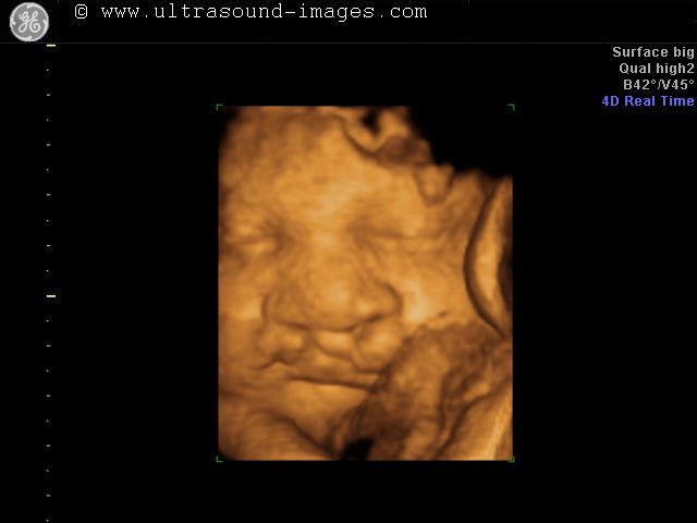
Fetus sucking thumb- 3D ultrasound images

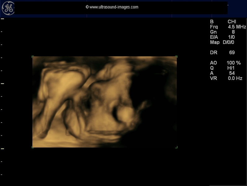
This 34 week fetus is seen sucking its thumb in these 3D ultrasound images.
cleft lip- 3-D ultrasound images
This is yet another example of cleft lip anomaly as seen in these 3-D ultrasound images taken on a GE-Voluson ultrasound system. Again it is surface rendering that brings out the anomaly. 3- D ultrasound imaging in such cases confirms the diagnosis and aids radiologist in viewing the extent of the cleft lip deformity.
fetal cataract
This was a 28 weeks old fetus which showed highly echogenic and thickened lens on ultrasound examination of the fetal eyes. These ultrasound images show crenation of the margins of the lenses which appear highly prominent in the fetal eyes. These ultrasound appearances are diagnostic of fetal cataract. Fetal cataract can be diagnosed as early as 15 weeks of gestation by transvaginal ultrasound. Among the causes of Fetal cataract are intrauterine infections such as TORCH. these images of Fetal cataract are courtesy of Dr. Amutha Csp, MD, India.
References:ultrasound imaging of Fetal cataract- free article
multiple cystic hygromas in neck and chest

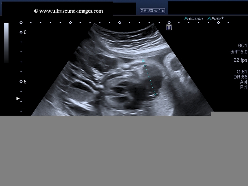
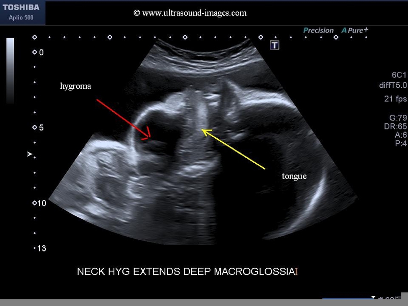
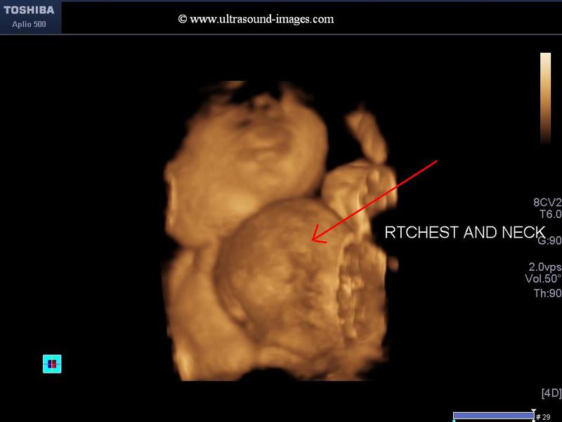
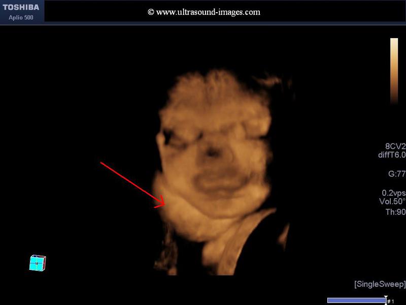
This 30 week fetus showed multiple cystic and septate lesions in the face, neck and chest- both along the soft tissue of the chest wall and within the thoracic cavity to the right of the heart, involving the right lung. Such extensive and multiple cystic lesions are diagnostic of severe, multiple cystic hygroma lesions (see ultrasound images above). The 3-D ultrasound images of this fetus show involvement of the soft tissues and muscles of the neck, producing protrusion of the tongue and associated macroglossia. This ultrasound imaging study of fetal multiple cystic hygromas of the fetus are courtesy of Dr. Durr-e-Sabih, FRCP. The ultrasound machine used here is the Toshiba Aplio 500 system.
Cystic hygromas are classified based on their location into:
1. Cervical- or neck- 75 % of cases
2. In the axilla- 20 % of cases
3. Retroperitoneal and abdominal- 2 % of cases
4. Limbs, bones, chest wall and scrotum etc.- 2% of cases
5. Cervico-mediastinal- 1%.
Our case above is primarily cervico-mediastinal with extensive involvement of the axilla and base of the tongue also and is extremely rare in occurence. The prognosis too is rather bleak in such cases.
References:Sonography of cervico-mediastinal and extensive multiple cystic hygroma
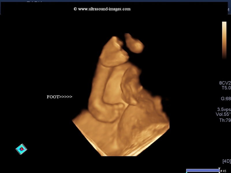
micrognathia with club foot
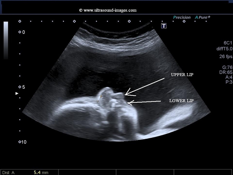
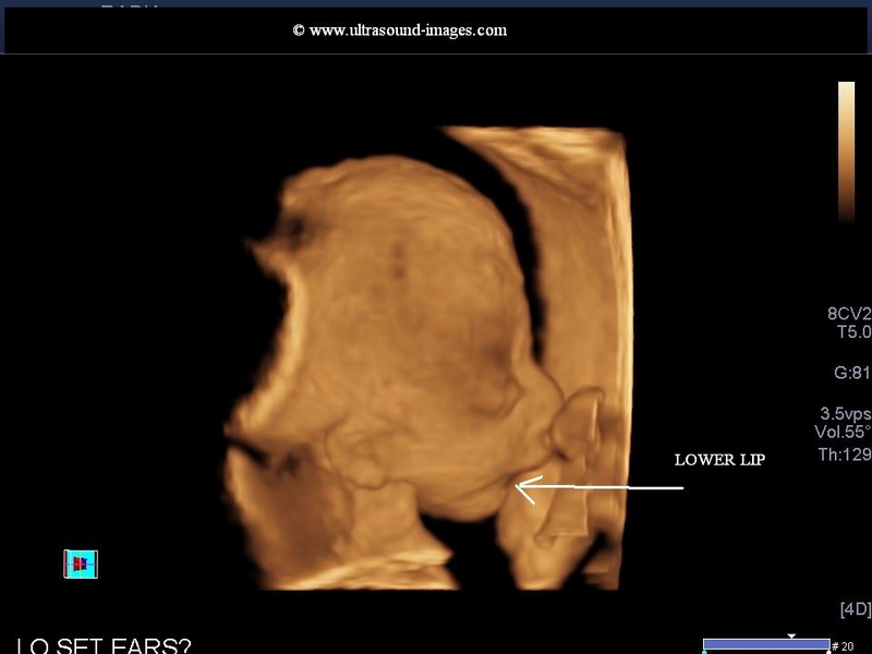
This 3rd trimester fetus (28 weeks) shows an anomaly of the fetal face; the chin and lower lip appear to be receded as compared to the upper lip and upper jaw. This ultrasound appearance seen best in a sagittal section of the fetal face can be appreciated in both 2-D and 3-D Ultrasound images shown above. these ultrasound features are diagnostic of a condition called micrognathia or a small lower jaw. Note the overhanging upper lip in these ultrasound images of micrognathia. Micrognathia is diagnosed usually in the third trimester because the lower jaw undergoes maximum growth in this time period of gestation. Associated with micrognathia, there is also an element of difficulty or improper Fetal swallowing resulting in polyhydramnios, which is also evident in these ultrasound images.
In addition the third ultrasound image also shows evidence of clubfoot or talipes equinovarus.
Micrognathia is the result of an abnormality in the development of the first branchial arch during early fetal life. micrognathia can be associated with a number of other fetal anomalies including aneuploidy and non-aneuploidic syndromes.These ultrasound images of micrognathia are courtesy of Dr Durr-e-Sabih, FRCP.
References:
1) http://www.jultrasoundmed.org/content/21/7/775.full.pdf
2) http://radiopaedia.org/articles/micrognathia
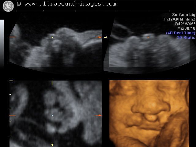
fetal eyelashes

Sometimes minute details of fetal anatomy can be imaged. This is one such example wherein the sonologist has managed to capture fetal eyelashes in excellent detail. The normal fetal eyelashes (see ultrasound images above) are seen as echogenic linear structures extending outward from the eyelids. Images are courtesy of Dr. Mayank Chowdhury, MD (ultrasound system here is the GE- Voluson E8).
3-D alobar holoprosencephaly

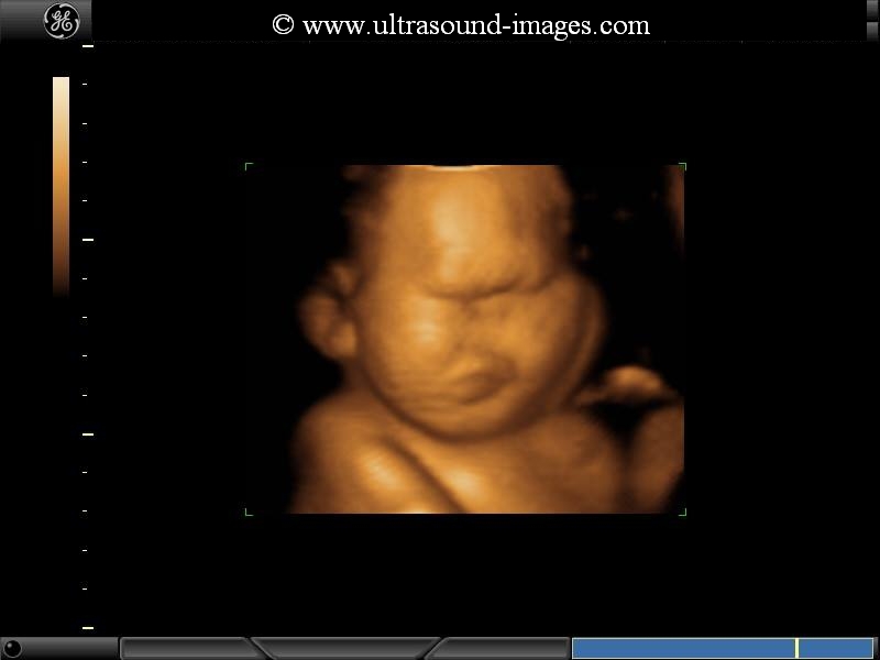

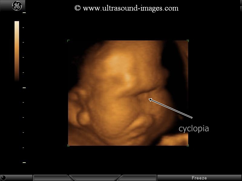
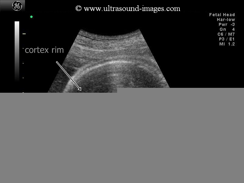
This second trimester fetus shows all the features of alobar holoprosencephaly. The 3-D ultrasound images of the fetal face show absence of the nose or arhinia. In addition, there is a rudimentary single eye seen lower down in the midline. This is known as cyclopia and the fetus is said to be a cyclops. In addition, the 2-D ultrasound image of the fetal brain shows a single common ventricle or what is known as a mono-ventricle running from one side to the other with absence of midline structures such as the falx cerebri. Besides, both the thalami appear to be fused forming a single structure. Also, surrounding the mono ventricle is a rim of cerebral cortical tissue. All these ultrasound features are suggestive of a severe case of holoprosencephaly also known as alobar holoprosencephaly. The above ultrasound images of alobar holoprosencephaly are courtesy of Dr. Rami Barakat, MD.
References:
1. Radiopedia article on radiology of alobar holoprosencephaly
2. Ultrasound imaging of alobar holoprosencephaly- sonoworld article
saddle-nose-with-cleft-lip-in-holoprosencephaly
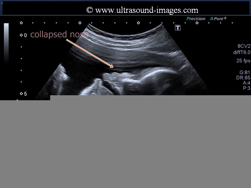
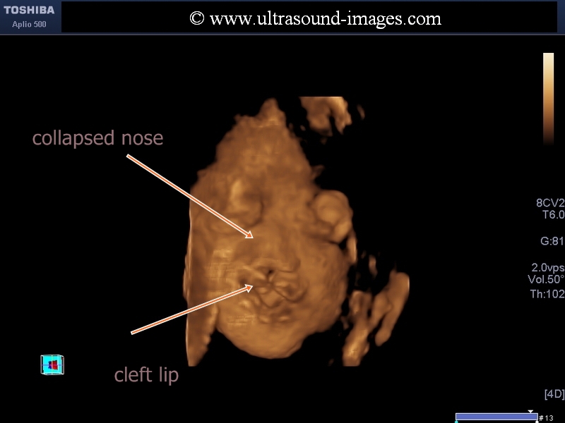
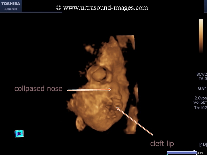
Fetal holoprosencephaly is often associated with multiple facial anomalies also. In this case, fetal ultrasound shows collapse of the nose aka saddle nose which is well appreciated in both sagittal B-mode ultrasound as well as in the 3-D ultrasound images. Along with this anomaly there is also cleft cleft lip deformity. Saddle nose deformity is also seen in chronic maternal alcoholism as well as in congenital syphilis.
These ultrasound images are courtesy of Durr-e-Sabih, MBBS, FRCP.
References:
1) 2D ultrasound and cranio-facial anomalies including saddle nose/ collapsed nose http://onlinelibrary.wiley.com/doi/10.1046/j.1469-0705.2002.00721.x/pdf
2) congenital syphilis and saddle nose
http://en.wikipedia.org/wiki/Congenital_syphilis
3) Article on low nasal bridge or saddle nose or collapsed nose http://www.healthline.com/health/low-nasal-bridge#Overview1
4) Article on congenital syphilis and saddle nose etc http://www.nlm.nih.gov/medlineplus/ency/article/001344.htm
5) causes of saddle nose
6) maternal alchohol intoxication and saddle nose http://www.ncbi.nlm.nih.gov/pubmed/1819957
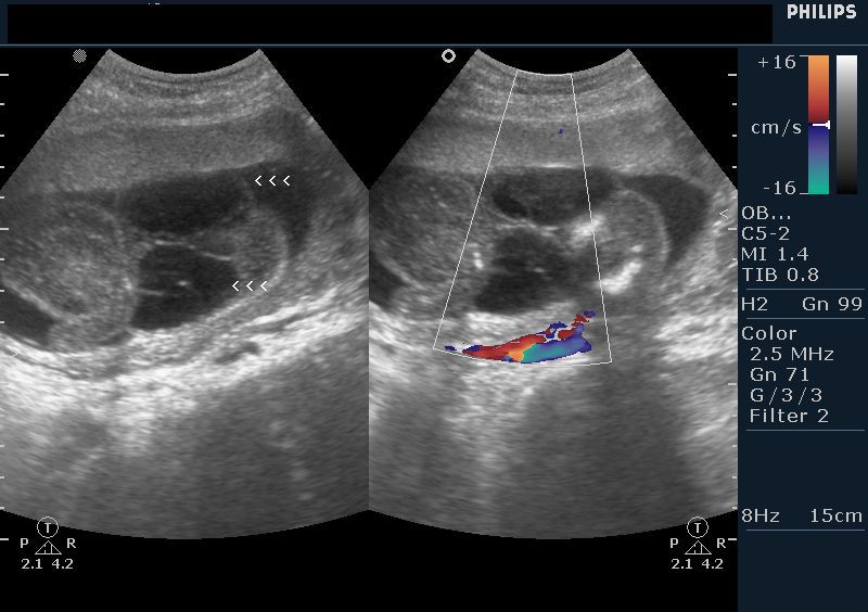
fetal-thyroid-goiter
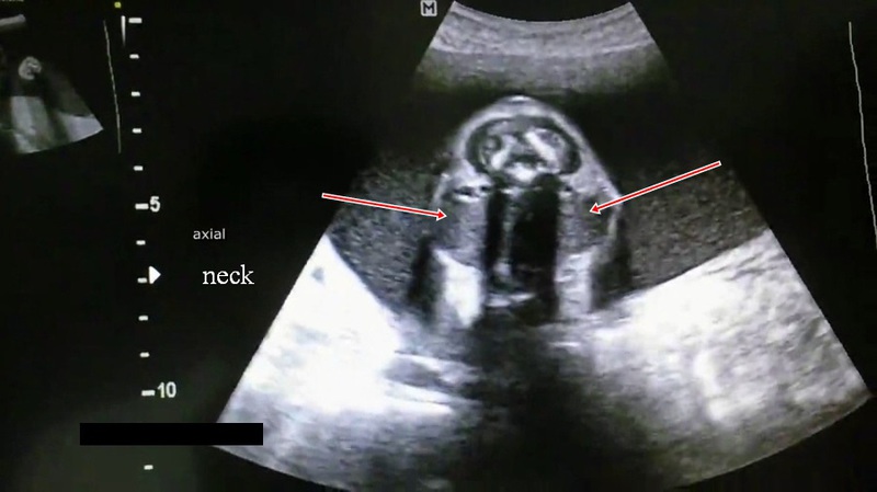
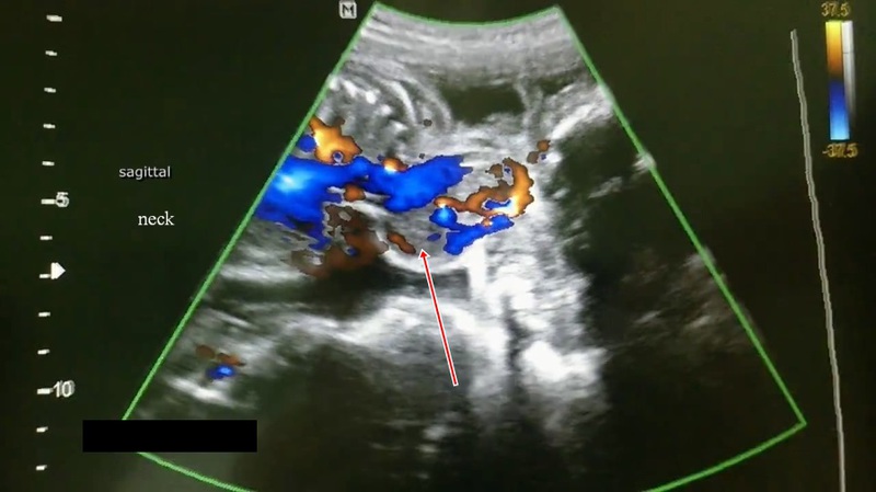
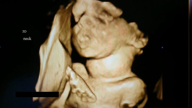
The above ultrasound images (both sagittal and axial sections of the Fetal neck) show a soft tissue mass anterior to the trachea which is homogenenous in echotexture and produces and anterior bulge of the Fetal neck. This is further confirmed on colour Doppler and 3-D ultrasound imaging of the same region. In addition, the images show moderate polyhydramnios. The location of the thyroid is in the exact same region as seen in the images above.In addition, the Fetal neck appears hyper extended. All these findings suggest a diagnosis of Fetal goiter. Fetal thyroid enlargement is usually the result of maternal hyperthyroidism which is being treated medically. Fetal thyroid enlargement or goiter can be due to hyperthyroidism or hypothyroidism. Here colour Doppler ultrasound is helpful in distinguishing both these conditions. In hyperthyroidism, there is significant internal vascularity of the fetal thyroid. However, as in the image above in hypothyroidism there is usually increased vascularity of the rim of the fetal thyroid. also added above is an ultrasound video ( courtesy of Vijay Hingra, MD) of the same case of Fetal thyroid goitre.
references:
1. Ultrasound imaging of Fetal thyroid goitre- radiopedia article
2. sonography of Fetal thyroid disease- Journal of ultrasound
© 2026, Joe Antony, MD All images and content in this site are copyrighted. www.ultrasound-images.com When visiting this site, we may use cookies to improve the performance of this site. Use of this site means you accept the use of cookies. Read our privacy policy at this page: https://www.ultrasound-images.com/privacy-policy/