
scrotal infections
Contents of this page
- Filariasis of scrotum
- Testicular abscess
- Pyocele with orchitis
- Tuberculous epididymitis and tuberculous epididymo-orchitis
- Acute epididymitis- case-2
- miliary tuberculosis of right testis-2
- fournier gangrene of scrotum
- Testicular abscess
- Pyocele with orchitis
- Tuberculous epididymitis and tuberculous epididymo-orchitis
- Acute epididymitis- case-2
- miliary tuberculosis of right testis-2
- fournier gangrene of scrotum
Filariasis of scrotum
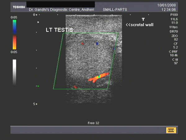
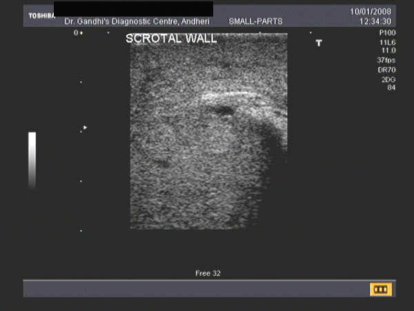
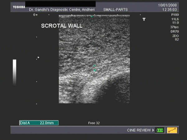
These sonographic images of the scrotum of a 65 yr. old male patient known to have filariasis reveal a markedly thickened scrotal wall. The testes itself appears normal. Multiple, hypoechoic tubular channels are seen in the scrotum. These ultrasound images suggest lymphedema of the scrotal wall following filariasis. Images taken using a Toshiba Nemio 30 color doppler machine, courtesy of Dr. Jaydeep Gandhi, Mumbai, India.
Below is an ultrasound video recording of live filarial parasites showing motion- the so called "filarial dance" within small fluid collection in the scrotum (video courtesy of Dr. Gunjan Puri, MD, India).
Reference:
1)http://www.jultrasoundmed.org/cgi/reprint/22/8/765(excellent free article and images)
2) Filariasis article (free article)
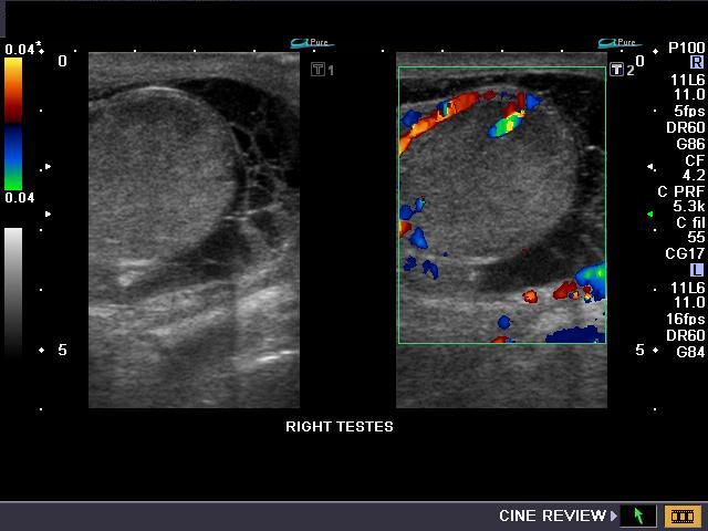
Testicular abscess
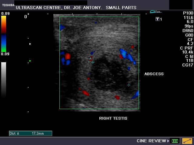
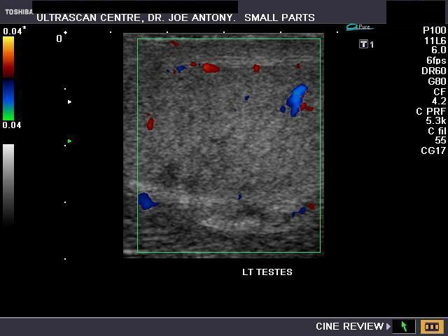
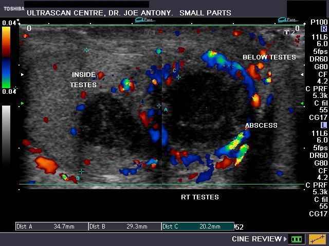
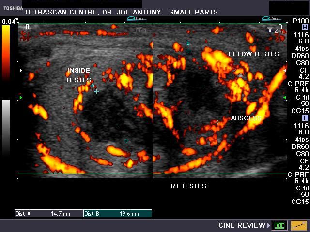
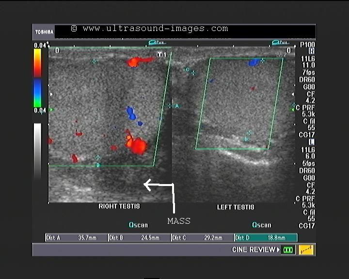
A case of abscess of the right testes.
This middle aged male patient had a history of drainage of scrotal abscess a few months back. he now presented with discharge, pain and swelling of the right half of the scrotum. Sonography of the scrotum revealed: a) a hypoechoic (almost cystic) lesion with shaggy walls within the right testes with b) another similar lesion extending from the first lesion. c) Color Doppler imaging/ and Power Doppler imaging confirm marked increase in vascularity around both lesions suggesting marked hyperemia and severe right orchitis. These ultrasound images are diagnostic of testicular abscess (right) with extension outside the testes. The left testes also shows evidence of mild orchitis.Ultrasound images taken by Joe Antony, MD, India, using a Toshiba Nemio - XG Color Doppler machine.
Pyocele with orchitis
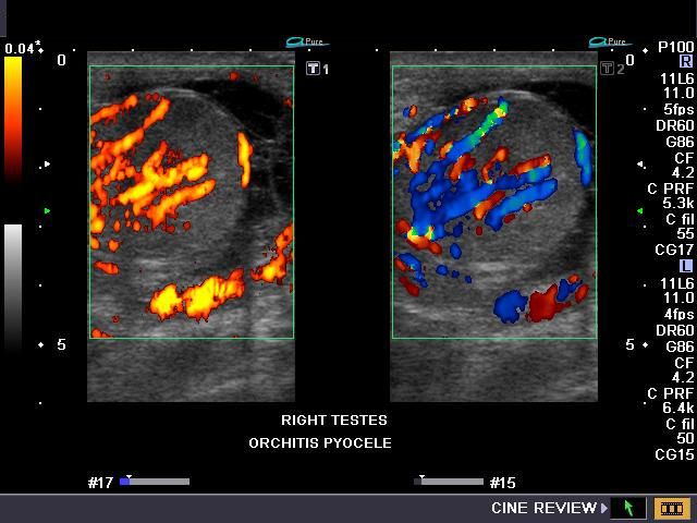
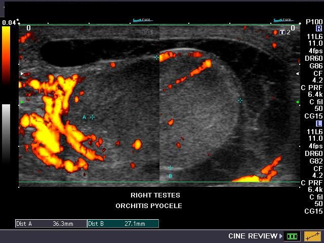
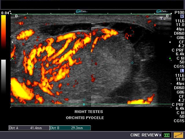
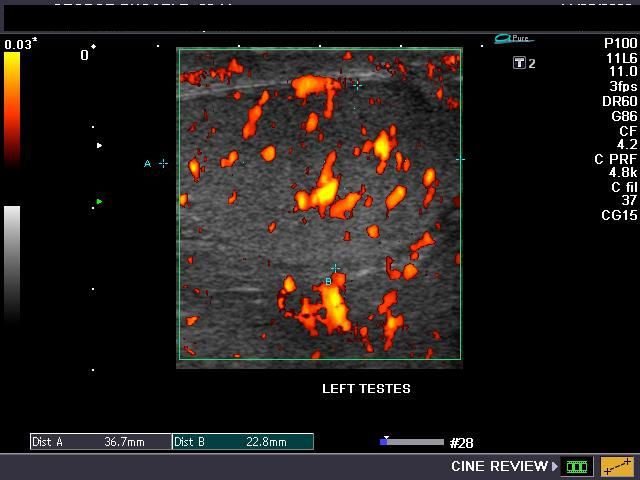
This patient presented with swollen scrotum with marked tenderness on the right testes. Ultrasound images show inhomogenous echotexture of the right testes with hypoechoic areas seen in lower part. Color Doppler and Power Doppler images show markedly increased vascularity in the right testes with moderate vascularity in the left testes. There is a large fluid collection around the right testes with multiple fibrous septae within the fluid s/o infected hydrocele (pyocele). These ultrasound and color Doppler images are diagnostic of Bilateral orchitis with pyocele on right side. Ultrasound images by Dr. Joe Antony, MD, India. These images were taken using a Nemio-XG Color Doppler system (Toshiba).
See a nice article on differential diagnosis of swelling of the scrotum:
http://www.squidoo.com/scrotum
Reference:
Tuberculous epididymitis and tuberculous epididymo-orchitis
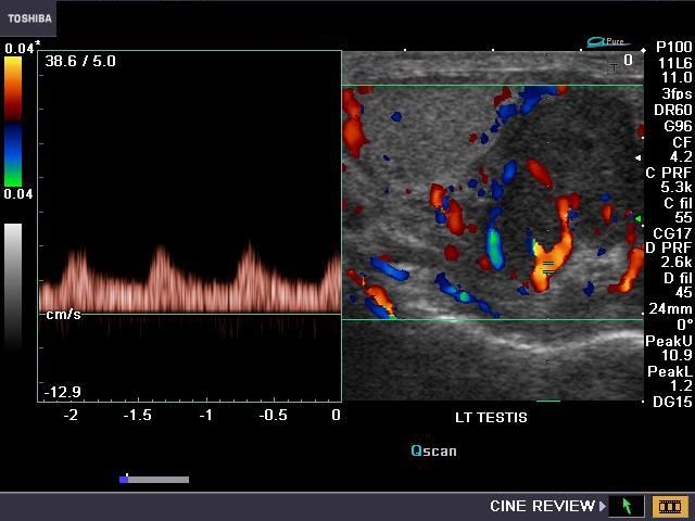
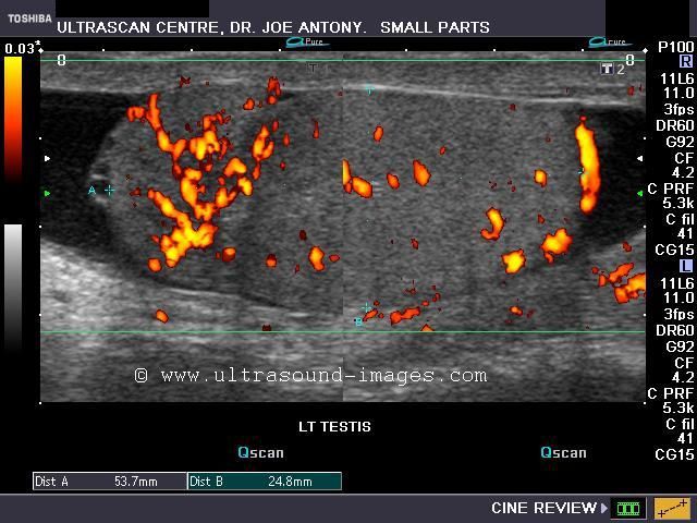
This adult, 48 yr. old male patient had a mildly painful mass in the left scrotum. Ultrasound image shows a hypoechoic, nodular mass of 3 x 2 cms. (appx.) below the left testis, involving the left epididymis (globus minor or tail) and the adjacent part of the left testis. The color and Power Doppler images (right) show considerable vascularity around and within the mass.Transverse sections (grey scale and color Doppler) images show a small hydrocele around the left testis.The scrotal skin also appears thickened.The right testis appears normal. Spectral Doppler imaging of the mass shows a low resistance flow within the vessels in this lesion. These color Doppler and ultrasound images show an inflammatory pathology of the left epididymis- typically these findings are those of left tuberculous epididymo-orchitis.
See:
Also:http://www.ajronline.org/cgi/reprint/176/6/1459
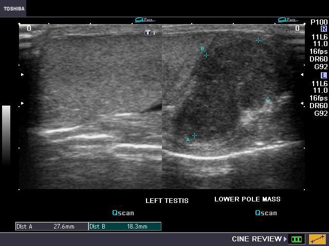
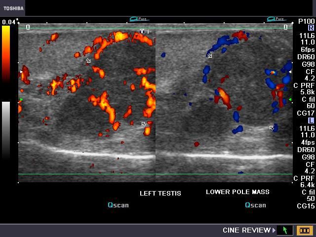
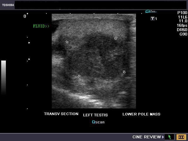
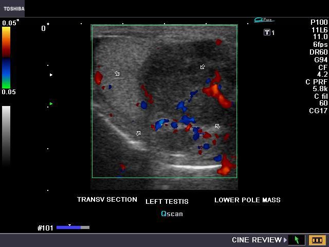
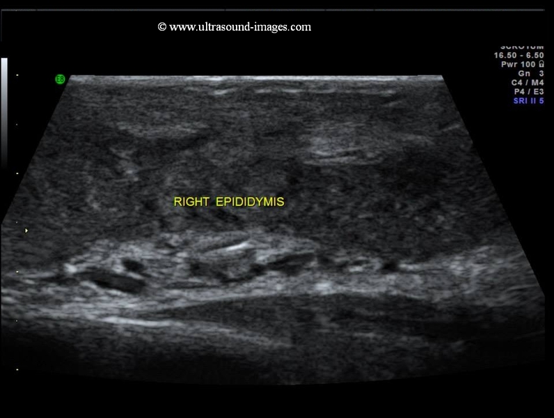
case-2-tuberculous epididymitis
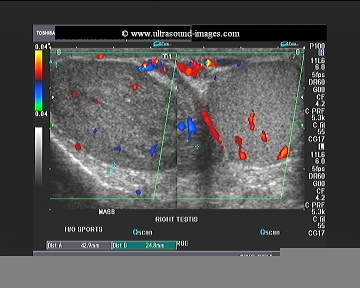

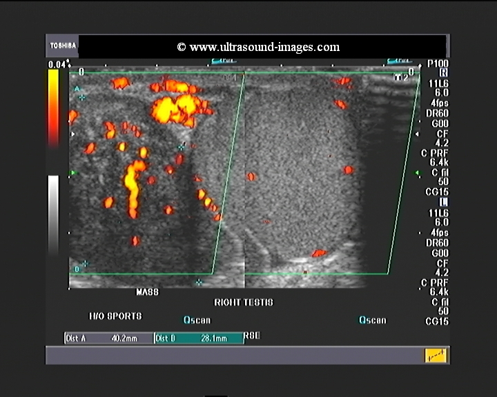
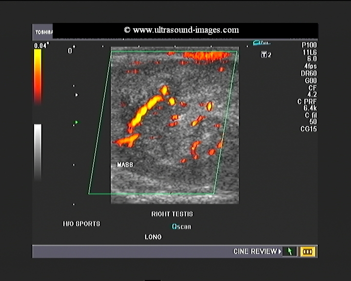
This is yet another case of tuberculous epididymitis. A large mass lesion is seen along the posterior and lateral aspect of the right testis. This lesion appears heterogeneous and moderately vascular. Such an appearance giving rise to the likeness of an extra testicular mass in this typical location is diagnostic of right epididymitis- and in this case a case of right tuberculous epididymitis. such an ultrasound appearance can often be confused with a testicular neoplasm. However, note that this lesion is well defined and located in the typical position of the epididymis. Thus, despite the large size of the lesion, the most common and likely possibility based on these ultrasound images, is tuberculous epididymitis. These ultrasound and colour Doppler images of tuberculous epididymitis were taken using a Toshiba Nemio XG ultrasound system. Dr. Joe Antony, MD
Acute epididymitis- case-2
Power Doppler image- left testis and epididymal head
B-mode image
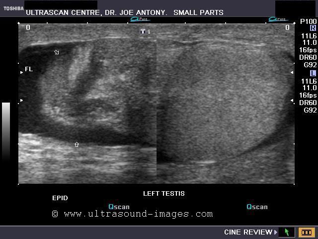
Both testes seen: Power Doppler
Color Doppler image - left testis
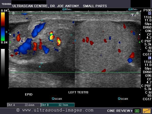
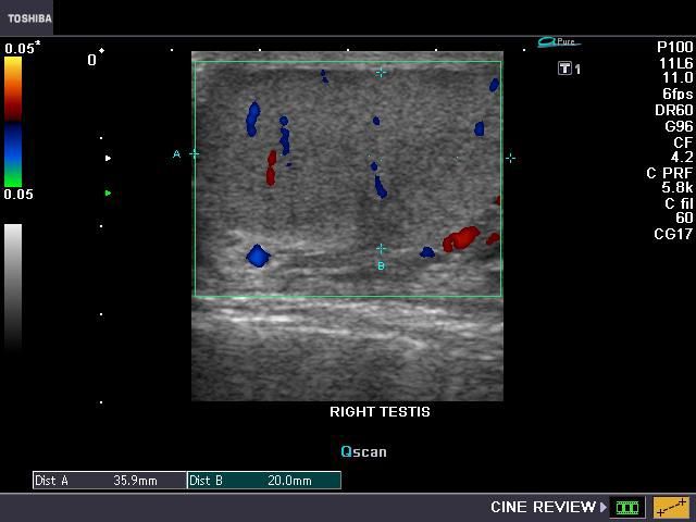
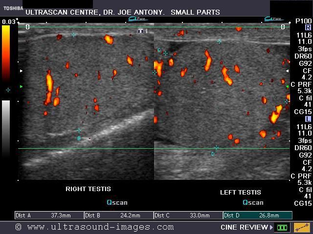
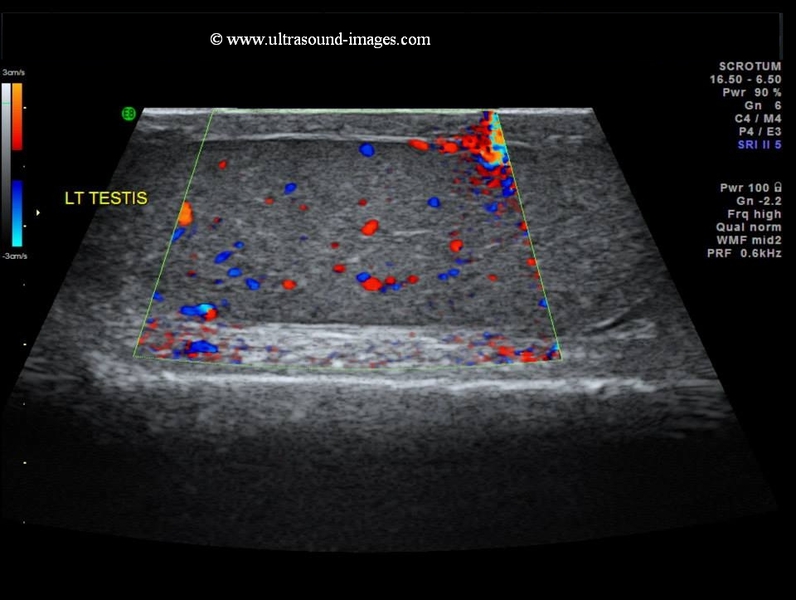
This patient had acute onset of swelling and pain with tenderness over the left scrotum. Ultrasound shows markedly enlarged left epididymis with marked vascularity of the head of epididymis on color Doppler scan. There is also a small left hydrocele with particulate matter within it suggestive of early purulent material within it. The left testis itself is relatively normal in appearance. This suggests a left acute epididymitis involving the epididymal head. The marked enlargement of the epididymal head can mimic a torsion of the left testis; however the presence of normal vascularity in the left testis goes against that diagnosis. This patient will benefit from conservative medical treatment, though surgical intervention may be needed to manage the hydrocele.
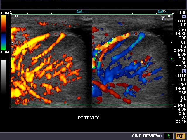
miliary tuberculosis of right testis-2

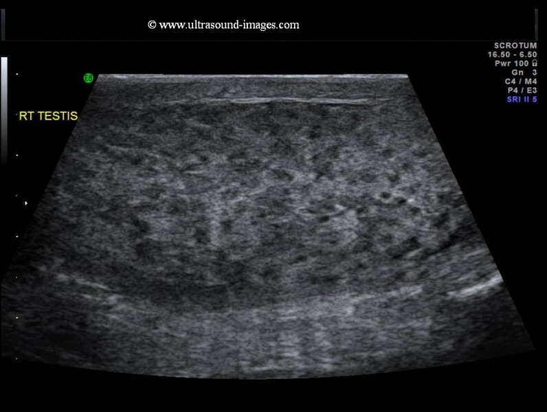
This young adult male presented with pain and swelling in the right scrotum. Ultrasound and colour Doppler sonography of the scrotum revealed multiple patchy hypoechoic nodules throughout the right testis. Ultrasound images of the right testis showed a diffusely enlarged and hypoechoic testis. These findings are diagnostic of miliary tuberculosis of the right testis. The right epididymis also showed diffusely enlarged hypoechoic right epididymis suggesting a tuberculous infection of this organ also. The left testis shows normal vascularity and echotexture. Final diagnosis confirmed by biopsy- tuberculous epididymo-orchitis (RT).
References:http://www.ajronline.org/doi/full/10.2214/ajr.176.6.1761459
These ultrasound images of miliary tuberculosis of the testis are courtesy of Dr. Ajay Garg, MD.
fournier gangrene of scrotum
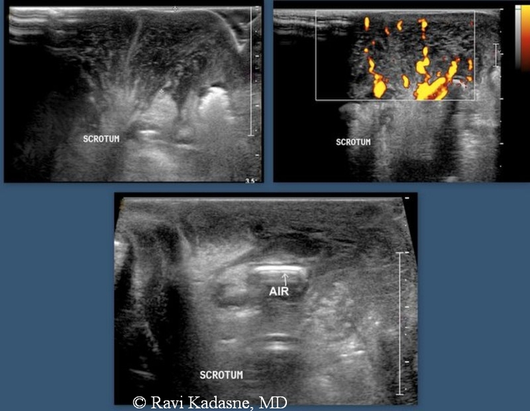
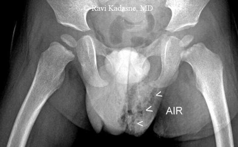
This two-year-old male baby had scrotal swelling. Ultrasound imaging of the scrotum showed marked thickening of the scrotal wall with hyperemia of the scrotum seen as increased vascularity on colour Doppler ultrasound imaging. In addition, the ultrasound images show echogenic foci within the scrotal wall suggestive of gas within the scrotum, with dirty acoustic shadowing. x-ray of the scrotum shows presence of gas within the scrotum and confirms the above-mentioned findings. Thus, these ultrasound images, colour Doppler imaging and x-ray findings all confirm gangrenenous infection of the scrotum- or Fournier gangrene. Fournier gangrene of the scrotum is often seen in adult male patients; very rarely this condition is seen in the paediatric age group. In this case, the testes were well preserved and showed normal ultrasound appearances. These ultrasound and X-ray images of Fournier gangrene are courtesy of Ravi Kadasne, MD, UAE.
References:Radiology article on Fournier gangrene of scrotum
© 2026, Joe Antony, MD All images and content in this site are copyrighted. www.ultrasound-images.com When visiting this site, we may use cookies to improve the performance of this site. Use of this site means you accept the use of cookies. Read our privacy policy at this page: https://www.ultrasound-images.com/privacy-policy/