
Normal anatomical variants of Appendix
Contents of this page
- Horseshoe shaped appendix
- Acute appendicitis with appendicolith
- Perforated appendix or appendiceal perforation
- Acute appendicitis- case-2
- acute appendicitis- case3
- appendix-3D-ultrasound
- evolution-of-subacute-appendicitis
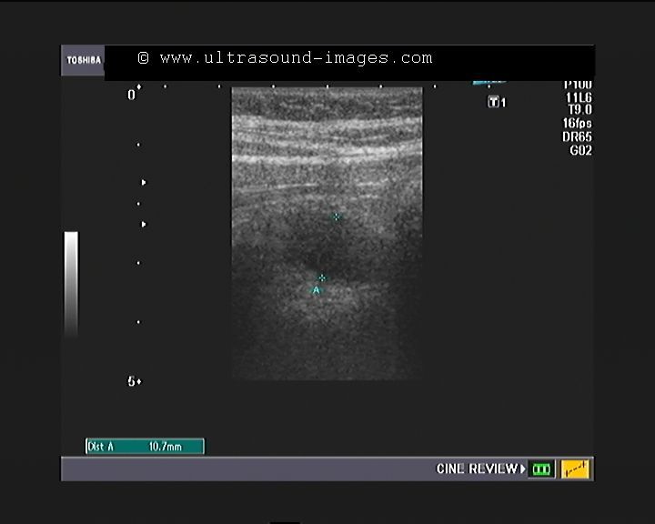
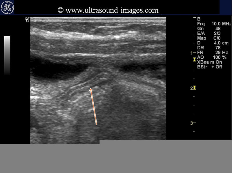
Horseshoe shaped appendix

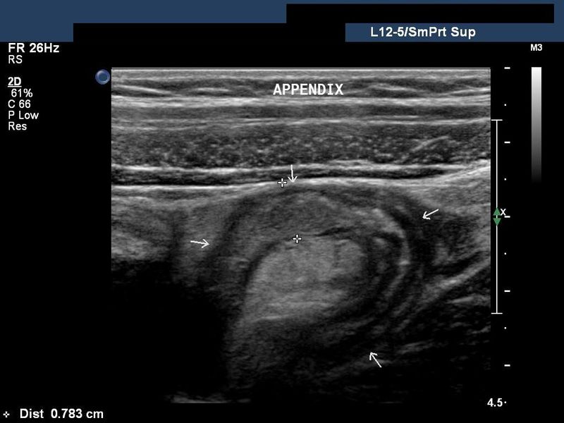
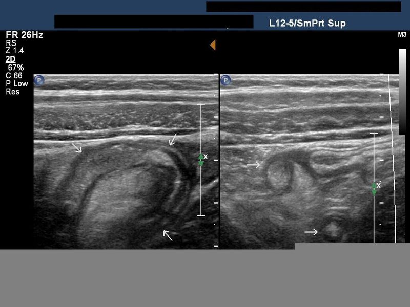
The horseshoe anomaly of the appendix is a rare, normal anatomical variant of the appendix. These ultrasound images show the horsehoe shaped inflammed appendix (forming a sort of inverted U -shape). Note the swollen appendix with echogenic content within it s/o appendicitis (diameter > 6mm.). Ultrasound Images taken using a Philips iU 22 ultrasound machine, courtesy of Dr. Ravi Kadasne, MD, UAE.
Reference: http://www.ncbi.nlm.nih.gov/pubmed/2772830(abstract- free).
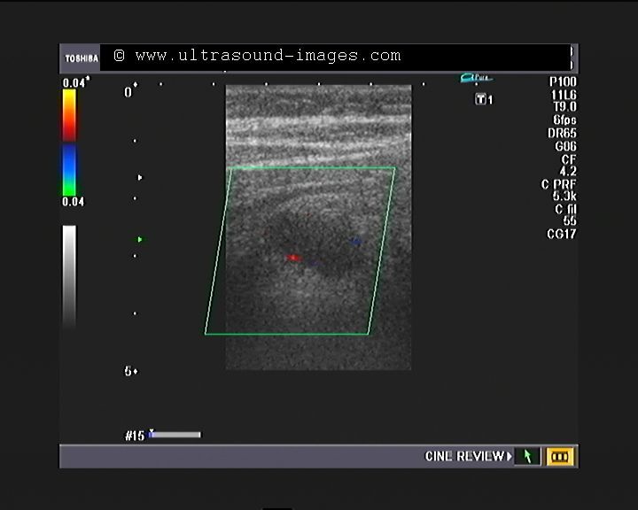
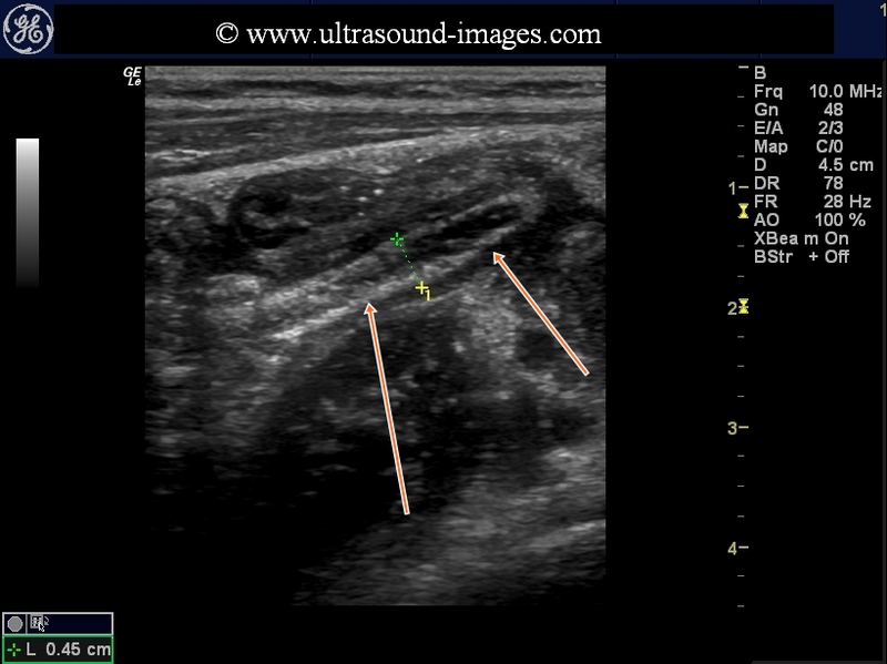
Acute appendicitis with appendicolith
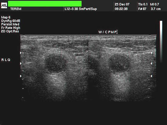
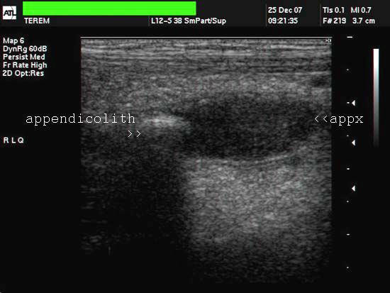
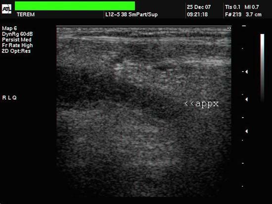

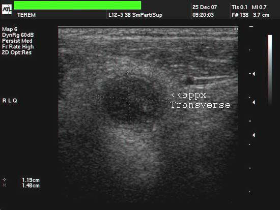
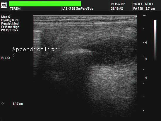
The above ultrasound images are of a young child (10yrs. old) with pain in the right iliac fossa. These images reveal a typical tubular structure with blind end, showing total diameter more than 6mm.(12 x 15 mm.). These sonographic images reveal hypoechoic content (purulent material) distending the appendix with an echogenic focus floating within it. The appendix was also non-compressible. Color flow imaging shows typical hyervascularity around the organ. These ultrasound images are diagnostic of acute appendicitis with appendicolith.( Images taken with a ATL (Philips HDI 3000 color doppler machine, courtesy of Mr. Shlomo Gobi, Israel).
Reference: http://www.emedicine.com/emerg/topic41.htm
Perforated appendix or appendiceal perforation
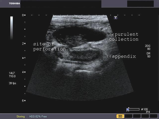
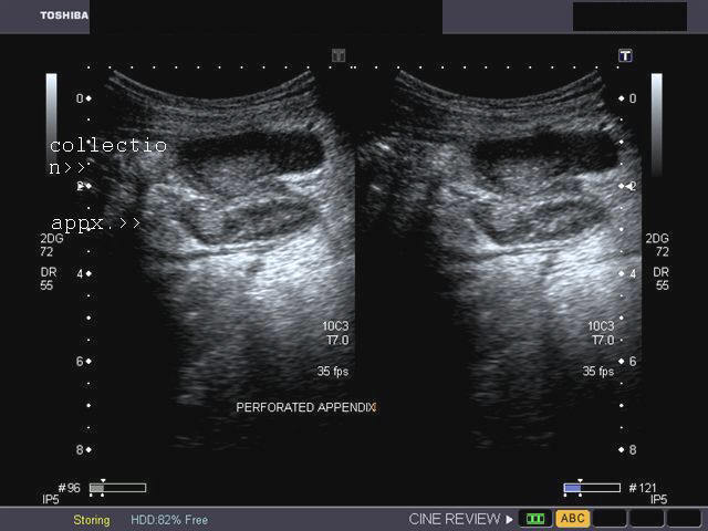
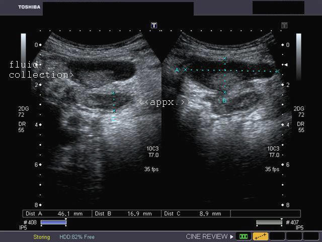
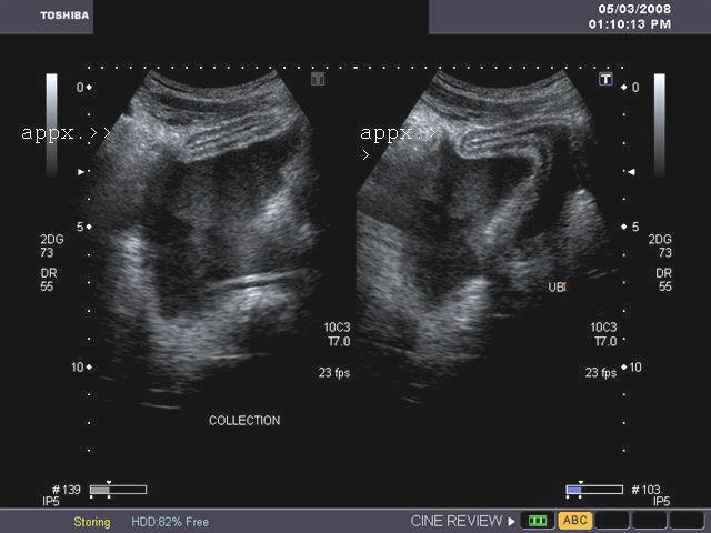
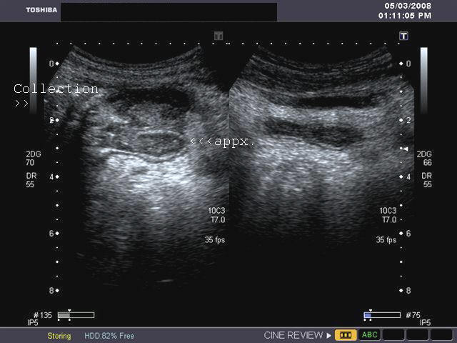
Sonography of ruptured appendix. Sonography of the right iliac fossa reveals 1) distended swollen appendix containing hypoechoic (purulent) fluid with 2) anechoic (fluid) collection anterior to the appendix, with 3)echogenic material (possibly phlegmon) within the fluid. 4) There is evidence of rupture of the proximal part of the appendicular wall. These ultrasound images suggest rupture or perforation of the acutely inflamed appendix. These sonographic images were taken with a Toshiba Xario ultrasound machine, courtesy of Dr. Gunjan Puri, Surat, India.
Reference:
1)http://www.medicinenet.com/appendicitis/article.htm(free article and pictures)
2)http://www.aafp.org/afp/20050101/71.html(free article and images).. rated excellent
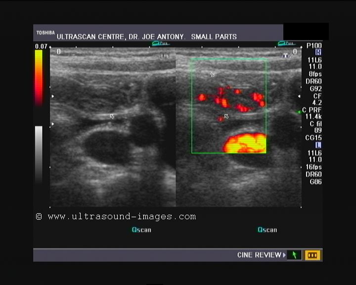
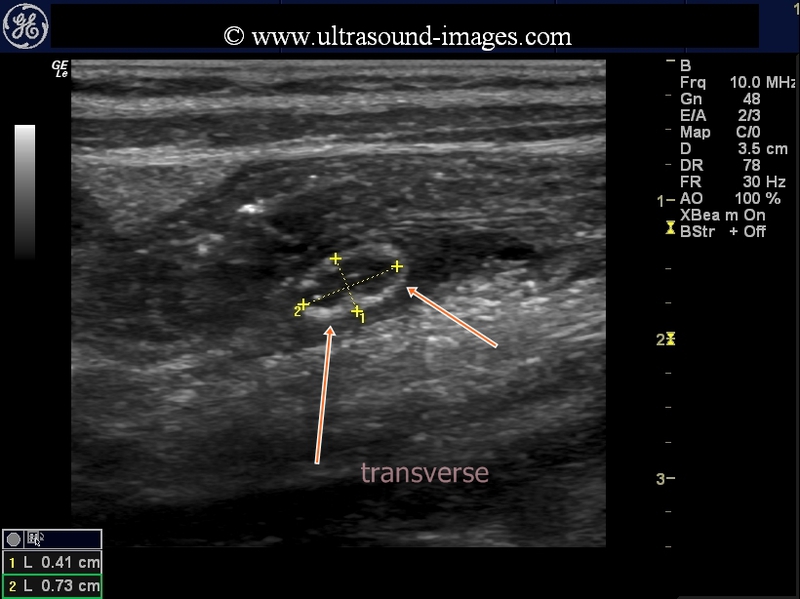
Acute appendicitis- case-2
Long section ultrasound/ Power Doppler images of appendix

Increased vascularity of the appendix- long section

Oblique section Power Doppler image
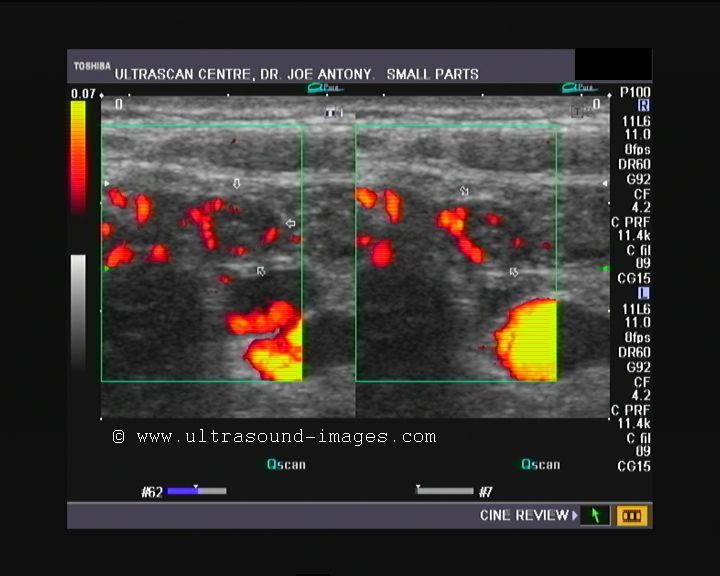
Cross section (transverse section)- Markedly vascular appendix

This male child has a markedly vascular and swollen appendix at 5 to 7 mm. width. Using graded compression it was possible to visualize the acutely inflamed appendix in its entirety. These ultrasound and Power Doppler images show a typical case of acute appendicitis. The Power Doppler ultrasound image at bottom shows a literal ring of fire within the cross section of the inflamed appenidx. Such a case may or may not respond to conservative medical treatment and surgery may be the final option. Fortunately for the patient there is no evidence of impending rupture of the appendix at the present stage. There is no evidence of collection of fluid (phlegmon) around the appendix.
Here is the ultrasound and Power Doppler video clip of this case:
acute appendicitis- case3

this young girl had severe pain and tenderness in right iliac fossa. Ultrasound shows a markedly inflamed appendix, measuring 11 mm in diameter. Even though there is only mild vascularity evident in these ultrasound and colour Doppler images, this appears to be a case of severe acute appendicitis. The danger of an impending appendicular perforation is very real in the child.
appendix-3D-ultrasound

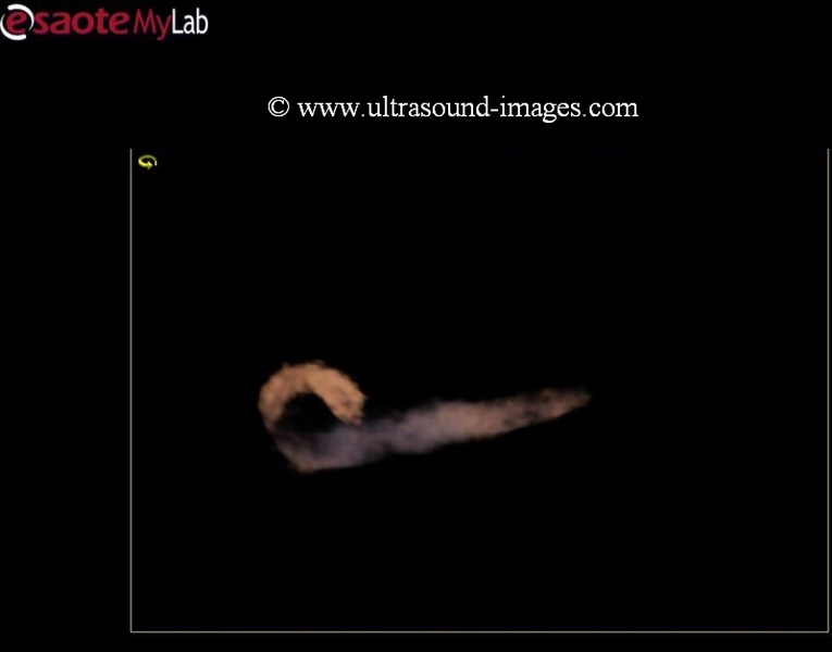

Sonography of the appendix is now well established as a key diagnostic tool in the diagnosis of acute appendicitis. But now with the advent of 3D ultrasound imaging, the appendix can be visualized with even greater resolution and detail. The above 3D ultrasound images (courtesy of Jorge Hernandez, MD) show the appendix in striking detail.
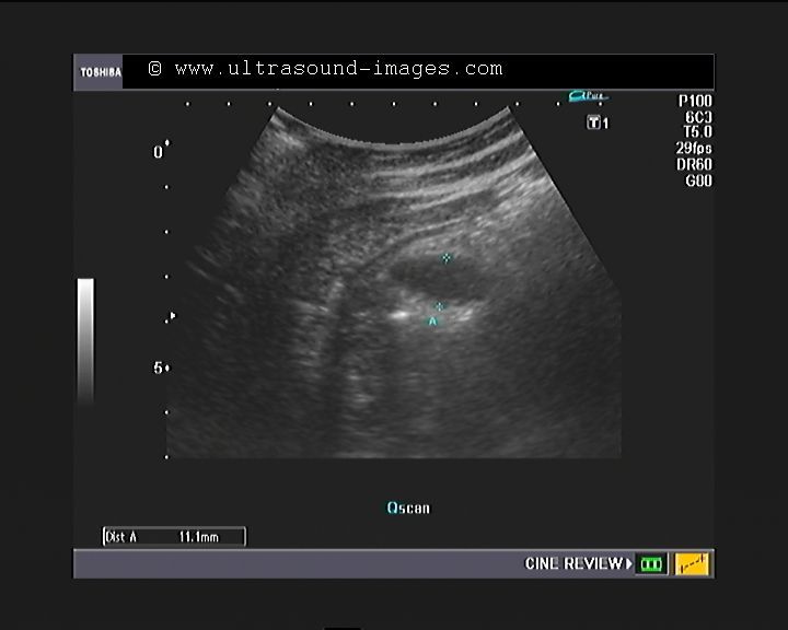
evolution-of-subacute-appendicitis

This young child has history of non-specific abdominal pain. Ultrasound shows ( see images above) a very early stage of subacute appendicitis with minimal dilation of the lumen of the appendix. There is no evidence of significant inflammation at this stage ( as seen from the ultra sound images above). In fact, the thread-like appendix can be mistaken for roundworm infestation within the small bowel.
Follow up of this case is shown below.
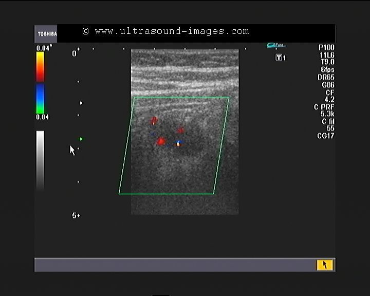
subacute-appendicitis-follow-up

Follow up of the same patient two days later:
However, two days later, the same structure ( the appendix) appears moderately distended with distension of the distal half of the apppendicular lumen with fluid and a suggestion of small amount of inflammation surrounding the appendix. This now qualifies for the term subacute appendicitis.
© 2026, Joe Antony, MD All images and content in this site are copyrighted. www.ultrasound-images.com When visiting this site, we may use cookies to improve the performance of this site. Use of this site means you accept the use of cookies. Read our privacy policy at this page: https://www.ultrasound-images.com/privacy-policy/