
GIT
Contents of this page
- Foreign body in the stomach
- Hypertrophic pyloric stenosis
- Case-2: Congenital hypertrophic pyloric stenosis
- Normal esophagus (also called oesophagus)
- Jejunal obstruction
- Gastric band (gastric banding in obesity)
- Intussusception
- ascariasis of small intestine
- henoch-schonlein-purpura
Foreign body in the stomach

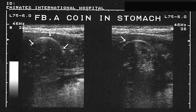
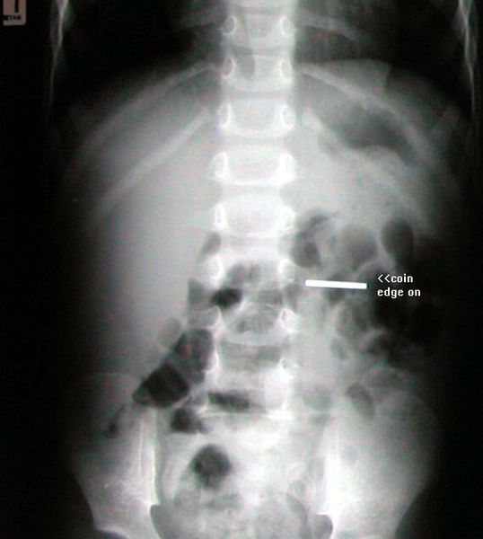
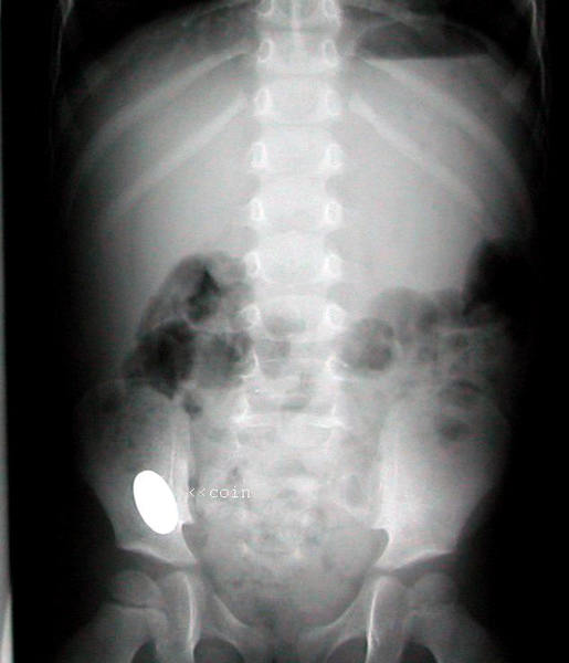
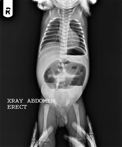
Sonography of the stomach revealed a linear echogenic structure with posterior acoustic shadowing. On changing the probe angle the edge appears to be a semicircle. Plain X-ray of the abdomen revealed the structure to be coin (seen edge-on in the left hypochondrium). Images courtesy of Dr. Ravi Kadasne, UAE.
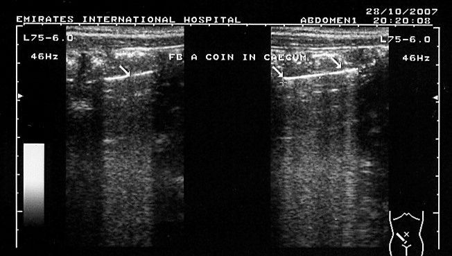
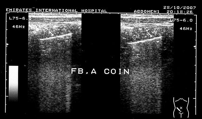
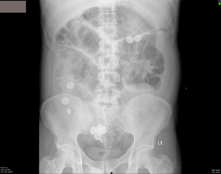
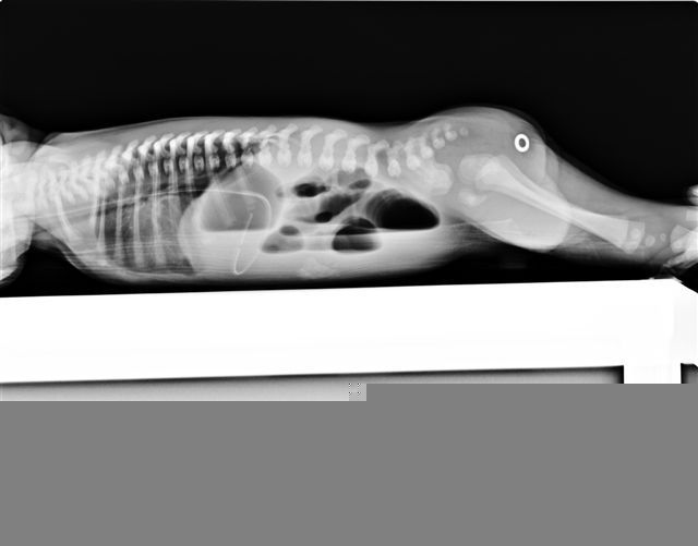
Here are the follow-up ultrasound scan and X-ray images of this patient, a young child. The coin, seen in the stomach (above images) has reached the caecum. (Images courtesy Dr. Ravi Kadasne).
Case-2:
Foreign body in GIT (X ray)
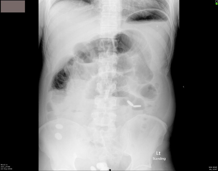
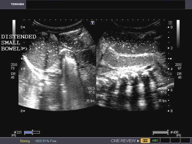
This patient presented with abdominal pain and distension of abdomen. Plain X-ray abdomen show multiple radio-opaque objects in the stomach and small intestine. These x-ray appearances are consistent with foreign bodies (pills) in the GI tract. The patient had a history of taking tablets (pills) orally just before the X-rays. There is also evidence of intestinal obstruction. Note the gas distended small bowel and air fluid levels. Both images are courtesy of Shlomo Gobi, Israel.
Hypertrophic pyloric stenosis
Pyloric stenosis in neonate.
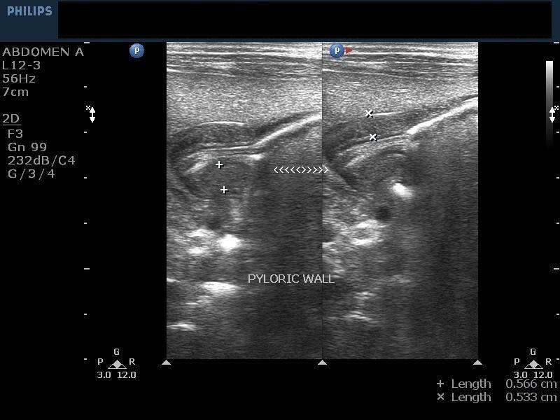
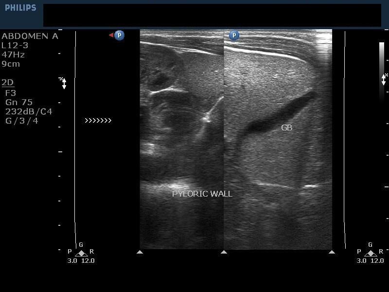
This neonate underwent sonography for persistent vomiting. Ultrasound images reveal a) marked thickening of the wall of the pyloric canal (5.5 mm.) b) elongation of the of the pyloric canal (the cervix sign) c) shifting of the pylorus bringing it in close proximity to the right kidney (antero-medial to right kidney) and gall bladder. d) distended stomach e) mucosal prolapse producing the antral nipple sign is also seen. These ultrasound images thus suggest a diagnosis of hypertrophic pyloric stenosis. Sonographic images courtesy of Dr. V. Ganesan, Bahrain. Images taken using a Philips IU22 ultrasound machine.
Reference:http://www.emedicine.com/radio/topic358.htm(free article and images)
Case-2: Congenital hypertrophic pyloric stenosis
Long section of the pylorus

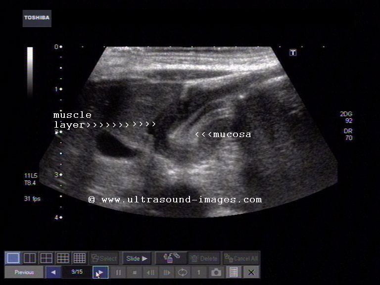
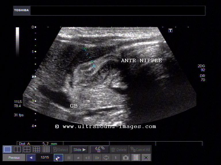

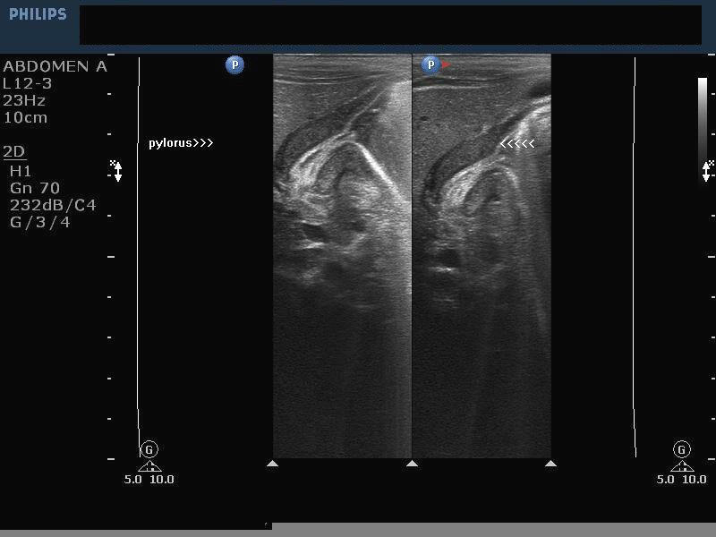
This neonate had severe vomiting and underwent sonography. Ultrasound images show markedly thickened pyloric muscle layer (5 to 6.4 mm.) and elongated pylorus (17mm.). Medical sonographic criteria of hypertrophic pyloric stenosis suggest that a muscle wall width of more than 3 mm and pyloric length of 17 mm. or more indicate congenital hypertrophic pyloric stenosis. In addition, the axial section of the pylorus shows the typical target sign or doughnut sign indicating hypertrophic pyloric stenosis. This sign is produced by the echogenic mucosa within the pylorus surrounded by the thickened hypoechoic muscle wall. In addition note the antral nipple sign (image in bottom left), produced by the prolapsing pyloric mucosa into the antrum. Ultrasound images of congenital hypertrophic pyloric stenosis taken using a Toshiba Xario Ultrasound system.
Reference: Sonography of hypertrophic pyloric stenosis
Normal esophagus (also called oesophagus)

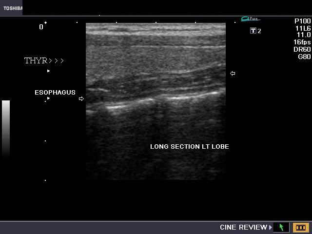
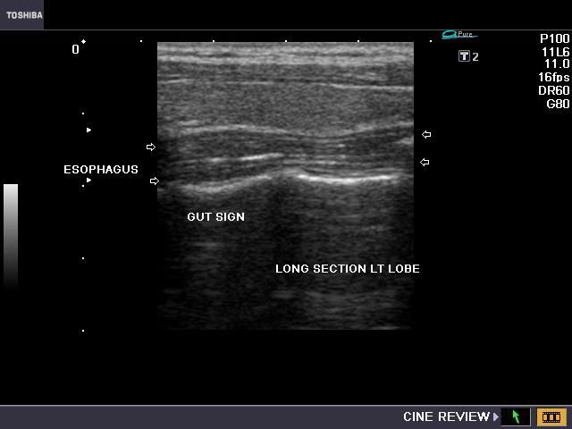
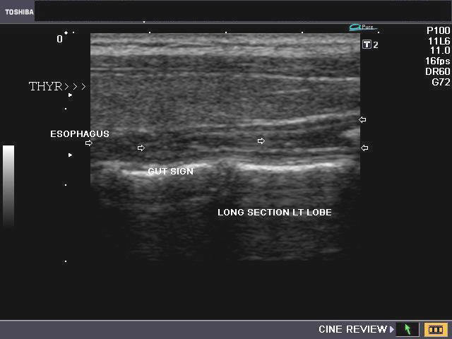
Sonography of the normal esophagus
The normal esophagus (also spelt oesophagus or food pipe) is seen in this ultrasound image, in both transverse and longitudinal sections, to lie posterior to the left lobe of thyroid. Usually, the esophagus lies postero-medial to the left lobe. Sonography of the esophagus often reveals a prominent tubular structure, with the typical gut signature, as in this case. Here it lies posterior and appears rather flattened (side to side).
Jejunal obstruction
Sonography of small bowel obstruction in neonate
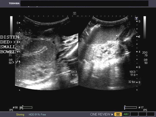
This neonate had severe vomiting and had inability to pass stool. Sonography of the abdomen showed multiple, distended loops of small bowel (jejunum) with collapsed thin large bowel (colon). X-ray images showed dilatation of the small bowel with multiple air fluid levels in the jejunum. There was no evidence of ascites. These ultrasound and X-ray images suggest a diagnosis of small bowel (jejunal) obstruction. The commonest cause of small bowel obstruction in fetus or neonate is meconium impaction (or meconium stones). Another important cause is jejunal atresia. This case is courtesy of Dr. Gunjan Puri, MD, India.
Reference:http://www.thefetus.net/page.php?id=2730(free article and images).
Gastric band (gastric banding in obesity)

The above ultrasound images show linear echogenic structures in the left hypochondrium. This was a patient of obesity who underwent gastric banding. A Gastric band is an adjustable silicon band placed around the upper part of the stomach to reduce the capacity of the stomach. The artificial pouch created by the surgically placed gastric band, gives the patient of a full stomach, after just about 1/2 cup of food, as compared to the 6 cups of food needed to satisfy hunger in a normal patient (the full capacity of the normal stomach). Gastric banding is the safest and most convenient of the weight loss or bariatric surgeries. Ultrasound images of gastric band are courtesy of Mr. Shlomo Gobi, Israel.
References:
http://www.webmd.com/diet/weight-loss-surgery/gastric-banding-surgery-for-weight-loss
http://en.wikipedia.org/wiki/Adjustable_gastric_band
Intussusception
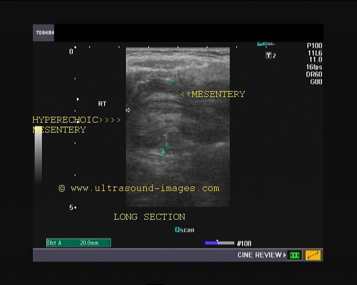
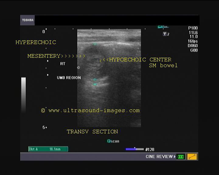

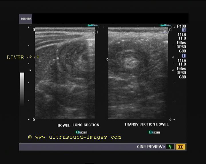
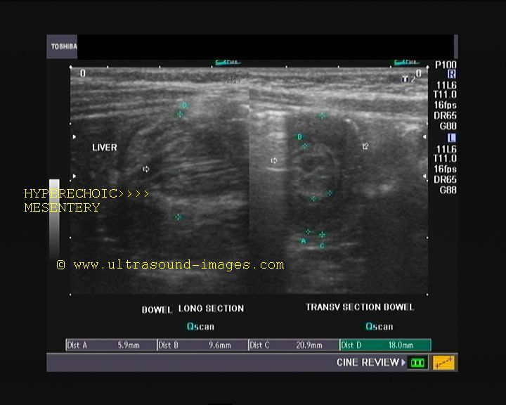
This 1 year old infant presented with severe vomiting and pain in abdomen with excessive crying. Ultrasound images of the abdomen show a mass just below the liver which shows concentric rings on transverse section. The longitudinal section of the mass appears multilayered too with a somewhat "sandwich" appearance. These ultrasound images are typical of intussusception of the small bowel into the colon (ileo-colic type). Intussusception is a condition wherein the small intestine is telescoped or herniated into the adjacent part of the intestine; usually the small intestine herniates into the colon (ileocolic intussuception) or starts as ileo-ileal and later proceeds to become ileocolic in nature. The sonographic images above show echogenic layers (mesentery) (on longitudinal section) on both sides of the hypoechoic central part (small intestine wall with mucosa); these layers form the intussusceptum or part of the intestine that enters into the lumen of the colon (the intussuscepiens) which surrounds the intussusceptum. Thus the intussuceptum is the donor loop of small intestine and is formed of hypoechoic central part surrounded by echogenic mesentery' the intussuscepiens is the receiving loop and surrounds the donor loop or intussusceptum. If echogenic mesentery is seen on only one side of the intussusceptum, the longitudinal section appearance is that of a "pseudokidney". In the images shown above, the echogenic mesentery is seen on both sides resulting in a sandwich appearance or sign. Intussusceptions are typically seen in children from 3 months to 2 years age. On transverse section this produces the sonographic "doughnut sign" or "crescent in doughnut sign".
References: Intussusception in children- Ultrasound imaging
Emedicine article on intussusception imaging
ascariasis of small intestine
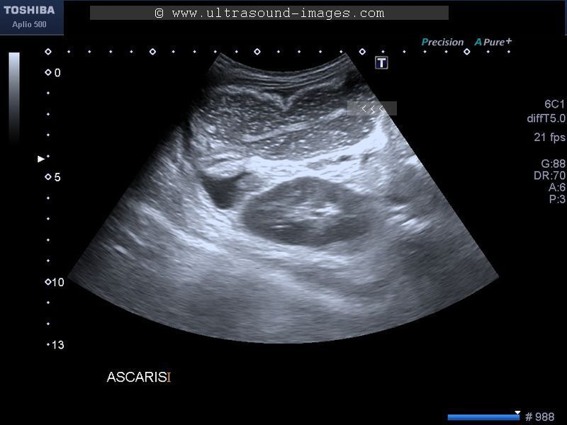
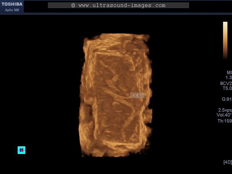
Ascariasis or round worm infestation is known to cause intestinal obstruction. This child has multiple round worms, distributed over a wide part of the small intestine. Ultrasound images show echogenic linear structures with hypoechoic central line within, a characteristic finding in round worm infestation or ascariasis. These B-mode and 3D ultrasound images of ascariasis are courtesy of Dr. Durr-e-Sabih, MBBS, FRCP.
References: Sonography in biliary ascariasis- sonoworld
henoch-schonlein-purpura
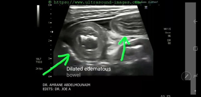
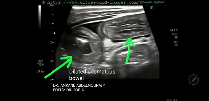

Henoch Schonlein purpura is an immune mediated (auto-immune) vasculitis or inflammation of the small arteries resulting in hemorrhage.
= it can affect the skin, GIT (gastro-intestinal tract), kidneys and joints.
= the ultrasound images and video above show a case of Henoch-Schonlein-purpura involving the GIT (in this case the small bowel) in a 7 year old child
= age of involvement is usually 3 to 10 years.
= the ultrasound images show bowel wall edema and thickening
= sonography of the bowel shows dilated bowel loops with loss of peristalsis (a typical feature of Henoch-Schonlein-purpura)
= another feature of Henoch-Schonlein-purpura is significant increase in vascularity of the small bowel (see the color Doppler ultrasound image).
= Intussusception is a known complication in cases of Henoch-Schonlein-purpura.
This ultrasound video of Henoch-Schonlein-purpura is courtesy of Dr. Amrane Abdenmounaim, MD, Bordj-bou arreridj Medical imaging center, Algeria.
Edits by me Joe Antony, MD.
References: sonography of GIT and bowel in Henoch-Schonlein-purpura
© 2026, Joe Antony, MD All images and content in this site are copyrighted. www.ultrasound-images.com When visiting this site, we may use cookies to improve the performance of this site. Use of this site means you accept the use of cookies. Read our privacy policy at this page: https://www.ultrasound-images.com/privacy-policy/