
Ultrasound images of diseases of the spleen
Contents of this page
- Ectopic spleen
- Splenosis
- Rupture of spleen and treatment of hemorrhage using embolization coils
- splenunculus-accessory-spleen
Ectopic spleen
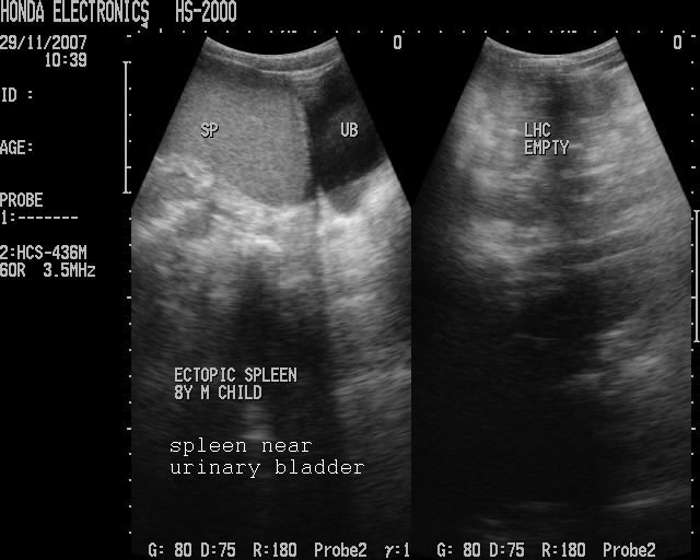
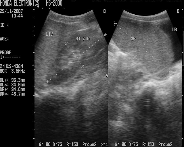
This patient was a young male child who presented with pain in the left iliac fossa. Ultrasound images reveal absence of the spleen and left kidney in left hypochondrium. A structure showing echogenicity and texture similiar to the spleen was seen in left iliac fossa next to the urinary bladder. These ultrasound images are diagnostic of pelvic spleen (ectopic spleen). The left kidney was absent in this case. The right kidney appears mildly enlarged and is seen in the right hypochondrium. Images taken using the HS2000 (Honda Electronics). Sonographic images courtessy of Dr. Tariq Mahmood, Pakistan.
Reference:
1) http://jdm.sagepub.com/cgi/content/abstract/8/1/25(abstract)
2) http://rad.usuhs.edu/medpix/medpix.html?mode=single&recnum=2007(free article and images)
Splenosis

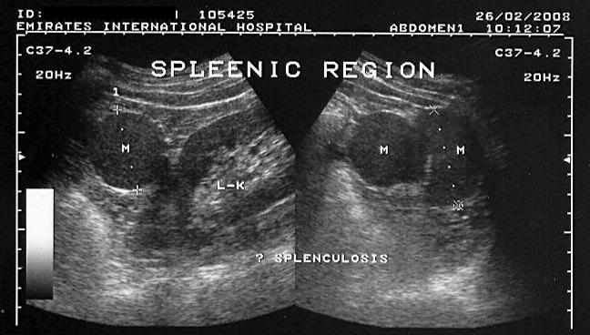
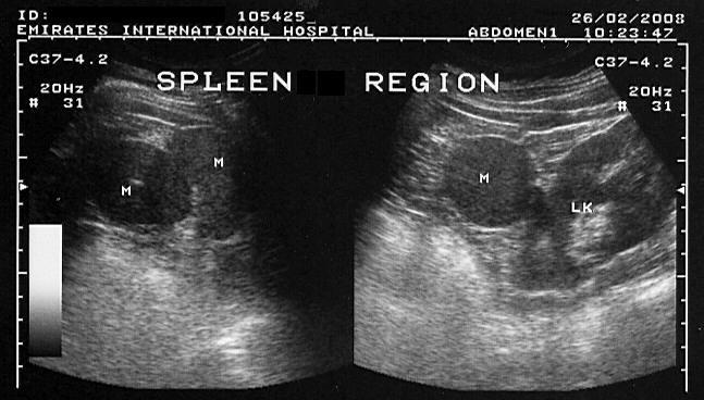
Splenosis following trauma
This patient had splenic rupture following severe trauma, for which splenectomy was done. Subsequent ultrasound imaging showed a large, rounded, well defined mass lesion in the left hypochondrium close to the left kidney. The lesion measured about 4 cms. in size. These ultrasound images suggest splenosis. Splenosis is the autotransplantaion of splenic tissue following splenic injury. Images courtesy of Dr. Ravi Kadasne, UAE (using a Toshiba Powervision Machine).
Reference: http://www.pubmedcentral.nih.gov/articlerender.fcgi?artid=1451006 (free article)
Rupture of spleen and treatment of hemorrhage using embolization coils
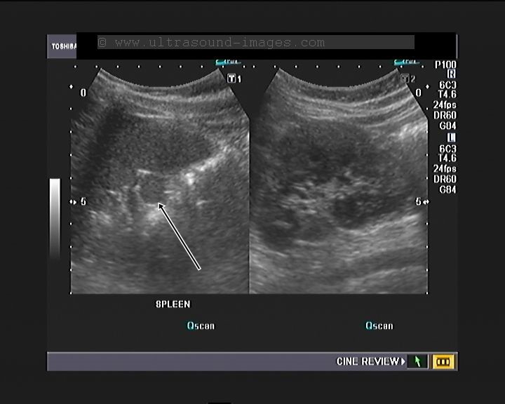
Ultrasound images showing splenic hematoma before coil embolization
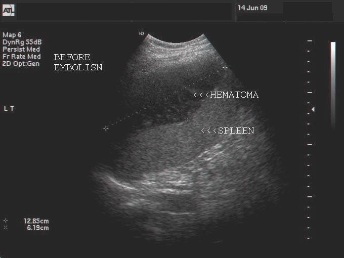
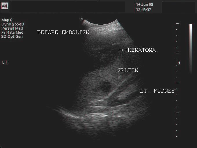
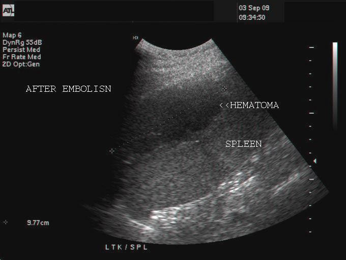
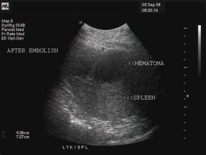
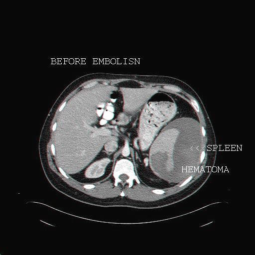
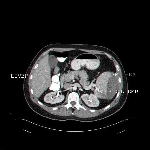
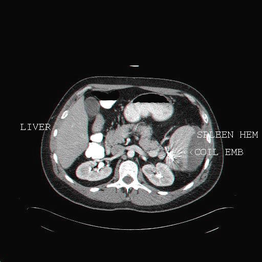
This patient had a rupture of spleen with large hematoma in the spleen. This is seen in the ultrasound images of the spleni hematoma (images on topmost row). The splenic hematoma measured almost 12 x 6 cms. Coil embolization of the splenic artery was done to control the bleeding.Sonography of the spleen shows (2nd row from top) after embolization shows reduction in the size of hematoma. CT scan (also called CAT scan) images of the splenic hematoma also show reduction in size of the hematoma. The coil is seen as a markedly hyperdense object in the splenic hilum (arrowhead- COIL EMB in CT images). There are 2 types of coils used for arterial embolization, the microcoils and macrocoils. All above images are courtesy of Shlomo Gobi, Israel.
Reference: http://emedicine.medscape.com/article/419614-overview (free article and images).
splenunculus-accessory-spleen

This patient shows an incidental finding on routine sonography of the abdomen. There is a rounded "mass" of 14 mm. in the region of the splenic hilum. This is an accessory spleen or splenunculus as it is known and shows the same echogenicity and texture as the normal spleen seen adjacent to it. The splenunculus is a normal variant and is the result of the failure of fusion of the multiple islands of splenic tissue into one organ (the spleen) during fetal life.
© 2026, Joe Antony, MD All images and content in this site are copyrighted. www.ultrasound-images.com When visiting this site, we may use cookies to improve the performance of this site. Use of this site means you accept the use of cookies. Read our privacy policy at this page: https://www.ultrasound-images.com/privacy-policy/