
Ultrasound images of diseases of the urinary bladder
Contents of this page
- Large urinary bladder calculus
- Multiple calculi in urinary bladder
- IUCD in urinary bladder
- Urinary bladder wall trabeculation in a case of Lower urinary tract obstruction
- Ureterocele
- Ureterocele seen on TRUS (Transrectal ultrasound) imaging
- Ureterocele with calculus
- Ureterocele - 3D ultrasound image
- Carcinoma of Urinary Bladder
- Case-2: Carcinoma of urinary bladder
- Diverticulum of urinary bladder
- Urostomy / Continent Pouch- Continent urinary diversion:Ultrasound study of Bladder replacement surgeries
- Bilharziasis (Schistosomiasis) of the urinary bladder
- Urachal cyst in urinary bladder
- Ultrasound images of polyp (inverted papilloma) in urinary bladder
- Cystitis cystica-sub mucosal cyst of bladder
- Endoscopic Teflon or Deflux gel treatment for Vesico-ureteral reflux
- Urinary bladder hernia
- Carcinoma urinary bladder- 3D ultrasound images
- normal urinary bladder-3d ultrasound
- calculus in female urethra
- 3D ultrasound- multiple bladder calculi
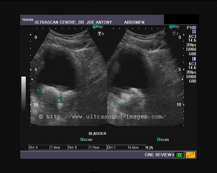
Large urinary bladder calculus
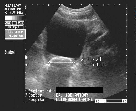
This 50 yr. old male patient complained of hematuria. Sonography reveals a large echogenic,oval object of 4.4 cms. in the urinary bladder. As the ultrasound images reveal, it is freely mobile with change in position, occupying the dependent part of the bladder. Diagnosis: large stone in the urinary bladder. Urinary bladder calculi are usually formed by migration of stones from the kidney or ureter. It may also be caused due to stasis of urine in the bladder, due to bladder outlet obstruction. Ultrasound images taken using Pie Scanner 100 Falco system.
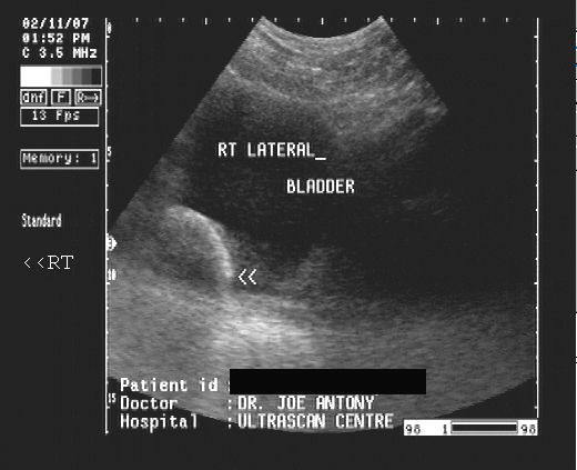
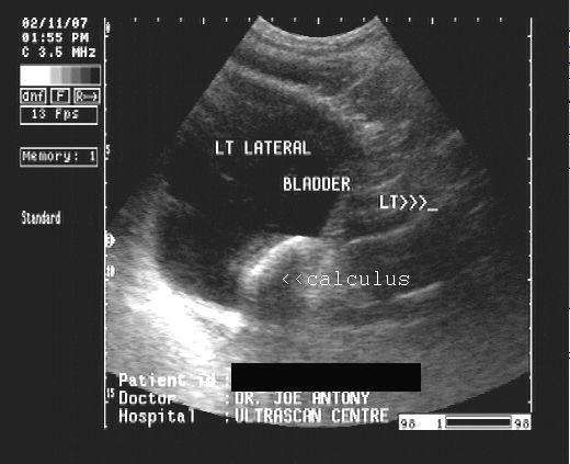
Case-2- huge urinary bladder calculus


This male patient has a huge bladder calculus, measuring more than 5 cm in diameter. The ultrasound images above show the convex external surface of the urinary bladder calculus. The vesical calculus is freely mobile and is seen to shift to the dependent area with change in posture. Such large bladder calculi often produce significant obstruction to the outflow of urine from the urinary bladder, as in this case too.
Multiple calculi in urinary bladder
Urinary bladder- multiple large stones
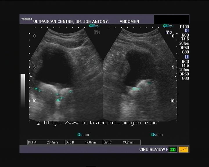
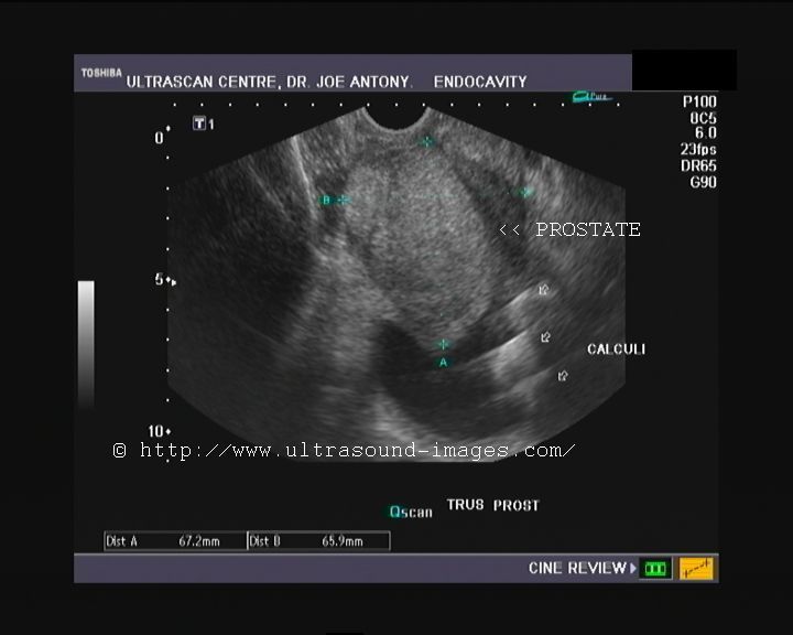
This patient was an elderly male with moderate prostatic symptoms. Transrectal sonography showed a severely enlarged prostate (benign prostatic hypertrophy). But at least part of the symptoms were due to multiple large stones in the urinary bladder. Both transabdominal and transrectal sonography show the bladder calculi gravitating to the dependent part of the urinary bladder. The cause of these multiple bladder calculi is obviously the persistent urinary tract obstruction due to the enlarged prostate, resulting in incomplete evacuation of the urine. The urinary bladder stones varied in size from 15 to 27 mm., and I could count at least 4 of them. The patient had severe urgency and incontinence.
View the videos of this case: Ultrasound video clip of multiple, large bladder stones
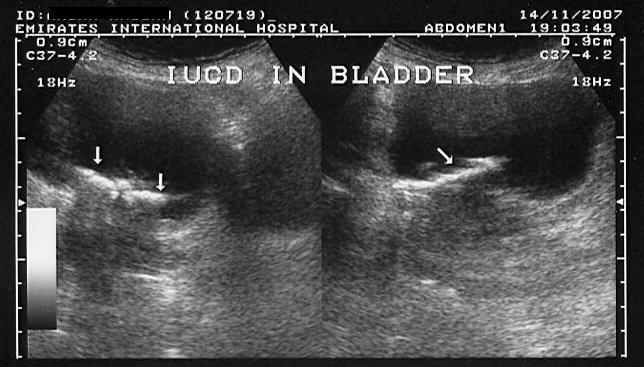
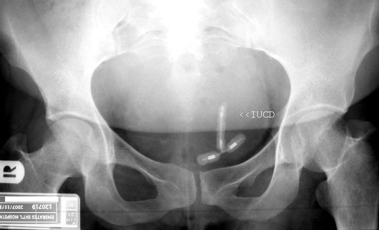
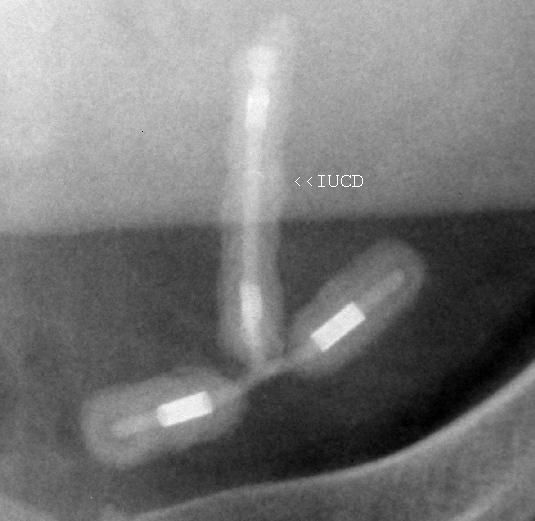
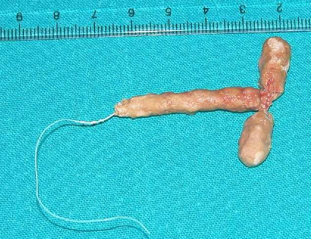
(Intrauterine contraceptive device in Urinary bladder)\
Sonography of the pelvis in this female patient revealed an echogenic linear object with posterior acoustic shadowing in the urinary bladder. X-ray images reveal the object to be a T-shaped structure. Ultrasound and X-ray images are diagnostic of a Copper-T (IUCD or intrauterine contraceptive device) which migrated to the urinary bladder. A snap of the specimen after removal is also seen. It appears to be coated with urinary sediment. Images courtesy of Dr. Ravi Kadasne, UAE.
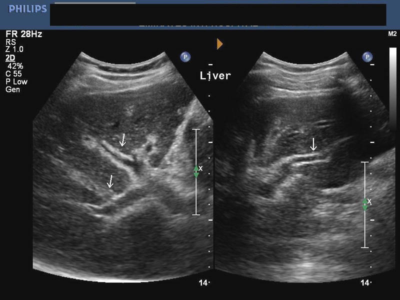
Urinary bladder wall trabeculation in a case of Lower urinary tract obstruction
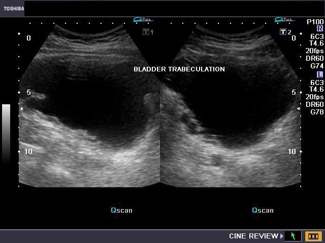
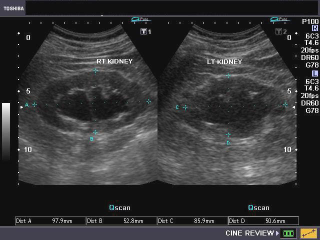
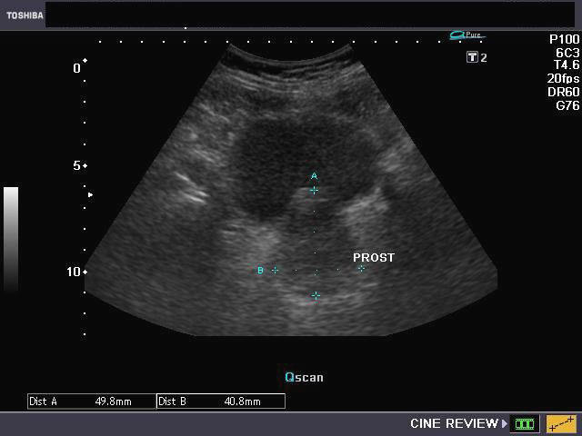
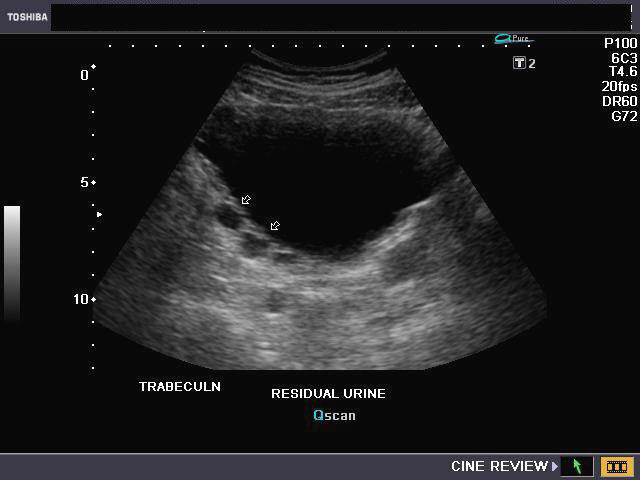
Sonography of the urinary system was done on this elderly male patient having lower urinary tract symptoms. Ultrasound images show evidence of trabeculation of the urinary bladder. This is seen as folds of hypertrophied bladder mucosa and bladder smooth muscle. There is also evidence of bilateral moderate hydronephrosis (image top right). The cause of Lower urinary tract obstruction appears to the enlarged prostate (benign prostatic hypertrophy) with intravesical enlargement of the median lobe (image on lower left). The fourth image shows significant post-voiding residual urine in the urinary bladder (Ultrasound image on lower right).
Bladder trabeculation has been graded from 0 to 3 as:
grade 0- no trabeculation.
grade1- mild: area affected is less than 1/2 of the bladder and depth of trabeculation less than 5 mm.
grade2- moderate: area affected is greater than 1/2 of the bladder and depth of trabeculation is 5 to 10 mm.
grade 3- severe: area affected is greater than 1/2 of the bladder and depth of trabeculation is greater than 10 mm.
All images by Joe Antony, MD, using a Toshiba Nemio-XG ultrasound system.
References:http://www.ncbi.nlm.nih.gov/pubmed/23452803
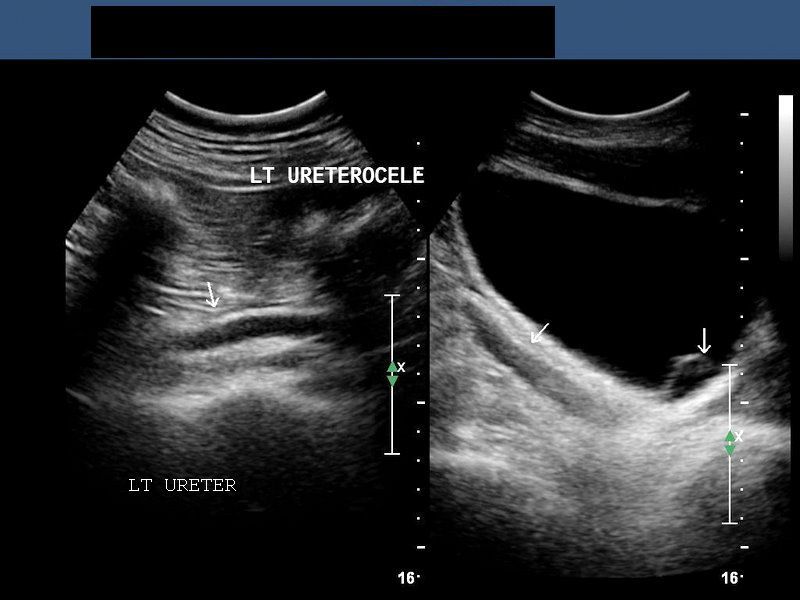
Ureterocele

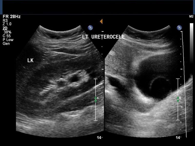

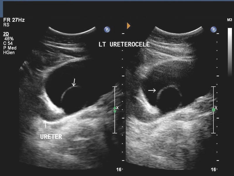
Sonography of the urinary bladder done on this patient, revealed a saccular outpouching of the distal end of the left ureter, into the distended urinary bladder. The left ureter also appears dilated (left hydroureter). These ultrasound images are diagnostic of left ureterocele. Ureteroceles are caused due to congenital obstruction of the ureter during the embryonic stage. Here the left kidney also shows back-pressure changes (left hydronephrosis). Observation of the orifice shows gradual distension of the membrane of the ureterocele sac and then collapse of the sac after evacuation of the contained urine into the bladder lumen. Ultrasound images courtesy of Dr. Ravi Kadasne, UAE.
Reference:
http://emedicine.medscape.com/article/451105-overview(free article)
http://radiographics.rsna.org/content/20/1/155.full(free article and images)
Ureterocele seen on TRUS (Transrectal ultrasound) imaging
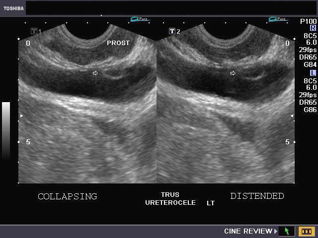
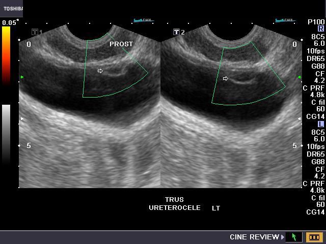
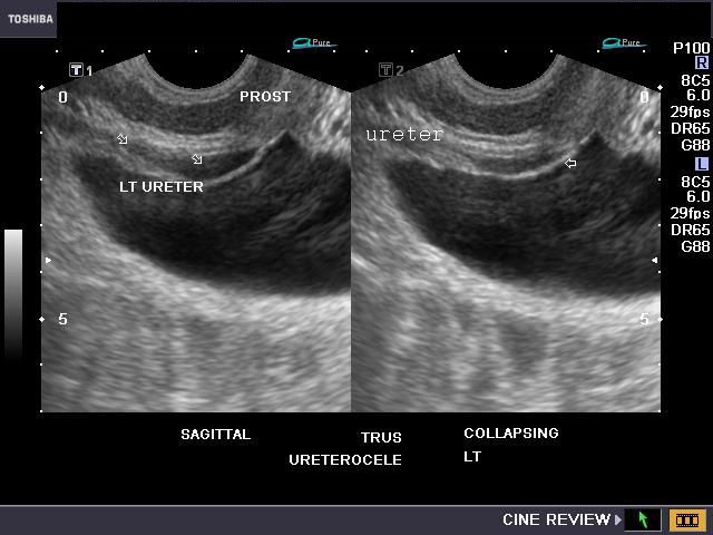
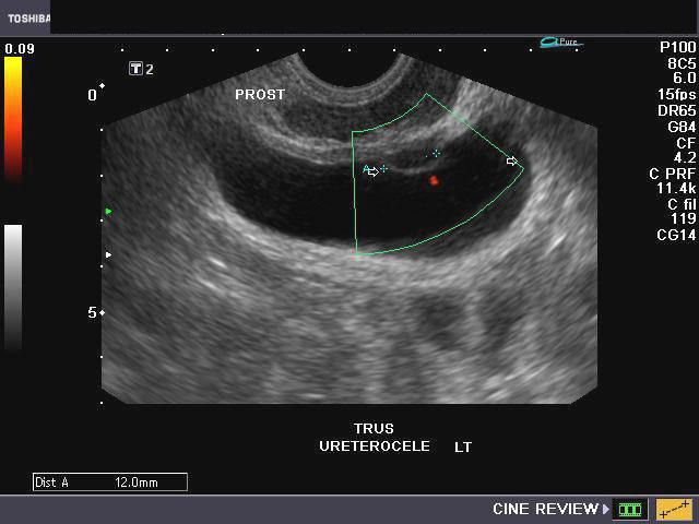
The above ultrasound images show transrectal imaging of the urinary bladder with a small left ureterocele visible. The left ureter also appears mildly dilated (hydroureter). The ureterocele is seen partially distended and also seen in the collapsing stage as the pressure builds up within the sac (of the ureterocele) with resultant evacuation of the urine into the bladder (seen on Power Doppler image- lower right). The jet of urine is seen emanating from the ureterocele sac. All 4 ultrasound images taken via TRUS study using Toshiba Nemio-XG system.
Ureterocele with calculus
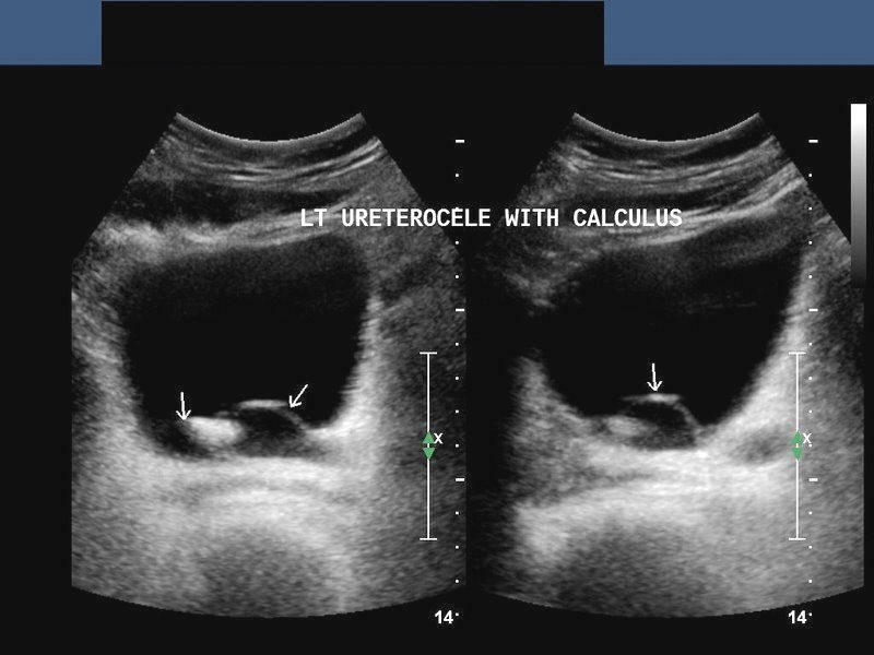
This patient underwent sonography of the abdomen for suspected calculus disease. Ultrasound images show left hydroureter with calculus within the left ureterocele. The image on left shows the dilated left ureter in long section. Ultrasound image on right shows the urinary bladder with the calculus impacted within the ureterocele. Both images are courtesy of Ravi Kadasne, MD, UAE.
Ureterocele - 3D ultrasound image
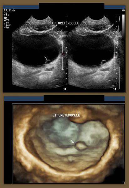
4D3D Ultrasound image (using Philips Matrix probe) showing left ureterocele (arrows). The Philips Matrix 3D system shows color coded 3D real time images. This image is courtesy of Dr. Ravi Kadasne, MD, UAE.
Case-2:
4D ultrasound video (courtesy of Dr. Vishal Kumat, MD) shows a left ureterocele (green arrows).
Carcinoma of Urinary Bladder
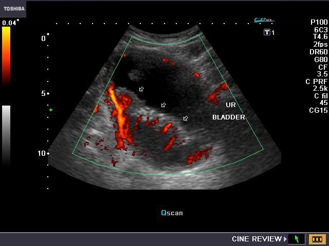

This elderly male patient presented with hematuria. Sonography of the urinary bladder showed: 1) a large cauliflower shaped mass producing thickening of the bladder wall (the lower half of the urinary bladder was affected). 2) Power Doppler images (top row) show considerable vascularity of the mass (arrows). Sonography of the kidneys show bilateral moderate hydronephrosis with bilateral hydroureter. This suggests obstruction of the distal ends of both ureters, probably at the level of both vesico-ureteric junctions. TRUS (transrectal ultrasound) imaging shows mild prostatic enlargement (benign prostatic hypertrophy). TRUS study also shows complete emptying of the urinary bladder with no post-voiding residual urine. However, the highly vascular mass is seen to involve a large portion of the urinary bladder (lower right). These ultrasound and Power Doppler images are suggestive of carcinoma of the urinary bladder. Images taken using a Nemio-XG ultrasound system by Joe Antony, MD, India.
Case-2: Carcinoma of urinary bladder
Ultrasound images of urinary bladder (mass) and both kidneys
Transabdominal ultrasound images show a polypoid mass in the bladder close to the bladder neck. Is this a bladder mass or an enlarged median lobe of the prostate? Faced with this dilemma we decided to perform a transrectal ultrasound scan (TRUS). The kidneys were almost normal but for a small calculus in left kidney.
TRUS ultrasound images of the prostate and bladder:


Coronal image Sagittal color Doppler images shows bladder mass: TRUS image showing relation of the masses to the prostate
Transrectal ultrasound showed not one but two contiguous masses on either side of the bladder neck and almost in close contact with the prostate. However, the prostate appeared non vascular and small in size compared to the irregular, polypoid masses which were very vascular. There does not appear to be any spread beyond the confines of the bladder wall. Final diagnosis- these are a pair of malignant masses of the urinary bladder- carcinoma of urinary bladder. This patient had severe urinary obstruction- a result of the location of the bladder tumors near the bladder neck. He had a history of undergoing surgery for bladder carcinoma. Clearly this was a case of recurrence of bladder malignancy. The video clip above shows the TRUS scan study of this patient. Note the clear separate nature of the masses from the prostate.
TRUS video clip of the bladder masses:
See above video clip of carcinoma bladder as seen on TRUS scan.
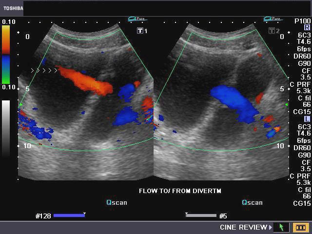
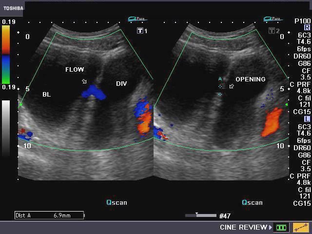
Diverticulum of urinary bladder

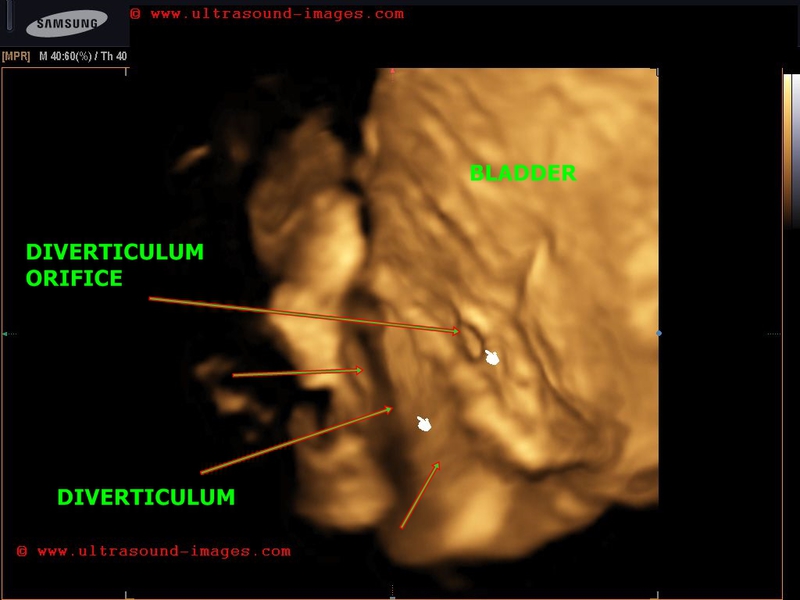
case-2-bladder-diverticulum-3D-ultrasound
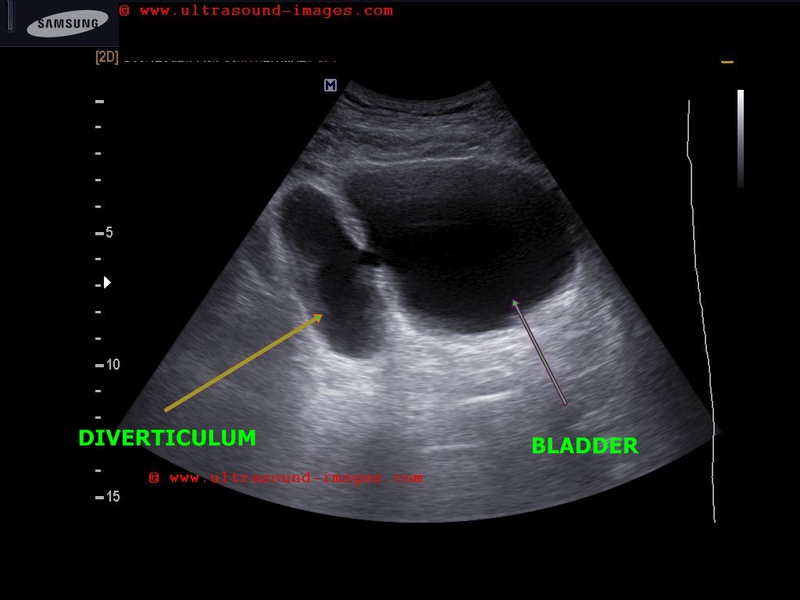

This elderly male patient had symptoms of lower urinary tract obstruction. Sonography of the abdomen shows a large sac like structure, communicating freely with the urinary bladder through a small orifice (6 mm. in size). These ultrasound images are diagnostic of diverticulum of the urinary bladder. Color Doppler images show to and fro flow across the orifice between the bladder and the diverticulum. Surprisingly, the kidneys appear normal in structure.
Case-2: shows 3D ultrasound images of bladder diverticulum with the orifice or neck leading to the diverticulum seen well.
Reference:
http://www.medcyclopaedia.com/library/topics/volume_iv_2/d/diverticulum_bladder.aspx
http://emedicine.medscape.com/article/1015329-overview
Urostomy / Continent Pouch- Continent urinary diversion:Ultrasound study of Bladder replacement surgeries
a) Artificial bladder made from small bowel

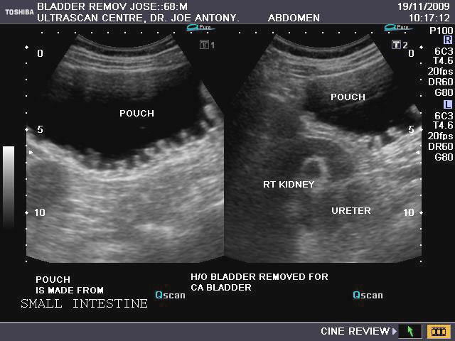
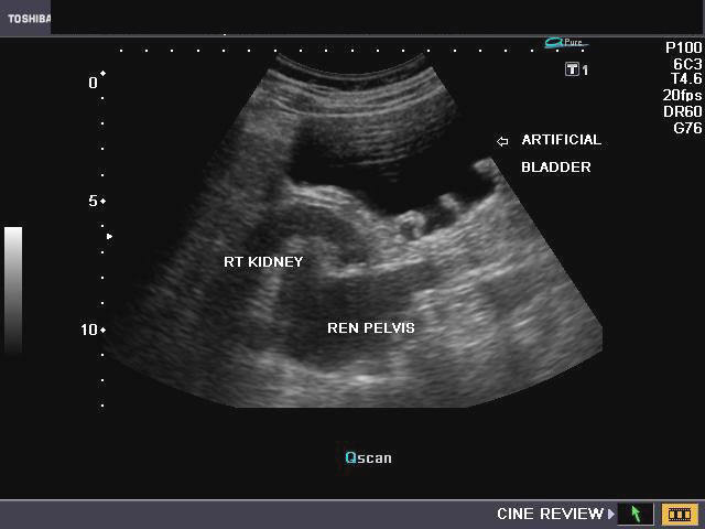
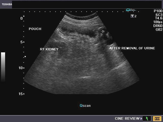
This elderly patient underwent surgical removal of the urinary bladder for malignant bladder mass (carcinoma). A urostomy was done with an artificial bladder (pouch) created using a segment of small bowel. The structure (pouch) is seen fully distended in the ultrasound images in the top row and bottom-left. The image on bottom right shows the pouch after evacuation of the urine via catheter inserted via stoma in the abdominal wall. Note the close relation of the pouch to the right kidney. Such a pouch is also called a continent cutaneous reservoir. Urine collects in this reservoir within the abdomen till it is full. Urine is drained via a stoma (opening in the abdomen connected to the continent cutaneous reservoir). Both kidneys (top-left) show mild pelvicalyceal dilation.
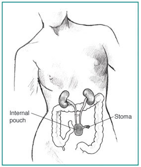
Anatomy of the continent cutaneous reservoir
b) Sigmoid pouch (sigma pouch) or uretero-sigmoidostomy
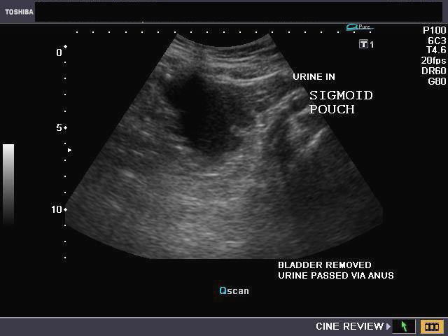
This male, elderly patient underwent cystectomy (surgical removal of urinary bladder) for carcinoma of the organ. A bladder replacement surgery in this case was done by creating an artificial pouch using the sgmoid colon (sigmoid pouch) with the ureters emptying in to this pouch (uretero-sigmoidostomy). Ultrasound images above show a partially (urine) filled sigmoid pouch in this study of the pelvis. It is important to do a follow up ultrasound study of the urinary tract to rule out pyelonephritis in such cases. Here the kidneys appear normal.
Reference:
http://www.ncbi.nlm.nih.gov/pubmed/16336348
http://www.moffitt.org/moffittapps/ccj/v3n6/a4.html
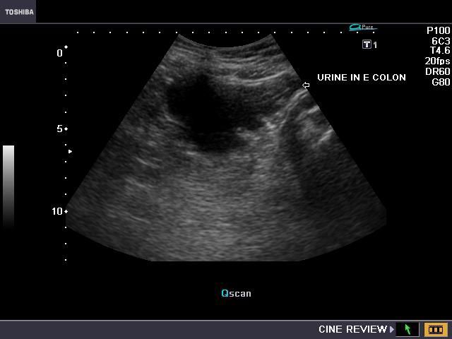
c) Orthotopic neobladder or neobladder to urethra diversion
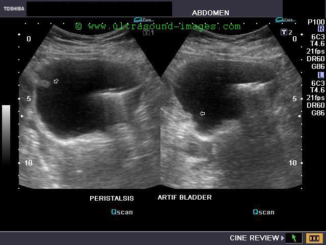

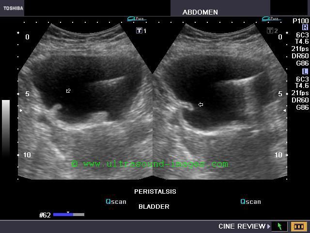
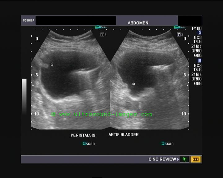
he above ultrasound pictures are those of a middle aged female patient with history of severe tuberculous infection of the urinary tract. Severe infection of the urinary bladder (cystitis) resulted in severe urinary complaints. The patient underwent surgical removal of the urinary bladder (cystectomy) with an artificial urinary bladder made out of the terminal ileum and caecum. The outlet of this ileo-caecal neobladder (artificial bladder) was connected to the patient's urethra. This type of surgery is called orthotopic neobladder as the new bladder is placed in the normal bladder location (in the lower pelvis). The ultrasound images of the neobladder to urethra diversion, show multiple peristaltic waves (arrows) caused by contractions within the distended urinary neobladder. This patient has to empty the bladder every 4 to 5 hours using a simple catheter, due to real possibility of rupture of the distended neobladder.
References: Article on types of urinary diversions (free excellent article)
Discussion on types of urinary diversions and neobladder options (for patients and laypersons)
Bilharziasis (Schistosomiasis) of the urinary bladder
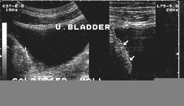
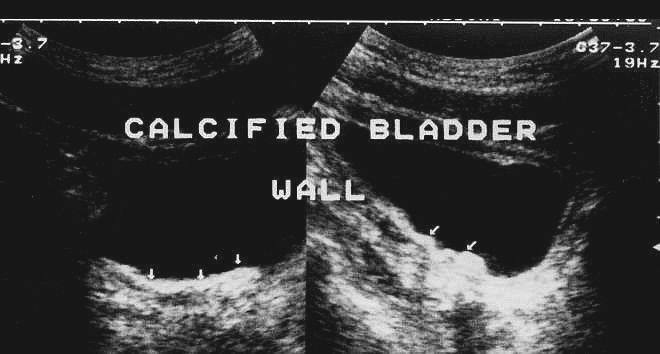
This patient presented with lower urinary symptoms, dysuria and hematuria. Sonography of the pelvis showed thickening of the wall of the urinary bladder with extensive calcification. These ultrasound images suggest a diagnosis of schistosomiasis or bilharziasis of the wall of the urinary bladder. Bilharziasis is a parasitic infestation which primarily involves the urinary bladder, though the liver and spleen may also be affected. The disease is caused by contact with water infested with the parasite- schistosoma and is endemic in parts of Africa (Egypt and Sudan). Both above images are courtesy of Ravi Kadasne, MD, UAE.
CT Scan imaging in Bilharziasis
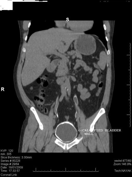

The above CT (computerized tomography) images (another patient) show hyperdense, extensive involvement of the urinary bladder suggesting calcification of the bladder wall. This is a typical appearance of Schistosomiasis of the urinary bladder and is caused by calcification of the dead schistosoma parasites or their eggs. CAT scan images are courtesy of Nirmali Dutta, MD, UAE.
Liver/ Spleen involvement in Schistosomiasis
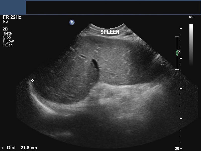
his patient is a known case of Bilharziasis and ultrasound showed hepatosplenomegaly with increased echogenicity of the periportal regions of the portal veins suggesting periportal fibrosis. Fibrosis of the periportal regions of the liver is a known complication of hepatic involvement in schistosomiasis. Ultrasound images are courtesy of Ravi Kadasne, MD, UAE.
Reference:
1)http://emedicine.medscape.com/article/377318-overview
2)http://en.wikipedia.org/wiki/Schistosomiasis
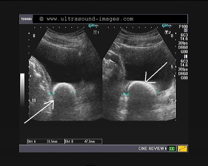
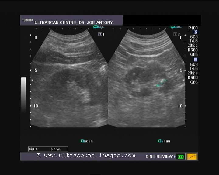
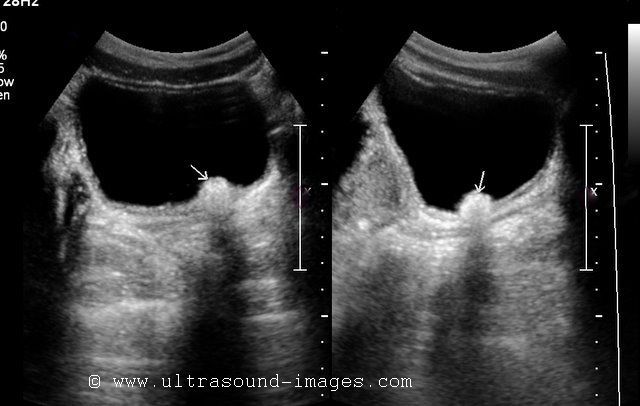
Urachal cyst in urinary bladder
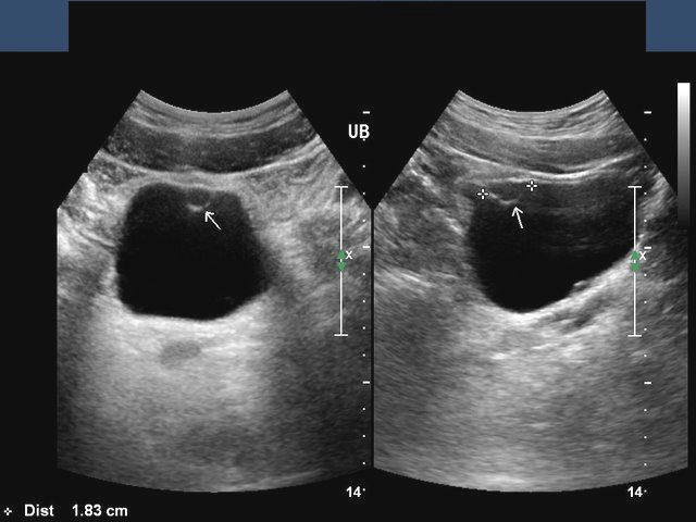
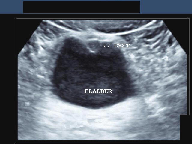
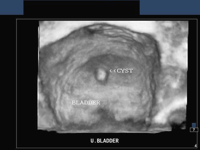
This patient underwent sonography of the urinary bladder which revealed a small cystic structure with well defined wall and turbid fluid content, protruding into the lumen of the bladder, from its dome. The ultrasound images at bottom and on top-right show 3-D display of the cyst (the image at bottom row shows a surface rendering 3-D image of the lesion). These ultrasound appearances are suggestive of urachal cyst involving the urinary bladder. A urachal cyst can occur anywhere between the ends of the obliterated part of the urachus. Most urachal cysts occur close to the umbilicus, ie: the upper part of the urachus. Images are courtesy of Ravi Kadasne, MD, UAE.
Reference: http://emedicine.medscape.com/article/1015329-overview(free article)
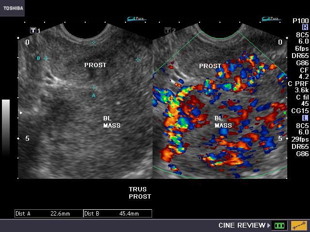
Ultrasound images of polyp (inverted papilloma) in urinary bladder
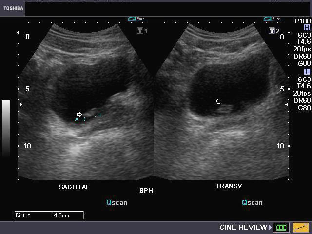

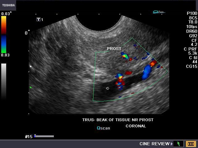
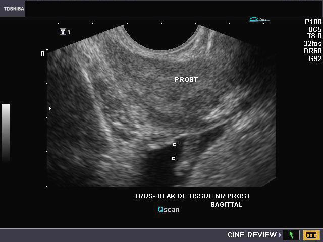
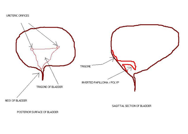
This polyp like mass was seen close to the bladder neck on transabdominal ultrasound imaging (image on top row-left). It measured 14 x 5 mm. and showed considerable vascularity on color Doppler imaging (see image in bottom -left). The TRUS ultrasound images (Transrectal ultrasound) show the polyp clearly arising from the urinary bladder mucosa (lower part of trigone), close to the prostate. Such polyps can give rise to obstructive voiding symptoms (as in this patient) due to its tendency to block the adjacent bladder neck by a valvular mechanism. This patient underwent a biopsy of the polyp to rule out malignancy of this mass. This was found to be an inverted papilloma of the urinary bladder on cystoscopy, and was confirmed by histopathological study. The sketch (bottom row) shows the trigone of the bladder with the inverted papilloma arising from the lower part of the trigone, producing obstruction at the bladder neck.
Reference:
http://emedicine.medscape.com/article/1788400-overview
http://www.ajronline.org/cgi/reprint/159/1/93.pdf( excellent article-free)
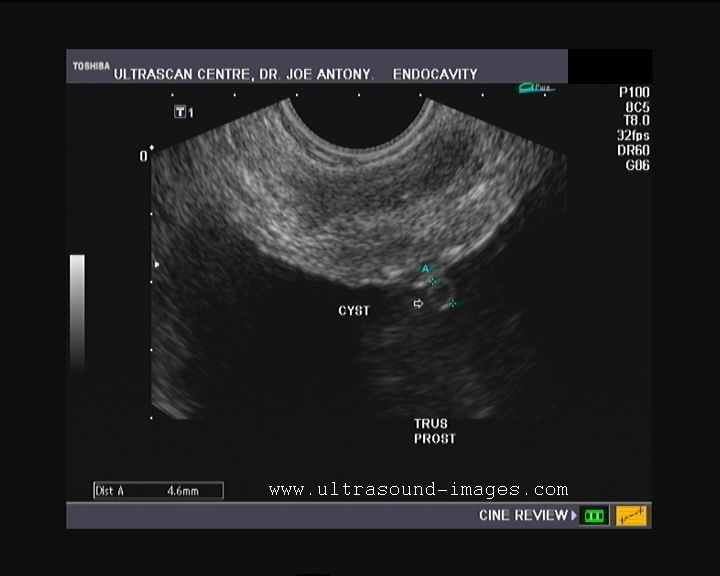
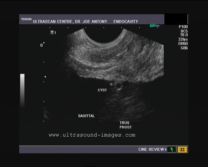
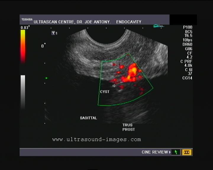
Cystitis cystica-sub mucosal cyst of bladder

Coronal TRUS Sagittal TRUS
This elderly male patient shows a distinct, small cyst on the mucosal surface of the bladder just above the neck of bladder, and in close proximity to the upper surface of the prostate in this TRUS image of the urinary bladder and prostate. Transrectal ultrasound was done to evaluate the prostate following treatment for proven carcinoma of the prostate. Presently this patient has a few episodes of hematuria.
The above images are TRUS studies of the prostate; Power Doppler image on right shows the lack of significant vascularity to the cystic lesion in the submucosal region of the urinary bladder. The urinary bladder cyst in this case measures just 4.5 mm. in size and is thin walled, almost sac like. These ultrasound images and appearances of this submucosal cyst of the urinary bladder are highly suggestive of cystitis cystica. Cystitis cystica is a relatively rare and poorly understood lesion of the urinary bladder mucosa resulting from cyst formation within hypertrophied clusters of bladder mucosal cells. There are 2 schools of thought regarding the significance of cystitis cystica of the urinary bladder. Some believe this lesion to be a pre-malignant condition whilst others maintain, that this (cystitis cystica) is just an indientaly finding of little significance. Other differential diagnoses in this case include retention cysts of the prostate or urinary bladder.
See ultrasound video of this case at:
http://ultrasound-videos.blogspot.com/2011/06/cystitis-cystica-transrectal-ultrasound.html
References:
http://www.ispub.com/ostia/index.php?xmlFilePath=journals/iju/vol4n2/cystitis.xml
http://www.ncbi.nlm.nih.gov/pmc/articles/PMC2006711/pdf/amjpathol00621-0065.pdf
http://vet.sagepub.com/content/18/1/113.full.pdf
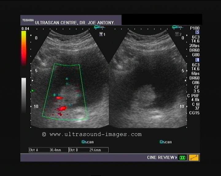
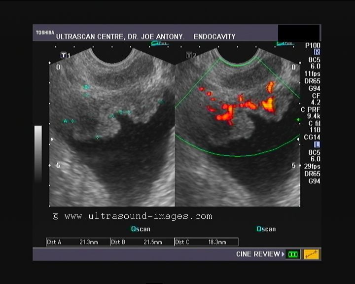
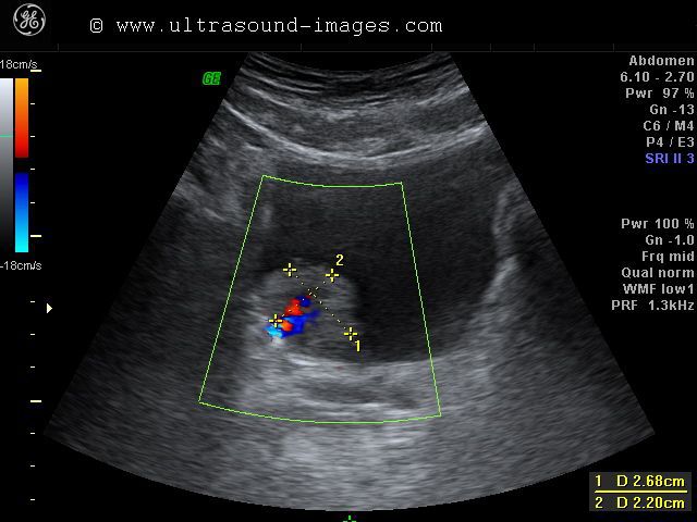
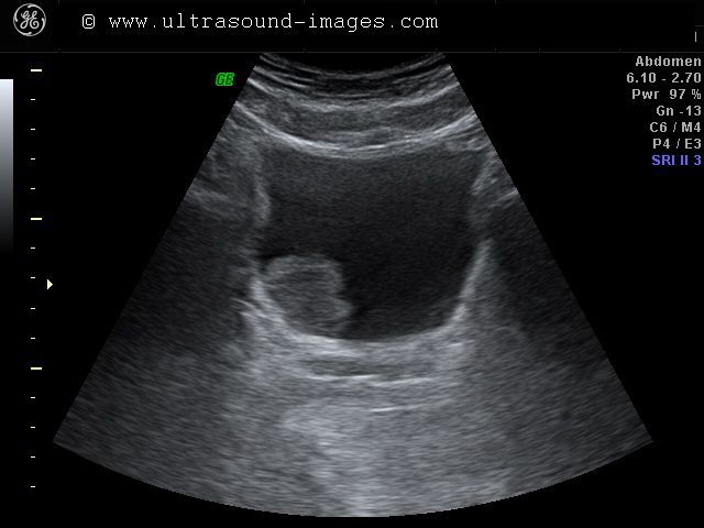
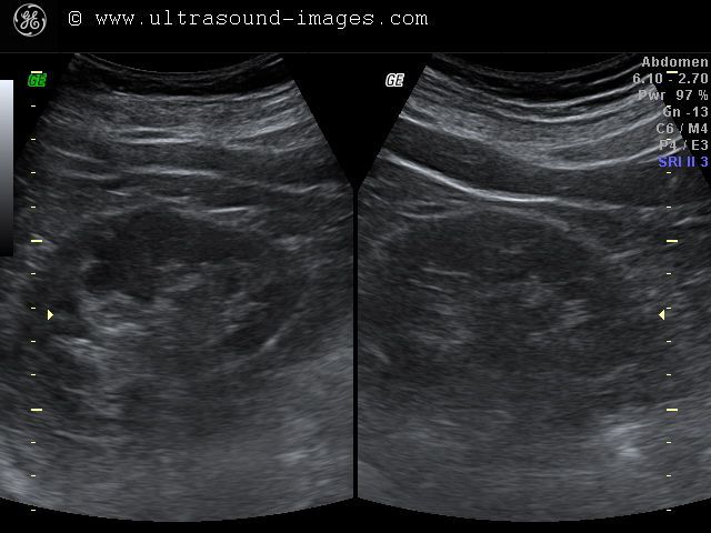
Endoscopic Teflon or Deflux gel treatment for Vesico-ureteral reflux
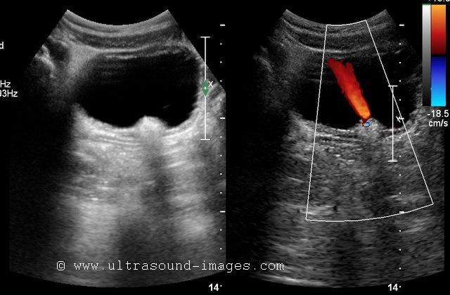

This patient shows an echogenic mound in the left vesico-ureteric junction. The Color Doppler image shows a ureteric jet emerging from this region suggesting that the left distal ureteric orifice is patent. This patient had a history of vesico-ureteric (vescio-ureteral) reflux. This was corrected by a Teflon gel injected in the submucosal part of the left VUJ (vesico-ureteral junction) via the endoscopic route. There are 5 grades of vescio-ureteral reflux. Grade-1: the VUR reaches below the renal pelvis. Grade-2: VUR reaches up to the renal pelvis without causing dilation of the pelvis. Grade-3: There is mild to moderate dilation of the renal pelvis and ureter. Grade-4: Moderate dilation of renal pelvis, ureter and calyces is present. Grade-5: Gross dilation of pelvicalyces with tortuous and dilated ureter. Endoscopic deflux or Teflon gel injection is used for correcting of VU reflux from grade-2 to grade-5. the gel causes a small mound to form in the submucosal part of the distal ureteral orifice resulting in a kind of valve formation preventing the reflux of urine up the ureter. Teflon is now being replaced by Deflux gel as the preferred material for this procedure. Ultrasound images of endoscopic Teflon gel injection are courtesy of Dr. ravi Kadasne, MD, UAE.
References:
http://urology.ucsf.edu/patientguides/pdf/pedUro/VUR_Endoscopic.pdf(simple article on the basics)
http://pednephrology.stanford.edu/secure/documents/Vesicoureteral-Reflux.pdf ( detailed description)
Urinary bladder hernia
Ultrasound images of urinary bladder herniation into the right scrotum

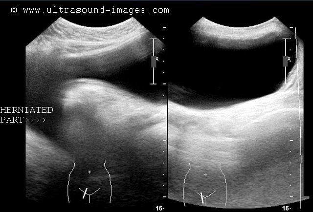

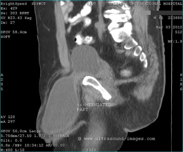
This 65 year old male presented with right inguino-scrotal swelling. Ultrasound images of the swelling showed a cystic fluid containing lesion with a prominent "beak" extending towards the top of the swelling/ lesion. Further, on sonographic imaging it was possible to show its continuity with the distended urinary bladder above. The lesion measured 18 x 9 cms. and showed reduction in size on emptying the urinary bladder. This sign is typical of the lesion/ swelling arising from the urinary bladder herniating through the inguinal canal into the scrotum. 3D ultrasound imaging further confirmed the internal surface of the swelling to be of the same texture as that of the urinary bladder. These images confirm that the swelling in question is an inguinal hernia of the urinary bladder. CT scan section further adds to the validity of the diagnosis. This case study is courtesy of Dr. Ravi Kadasne, MD, UAE. These ultrasound images were taken using a Philips IU 22 ultrasound system.
References: Radiology and sonography of urinary bladder hernia
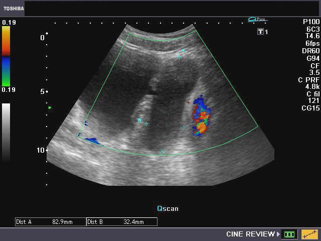
Carcinoma urinary bladder- 3D ultrasound images
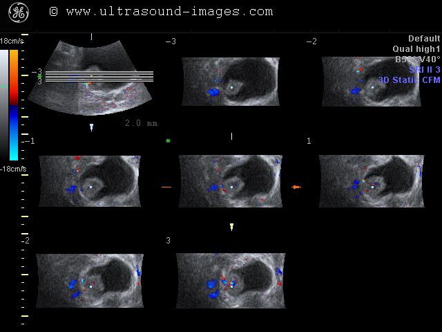

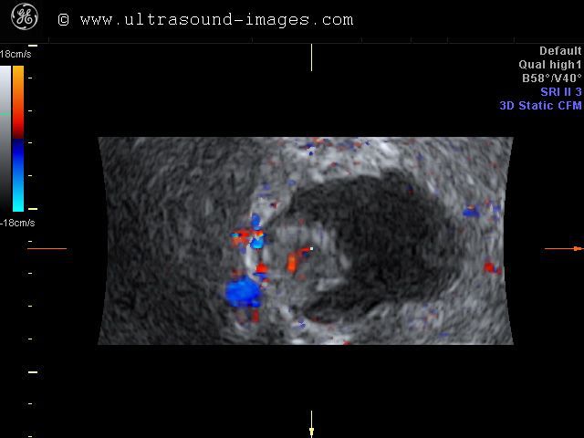
A case of carcinoma of the urinary bladder studied using 3D and 4D ultrasound imaging. The bladder mass is seen as a small cauliflower shaped lesion to the right of the bladder neck. 3D ultrasound and 3D color Doppler images show the surfac features and vascularity of the bladder mass in detail. The color Doppler images show the feeder vessels supplying the malginant bladder mass.
normal urinary bladder-3d ultrasound


what does the normal urinary bladder look like in a 3-D ultrasound image? These two 3-D ultrasound images of the urinary bladder show the inner surface of a male urinary bladder. The mucosa of bladder presents a fine cobblestone appearance in 3-D sonography. This can very well be appreciated in these images.
calculus in female urethra

This female patient complained of dysuria for which she underwent routine sonography. This ultrasound image shows the cause of the dysuria- a calculus lodged in the female urethra. Though calculi are often seen in the male urethra due to its length, it is very unusual to sonographically image a calculus in the female urethra. Despite its small size, such a calculus can produce significant dysuria. This ultrasound image is courtesy of Dr. Ravi Kadasne, MD, UAE.
3D ultrasound- multiple bladder calculi
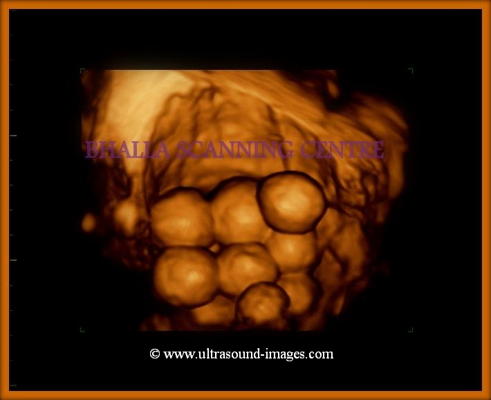
Dr. Sanjiv Bhalla, MD managed to capture this wonderful 3D ultrasound image of multiple urinary bladder calculi. It is rather unusual for a patient to have such a large number of bladder calculi; it is even more unusual to have all the bladder stones of almost the same size and captured in such breath-taking clarity on 3D sonography.
© 2026, Joe Antony, MD All images and content in this site are copyrighted. www.ultrasound-images.com When visiting this site, we may use cookies to improve the performance of this site. Use of this site means you accept the use of cookies. Read our privacy policy at this page: https://www.ultrasound-images.com/privacy-policy/