
Ultrasound images of pathology/ complications of the umbilical cord
Contents of this page
- Umbilical cord prolapse
- Umbilical vein varix
- Single umbilical artery (SUA) or 2 vessel umbilical cord
- Normal 3 vessel umbilical cord
- Nuchal cord or Umbilical cord around fetal neck
- Hypocoiled umbilical cord
- Case-2: hypocoiling of umbilical cord
- Case-3: Hypocoiled umbilical cord
- Normal coiling of the umbilical cord
- umbilical-cord-cysts
- multiple-umbilical-cord-cysts
- absent-ductus-venosus
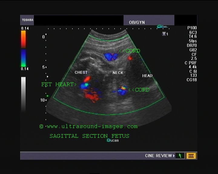
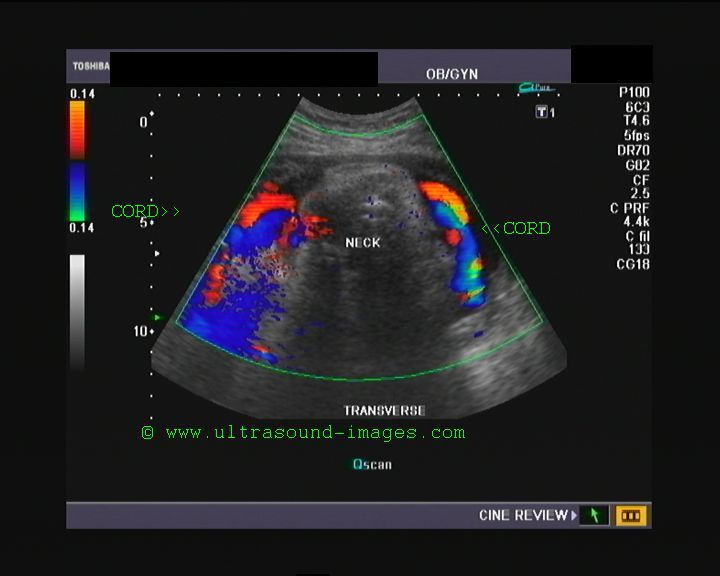
Umbilical cord prolapse
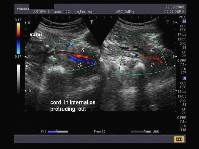
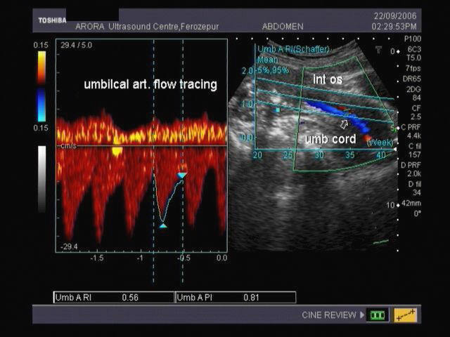
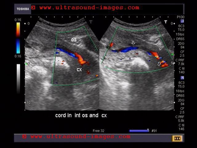
This 21 week gestation had labour contractions with leaking per vaginum. On color doppler scan, the internal os appears dilated with umbilical cord seen within the cervix. These ultrasound and color doppler images are diagnostic of umbilical cord prolapse. Images taken with a Nemio 30 (Toshiba) color doppler machine by Dr. Vikas Arora, Ferozepur, India.
Reference: http://www.emedicine.com/med/topic3276.htm
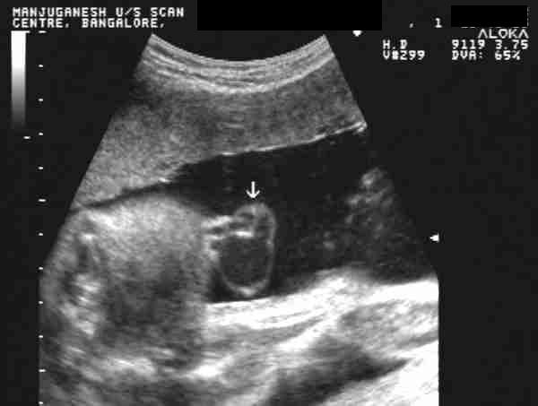
Umbilical vein varix
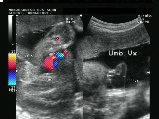
a) Extra-abdominal umbilical vein varix (varix inside the umbilical cord)
Download my E book for Amazon Kindle (download free Kindle reader for i Phone or Android)
Assorted ultrasound cases- by Joe Antony, MD
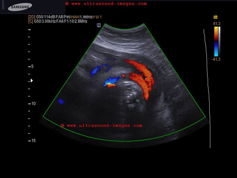
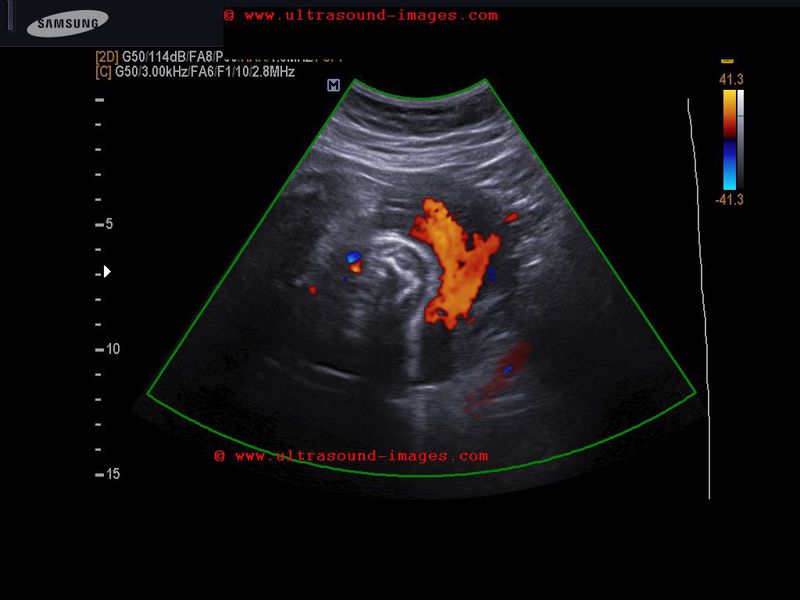
Routine obstetric sonography was done on this patient. Images reveal a cystic lesion in the umbilical cord close to the fetal abdomen. On color doppler scan, flow is present in the cystic area which shows continuity with the umbilical vein. These ultrasound and color doppler images suggest umbilical vein varix (within the cord). Images courtesy of Dr. Latha Natarajan, bangalore, India. She used an Aloka machine for these images.
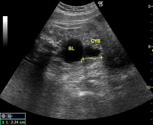
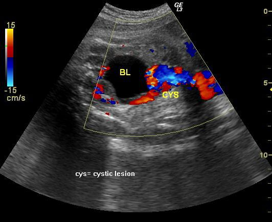
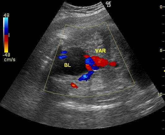

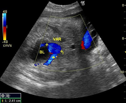
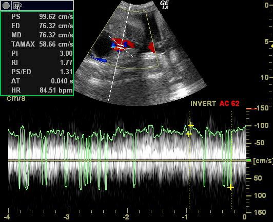
This 3rd trimester fetus shows a focal cystic lesion (2.2 x 2.4 cms.) close to the dome of the fetal urinary bladder. Using color doppler imaging, the lesion shows considerable vascularity, with spectral waveform suggestive of a venous flow. The vascular "cystic" lesion shows continuity with the fetal intra-abdominal umbilical vein suggesting a fetal intra-abdominal umbilical vein varix. No other fetal anomalies were seen.
Images taken using a Logiq 3 Color Doppler machine (GE) by Joe Antony, MD., Cochin, India.
Reference: 1) http://www.sonoworld.com/fetus/page.aspx?id=2838
(free article and images)
2)http://sonoworld.com/fetus/page.aspx?id=2193
(free article and images)
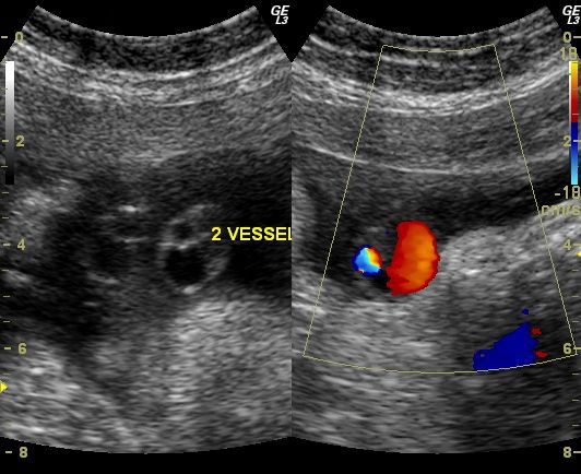
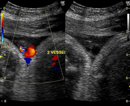
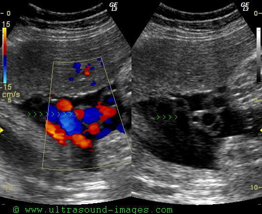
Single umbilical artery (SUA) or 2 vessel umbilical cord
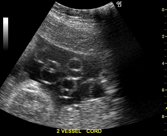
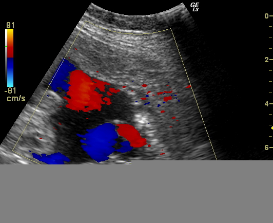
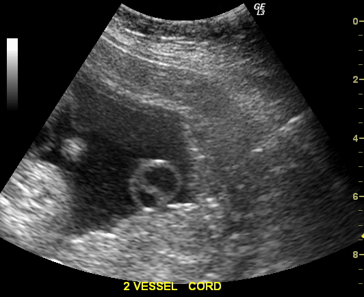
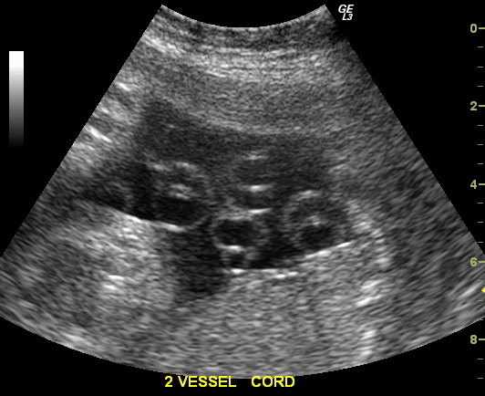
These (above) ultrasound and color Doppler images show a 3rd trimester pregnancy with an interesting anomaly of the umbilical cord. There are only 2 vessels seen in the umbilical cord. Normally, there are 2 umbilical arteries and 1 umbilical vein running within the umbilical cord. However, in this case, there is only one umbilical artery and an umbilical vein within this cord. This ultrasound finding is called a single umbilical artery (SUA) or 2 vessel cord and is associated with increased fetal morbidity including single/ multiple fetal congenital anomalies, still birth and premature delivery. However, no other anomaly was found in this fetus. The ultrasound images above were taken showing transverse sections of the cord along multiple levels to display the SUA (single umbilical artery). Ultrasound images were taken using a GE logiq 3 ultrasound machine.
Some more color Doppler images
References: http://www.medscape.com/viewarticle/453593
http://emedicine.medscape.com/article/262470-overview (free article and images)
Normal 3 vessel umbilical cord
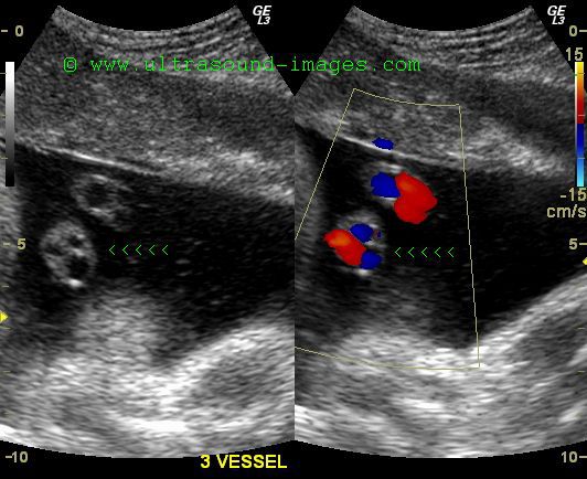
These ultrasound and color Doppler images show the normal umbilical cord with three vessels within it (compare with the 2 vessel cord in previous case). Cross section ultrasound images of the normal umbilical cord show 2 umbilical arteries (the smaller caliber vessels) and the larger caliber umbilical vein. The presence of sufficient liqor (amniotic fluid) helps to highlight the vessels, nicely.
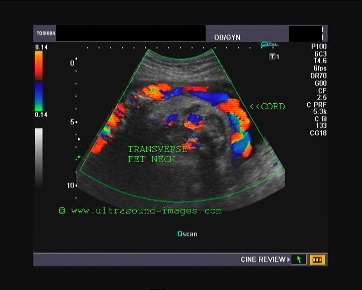
Nuchal cord or Umbilical cord around fetal neck
case-2-double-loop-nuchal-cord
The above color Doppler ultrasound images show a 35 weeks old fetus with a single loop of umbilical cord around the fetal neck. The images show a 360 degree loop around the neck and more, (see the sagittal section image), that there is only a single loop of umbilical cord ( arrowheads) around the neck. The sagittal section image shows a single cord loop posteriorly- meaning thereby that the nuchal cord, in this case, is not as significant and possibly safer than multiple umbilical cord loops around the fetal neck. Basically, there are two types of cord loops around the fetal neck-
Type A- where the umbilical nuchal cord encircles the fetal neck in a sliding manner (less dangerous) and,
Type B- where the nuchal cord encircles the neck in a locking manner (very dangerous). The color Doppler images of the case above suggest a single loop of cord in a sliding manner.
Color Doppler videos of this case are available at:
http://ultrasound-videos.blogspot.com/2011/03/nuchal-cord-in-late-3rd-trimester.html
References: http://sites.google.com/site/drjoea/gallery-fetal
http://en.wikipedia.org/wiki/Nuchal_cord
Case-2: (see above) shows 2 loops of cord around the fetal neck. The threat to the fetus is still low as the cord does not appear to be tight.
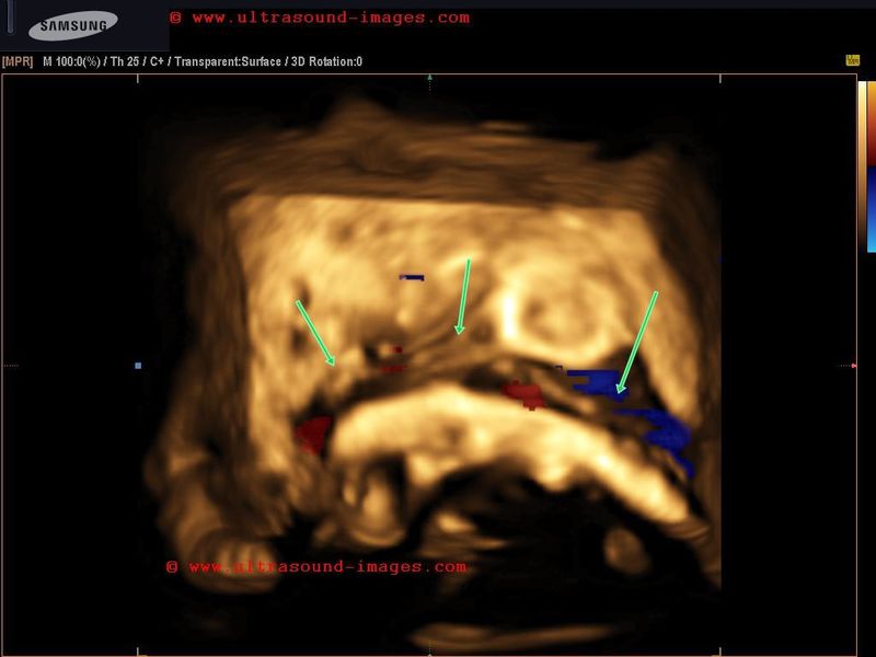
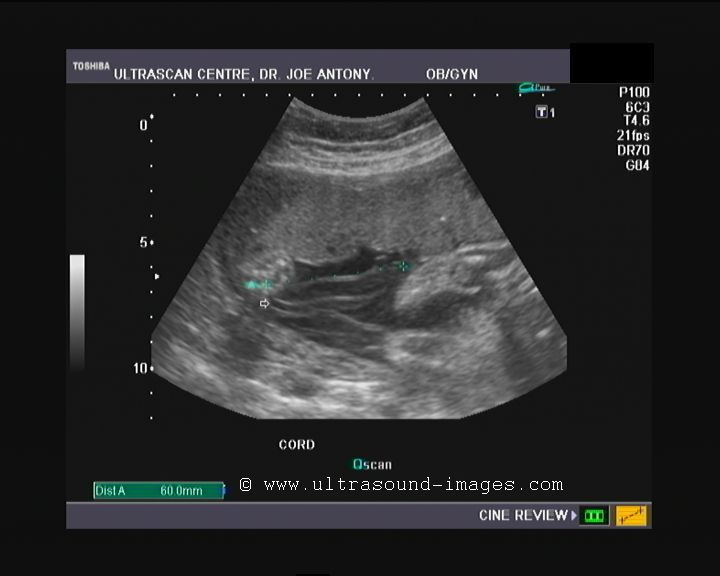
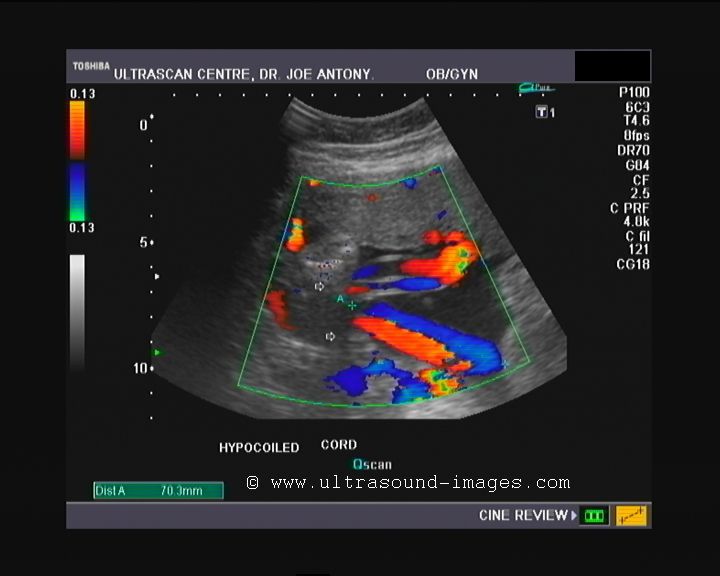
Hypocoiled umbilical cord
The normal umbilical cord extends from the placenta to the fetal umbilicus, twisting (coiling) as it traverses the distance between these two points. The umbilical cord contains two umbilical arteries and an umbilical vein which are normally coiled. Uncoiling or hypocoiled umbilical arteries are associated with increased fetal morbidity, including small for gestational age babies (fetuses). The above ultrasound and color Doppler images show an almost straight umbilical cord with almost no coils (hypocoiled cord). Such ultrasound findings are also associated with increased incidence (almost 64 %) of abnormal umbilical cord insertions, ie: marginal or velamentous. This case and images are courtesy of Dr. Durr -e-Sabih, Pakistan.
References: 1) http://www.ncbi.nlm.nih.gov/pubmed/10395130
http://www.ajog.org/article/S0002-9378%2805%2900068-2/abstract '
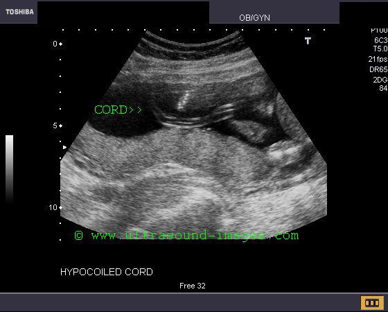
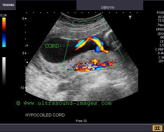
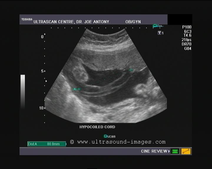
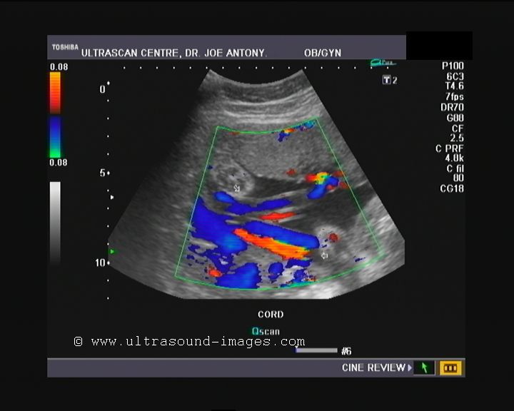
Case-2: hypocoiling of umbilical cord
This is yet another case of hypocoiled umbilical cord. The color Doppler image above shows an almost straight cord. The umbilical cord coiling index is calculated by the total length of the cord in centimeters divided by the total number of coils in the cord. Another method of calculating the umbilical coiling index is: 1/ distance in centimeters between a pair of coils (intercoil distance) of the cord. The normal umbilical coiling index varies from 0.19 to 0.21(based on studies by Ercal et al). Studies done on Indian pregnancies show normal UCI values ranging from- 0.13 to 0.21. Other studies have found that umbilical coiling index less than the 5th percentile is diagnostic of hypocoiled umbilical cord. This color Doppler ultrasound image is courtesy of Dr. Durr-e-Sabih, Pakistan.
Reference: http://medind.nic.in/jaq/t06/i4/jaqt06i4p315.pdf (free journal article- umbilical cord coiling index)
http://www.sirirajmedj.com/content.php?content_id=2539 (free article on method of umbilical cord coiling index and its use as an indicator of Trisomy 21). This article also has an image demonstrating the method of coiling index measurement.
http://www.hkmj.org/supplements/article_pdfs/hkm0702sp1p44.pdf (free article on umbilical cord morpholog and umbilical coiling index (UCI)).
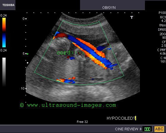
Case-3: Hypocoiled umbilical cord
This early 3rd trimester fetus showed a markedly hypocoiled cord. The intercoil distance here is more than 8 cms. Hence the UCI or umbilical coiling index is= 1/8 which is less than 0.12. (very low).
Normal coiling of the umbilical cord
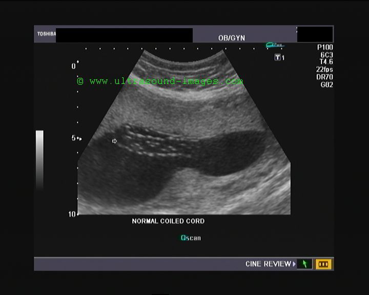
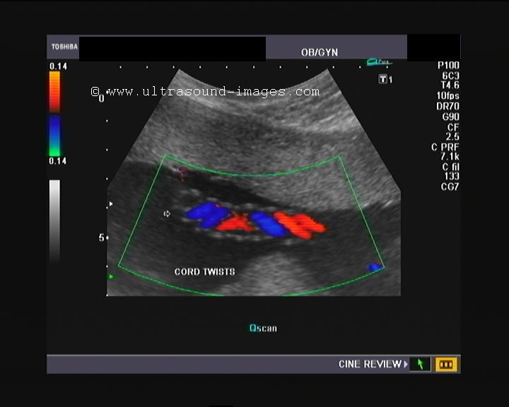
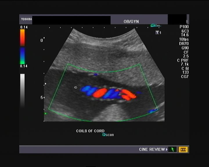
This 23 week old pregnancy shows the normal umbilical cord. The ultrasound and color Doppler sonography images show the normal twists in the umbilical cord (arrows). Contrast this cord to the previous case of hypocoiled umbilical cord. The difference is obvious.
Also see the color Doppler video clip of this case at: color Doppler video of normal umbilical cord
umbilical-cord-cysts
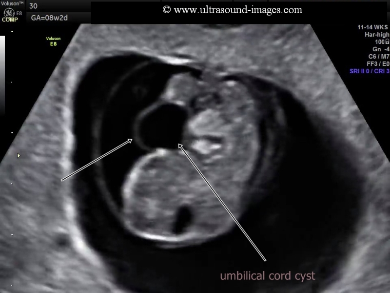
Umbilical cord cysts are cystic lesions within the umbilical cord.
Location: umbilical cord cysts are usually close to the fetus (fetal end) or close to the cord insertion site.
Types: umbilical cord cysts may be pseudycysts or true umbilical cord cysts. True umbilical cord cysts are remanants of the allantois also called allantoid cysts. Pseudocysts are umbilical cord cysts formed by edema of the wharton's jelly within the umbilical cord or due its liquifaction. Pseudocysts are said to be more common than true umbilical cord cysts. It is usually almost impossible to differentiate true umbilical cord cysts from pseudycysts of the cord.
Prognosis: generally most umbilical cord cysts disappear by the end of the 1st trimester. If they persist beyond 12 weeks or if the cysts are multiple, they are associated with a bad prognosis. Certain trisomies and fetal anomalies are linked to the presence of umbilical cord cysts.
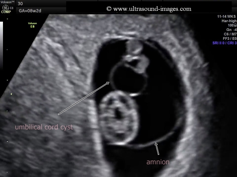
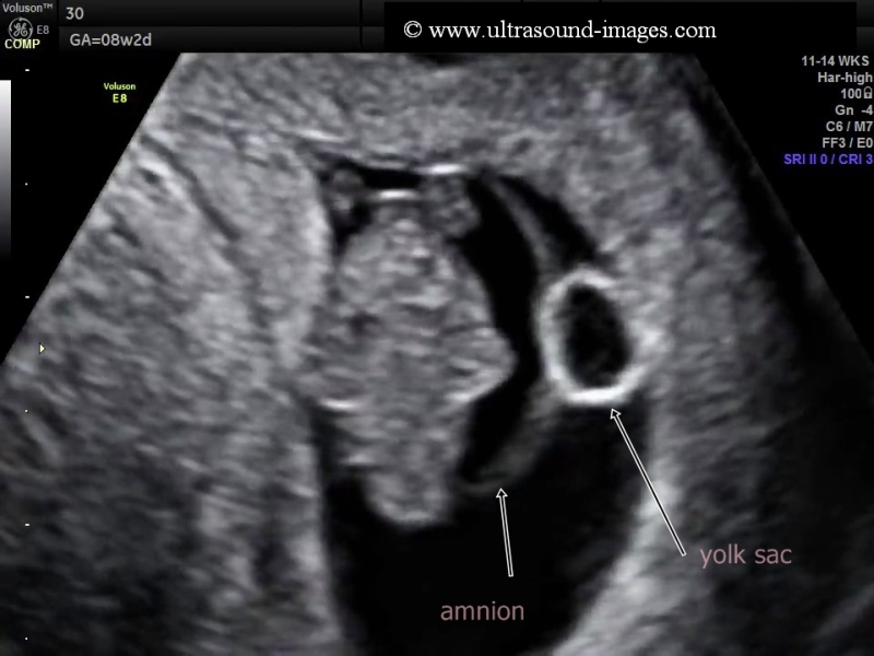
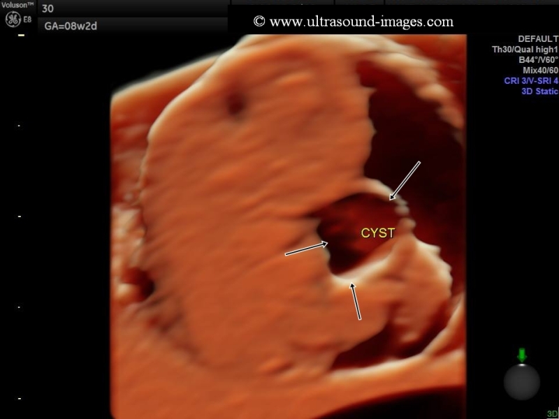
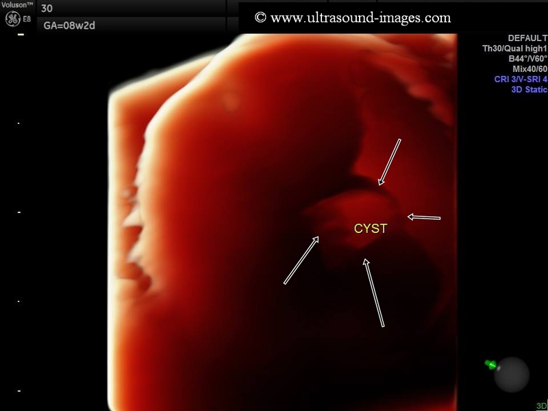
The B-mode and 3D ultrasound images above show a umbilical cord cyst near the fetal end of the cord in this 8 week fetus. Umbilical cord cysts may vary in size from 5 mm. to 50 mm. in size. The cord cyst must be differentiated from the more echogenic yolk sac which is seen outside the amnion. The amnion is seen surrounding the fetus in this case. The chief differential diagnosis in such cases are umbilical vein varix and umbilical hernia. Umbilical vein varix is easily identified by using color Doppler flow imaging as a varix would show flow signals within the cyst. Umbilical hernia and omentoceles would show bowel and abdominal viscera within the cystic lesion in the umbilical cord. These ultrasound and 3D images of the umbilical cord cyst are courtesy of Mayank Chowdhury, MD. The ultrasound system used here is the voluson E8 machine from GE.
References:
1. sonography of umbilical cord cysts
2. Ultrasound imaging of umbilical cord cysts
multiple-umbilical-cord-cysts
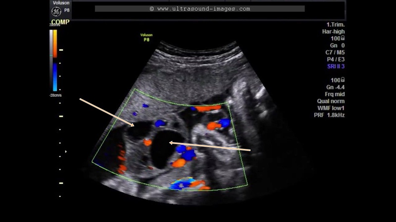
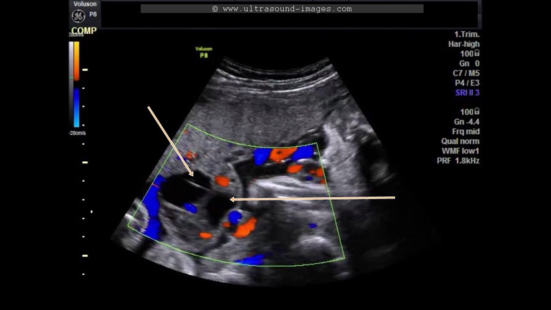
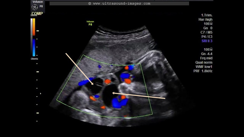
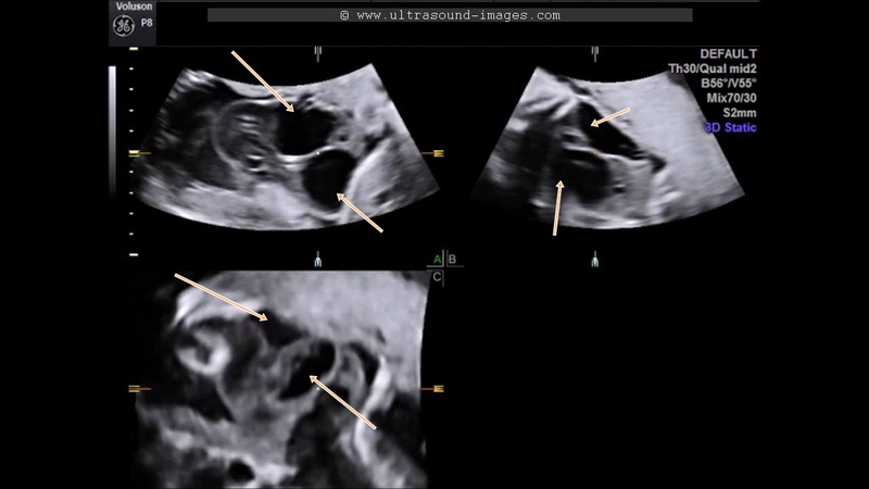
This second trimester pregnancy shows evidence of multiple umbilical cord cysts ( in this case, there are two umbilical cord cysts). The colour Doppler ultrasound images show anechoic cystic structures with absence of flow, suggesting umbilical cord cysts. The presence of multiple umbilical cord cysts is associated with increased morbidity and mortality for the fetus. Studies suggest that there is higher risk of aneuploidy in cases of multiple umbilical cord cysts. These images are courtesy of Firoz Bhuvar, MD). In the colour Doppler and 3-D ultrasound images above, the umbilical cord cysts are marked by arrows.
References:
imaging and significance of multiple umbilical cord cysts
See video of the case below:
absent-ductus-venosus
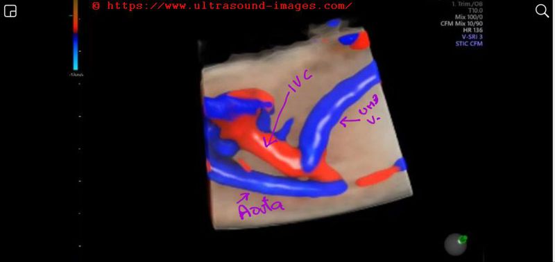
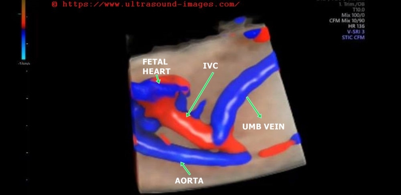
In this early 2nd trimester fetus, the umbilical vein drains directly to the fetal IVC (inferior vena cava) in the subhepatic location.
(see 4D color Doppler ultrasound video (STIC) and sonographic images above.
= absent ductus venosus is a very rare anomaly
= asbence of ductus venosus may be associated with other lethal fetal congenital anomalies, eg: absent fetal portal venous system, lethal cardiac anomalies, hydrops with serious morbidity and mortality
= in this fetus, absence of the ductus venosus was an isolated finding, hence having a better prognosis
= the absent ductus venosus may result in direct communication from the umbilical vein draining into the IVC (as is the case in this fetus), or may cause the umbilical vein draining to the portal veins
or communication with the fetal heart or fetal hepatic veins
The above 4D color Doppler ultrasound video of absent ductus venosus is courtesy of Firoz Bhuvar, MD. (edits by Joe Antony, MD)
To read more on the topic : sonography of absent ductus venosus
© 2026, Joe Antony, MD All images and content in this site are copyrighted. www.ultrasound-images.com When visiting this site, we may use cookies to improve the performance of this site. Use of this site means you accept the use of cookies. Read our privacy policy at this page: https://www.ultrasound-images.com/privacy-policy/