
Penis
Contents of this page
- Penile trauma
- Gun shot injury to the penis
- Insect bite injury to the penis
- Penile hematoma
- Penile fracture
- Low flow Priapism
- Peyronie disease
- Case-2: Peyronie disease
- Penile AVM (arterio-venous malformation)/ AVM in the glans of penis
- penile-urethra-calculus
- penile fracture case-2
- penile doppler
- normal-penile-doppler-after-oral-sildenafil
- penile-doppler-peyronies-disease
Penile trauma
Gun shot injury to the penis
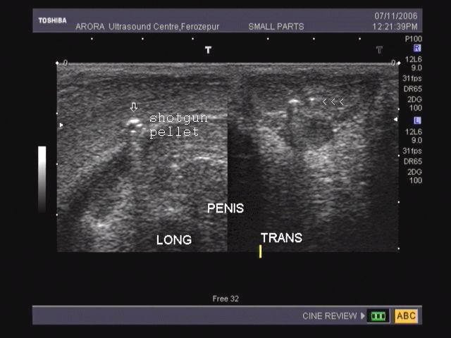
This ultrasound image shows a shotgun lodged deep in the corpus cavernosum of the penis. The pellets are seen as a bright echogenic structures (arrowed). Image courtesy of Dr. Vikas Arora, India.
Insect bite injury to the penis


Transverse section of penis Longitudinal section of penis
The 2 sonographic images above reveal gross edema of the dorsal skin and subcutaneous tissue as well as the corpus cavernosum of the penis following insect bite. Ultrasound images courtesy of Dr. Ravi Kadasne, UAE.
Penile hematoma
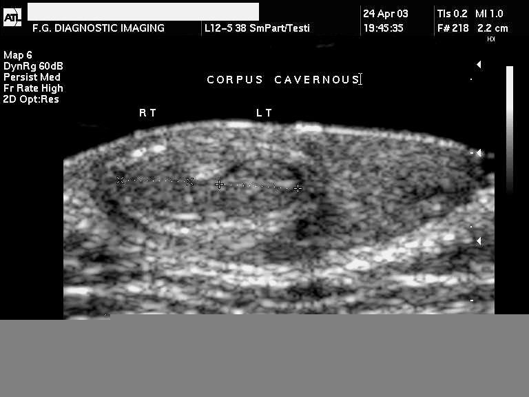

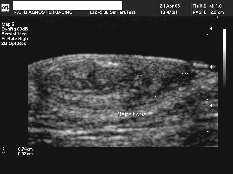
The above 3 ultrasound images reveal a hypoechoic collection lateral to the left corpus cavernosum of the penis following penile trauma. Color doppler image reveals a possible source of the hemorrhage. There is evidence of rupture of the lateral aspect of the tunica albuginea of the penis. Images reveal a transverse section of the affected part. Case and images courtesy of Mr. Shlomo Gobi, Israel.
Reference:http://www.jultrasoundmed.org/cgi/content/full/24/7/993 (free article..Rated--- excellent)
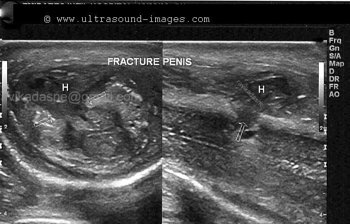
Penile fracture
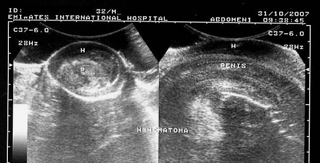
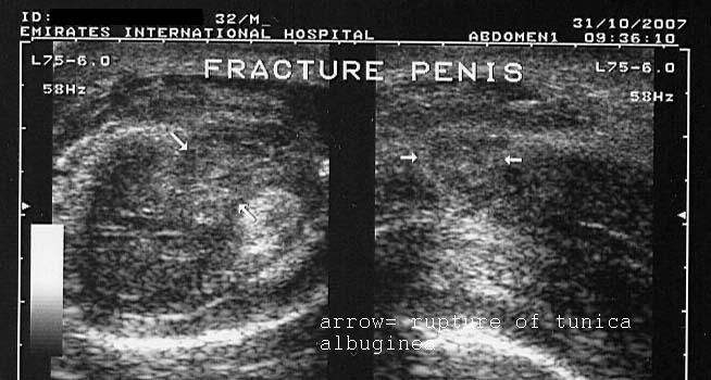
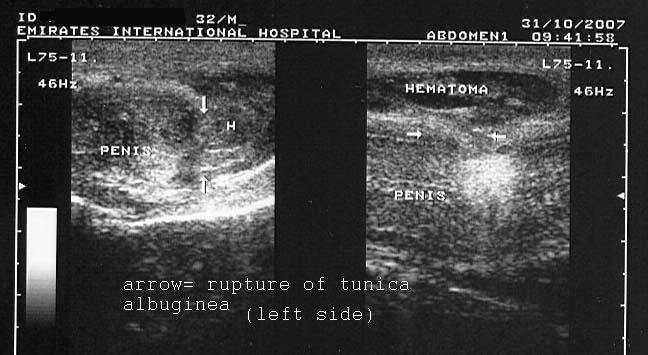
The above ultrasound images reveal a hypoechoic collection (hematoma) with rupture of the tunica albuginea covering the left corpus cavernosum. Fracture of the penis is caused by trauma to an erect penis, resulting in rupture of the corpus cavernosum and the tunica albuginea covering it. Usually, the left corpus cavernosum is affected. Images courtesy of Dr. Ravi Kadasne, UAE.
Reference: http://www.emedicine.com/med/topic3415.htm (free article).
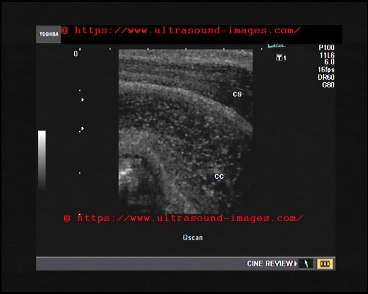
Low flow Priapism
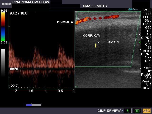

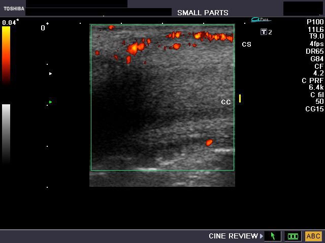
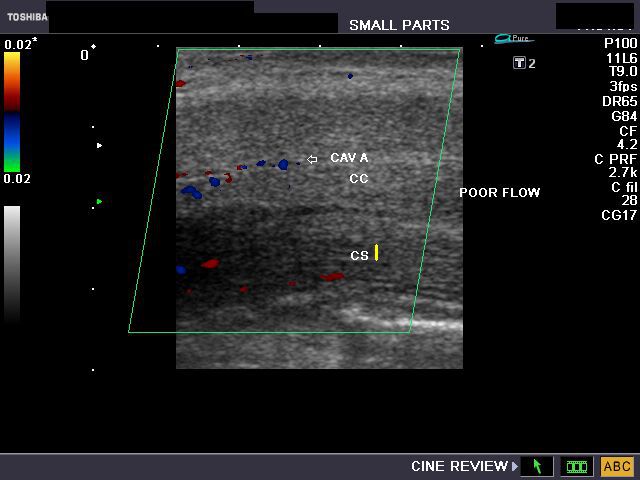
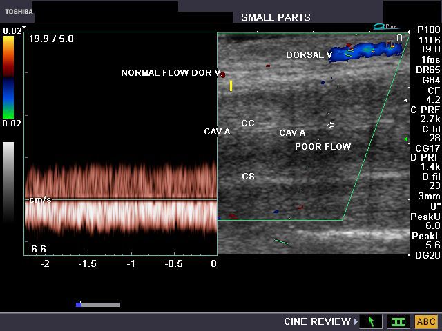
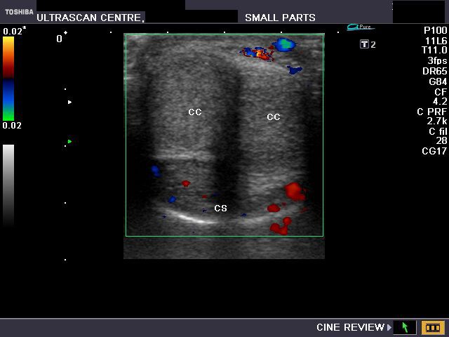
(CC= corpus cavernosum, CS= Corpus spongiosum, Dorsal V= dorsal vein of penis, Cav A= cavernosal artery)
This middle aged male patient presented with painful persistent erection (priapism) since 6 days. He did not have any history of trauma. Sonography, power doppler and color doppler imaging of the penis revealed poor flow or absent flow in the cavernosal artery of the penis with moderate flow in the dorsal artery and vein. There is also no flow within the corpora cavernosa of the penis. The ultrasound and color doppler images are diagnostic of low flow priapism. Images taken by Joe Antony, MD, India, using a Toshiba Nemio- XG ultrasound system.
Reference:
1) http://www.medscape.com/viewarticle/550324_6(good article- free images)
2)http://www.clevelandclinic.org/health/health-info/docs/2900/2908.asp
Peyronie disease

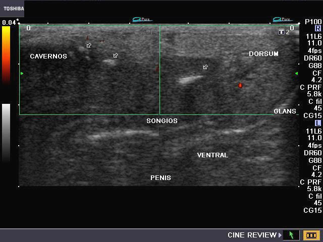
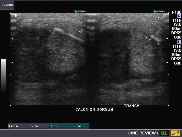
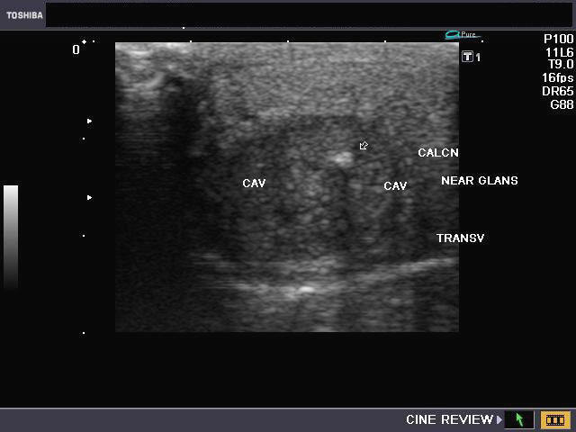
This 47 yr. old male patient presented with a palpable nodule on the dorsum of the penis. Clinically, he did not have significant erectile dysfunction but Peyronie disease was suspected. Sonography of the penis was done and showed multiple (4 in number) calcific plaques of the dorsum of the penis, involving the tunica albuginea and also the septum between the corpora cavernosa. There was evidence of significant acoustic shadowing beyond the calcific plaques. The plaques varied in size from 4 mm. to 10 mm. The calcific plaques are shown by arrows in the ultrasound images above. These ultrasound images are diagnostic of Peyronie disease of the penis. Power Doppler imaging (see middle row -left), did not show any altered vascular flow in the penis. Ultrasound images taken using a Toshiba, Nemio-XG system by Joe Antony, M.D., India.
Reference:
1)http://drjoea.googlepages.com/penis
2) http://www.jultrasoundmed.org/cgi/reprint/28/2/217(free article and images).
Case-2: Peyronie disease


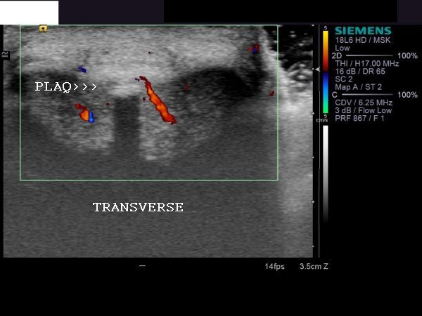
This patient showed a long calcific plaque of the tunica albuginea of the penis. The transverse section image (color Doppler) showed normal vascularity, but the penile plaque is clearly seen with acoustic shadowing. This is a typical apperance of Peyronie disease. (PLAQ= plaque). Ultrasound images courtesy of Shlomo Gobi, Israel.
Penile AVM (arterio-venous malformation)/ AVM in the glans of penis
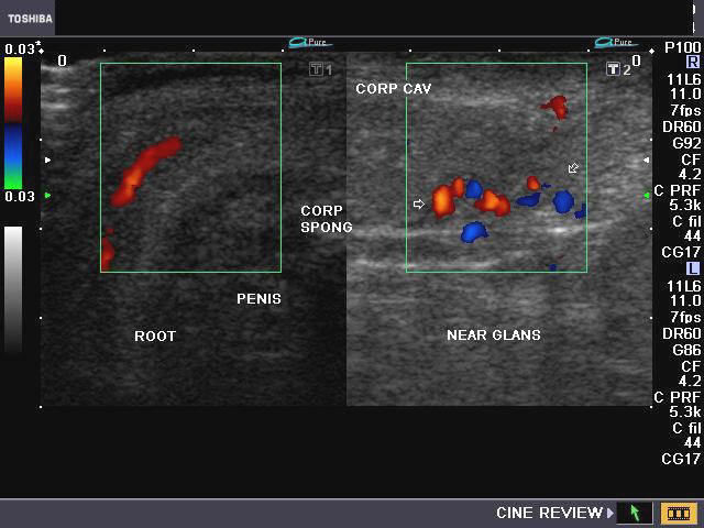
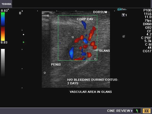
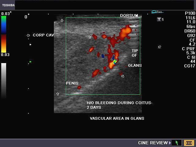
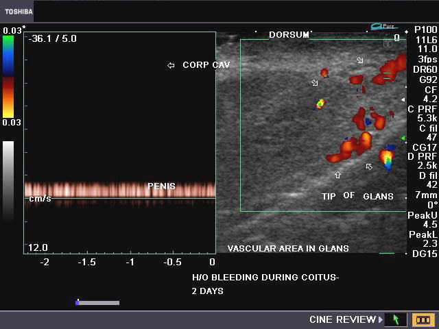
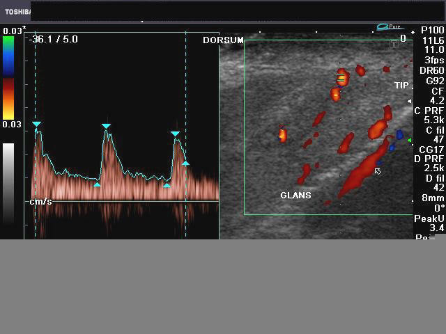
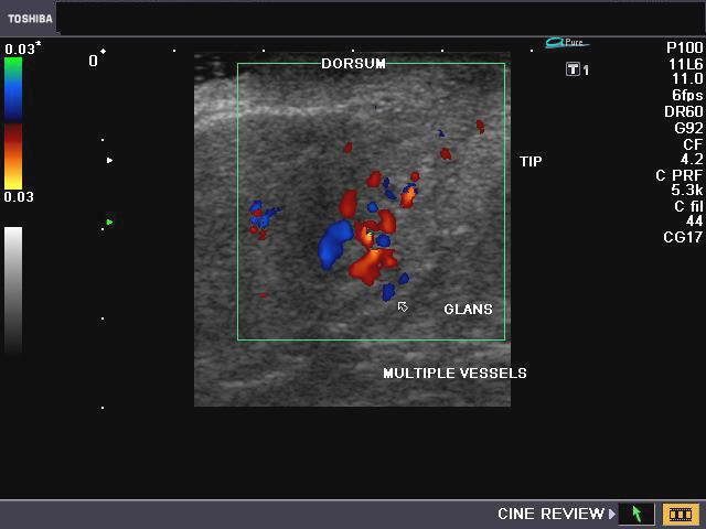
Ultrasound and Doppler images of vascular malformation of the glans penis
This male patient aged 50 yrs. presented with a bout of severe hemorrhage from the penile urethra during coitus for the past 2 days. Sonography and Color as well as Power Doppler imaging of the penis was done. Ultrasound/ Doppler flow images show marked vascularity along the glans of penis. Spectral Doppler waveforms show presence of both arterial and venous flow patterns in the vessels in and around the glans penis. These ultrasound, Power Doppler and Color Doppler images of the penis suggest arterio-venous malformation (AVM) in the glans penis. Trauma during coitus would have precipitated this episode of hemorrhage. The other differential diagnosis would be hemangioma of the glans penis. (CORP SPONG= corpus spongiosum; CORP AV= corpus cavernosus; Dorsum= dorsal part of the penis). AVM and hemangioma of the glans penis are extremely and hardly any cases are reported in medical literature.
References:1) Hemangioma of glans penis (free article)
2) Vascular malformations (free article)
penile-urethra-calculus

This calculus is launched at the distal end of the penile urethra (see ultrasound image of the calculus in the distal end of the penis). This patient complained of pain and difficulty in passing urine. Ultrasound image of penile urethral calculus is courtesy of Dr. Ravi Kadasne, MD, UAE.
penile fracture case-2
Another example of penile fracture. Here again, there is rupture of the tunica albuginea and the corpus cavernosum. Ultrasound image is courtesy of Dr. Ravi Kadasne, MD, UAE.
penile doppler
Penile doppler is increasingly playing a major role in the study of male sexual dysfunction. Perhaps the best links to information and reference material on penile doppler ultrasound are given below:
1) http://www.medscape.com/viewarticle/550323_5
2) http://radiopaedia.org/articles/penile-doppler-role-in-erectile-dysfunction-3
normal-penile-doppler-after-oral-sildenafil
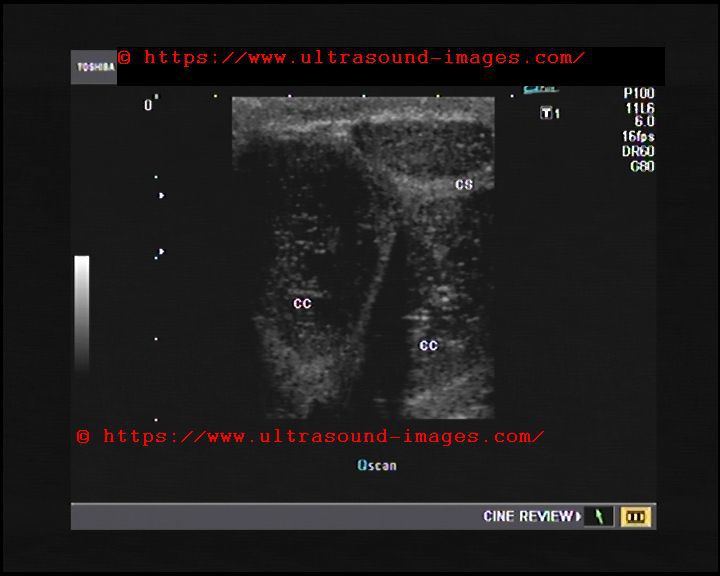


This 60 year old male patient underwent penile ultrasound and Doppler ultrasound imaging for mild erectile dysfunction:
Oral sildenafil (Manforce 50 mg tablet) was taken. Oral sildenafil (Viagra/ Manforce) is now being used instead of papaverine intracavernosal injection. Results with oral sildenafil are comparable and produce good erectile response within 1 hour.
Following findings are to be noted 1 hour after oral sildenafil:
Normal findings on penile Doppler ultrasound after 50 mg of oral sildenafil :
- Cavernosal artery measures 0.5 mm. (normal diameter = 0.3 to 0.5 mm. without sildenafil.
- After sildenafil stimulus > 75 % increase is normal.
- EDV (end diastolic velocity) < or = 5 cms/sec after sildenafil.
- AT (acceleration time) after sildenafil is normally < 0.11 sec.
- normal PSV cavernosal artery > 25 cms./sec after 1 hour of Sildenafil
- Phasic changes in PSV after oral sildenafil during 1 hour:a) gradual increase in PSV. b) Increase in diastolic velocity until 3rd phase of the scan when decrease in diastolic flow and later reversal in diastolic flow takes place.
Criteria: Abnormal or indicating arterial cause (CC artery) of erectile dysfunction if after 1 hour of oral sildenafil :
1)< 75 % increase in diameter of CC artery
2)PSV< 25 cms./sec
3)AT> 0.11 sec.
Also veno-occlusive disease of
4)EDV > 5 cms./sec
5)RI <0.85
Also Deep dorsal vein of penis flow velocity is normally < 3 cms.
If flow velocity in deep dorsal vein is > 20 cms. sec s/o veno-occlusive disease
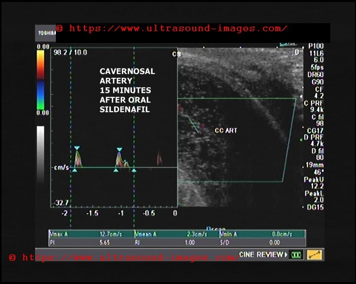
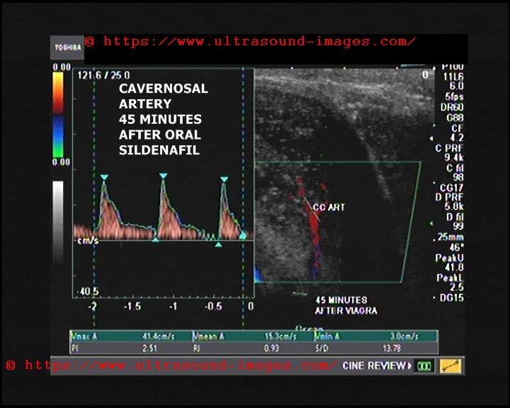
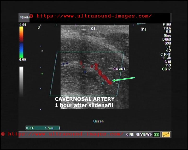
In above patient (see images above) cavernosal artery Doppler after oral sildenafil (after 1 hour) following observations were made:
- gradual increase in PSV (peak systolic velocity) from 20 cms./ sec to 42 cms./sec
- RI= 1.0 after 1 hour
- Increase in cavernosal artery diameter by more than 75 % to 1.7 mm. Thus this was a normal penile Doppler ultrasound study and the mild erectile dysfunction is because of psychogenic causes
References:
1) Penile doppler using oral sildenafil- Radiopedia article (excellent article)
2) Journal article on penile Doppler ultrasound using papverine (excellent article)
penile-doppler-peyronies-disease


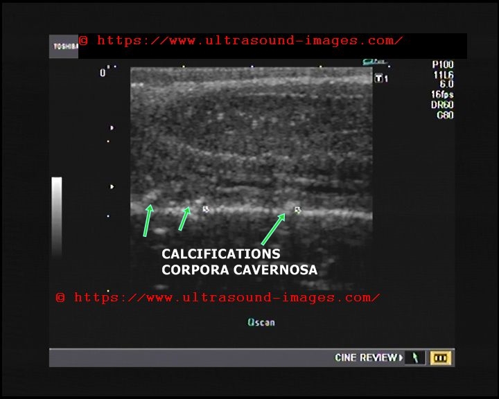
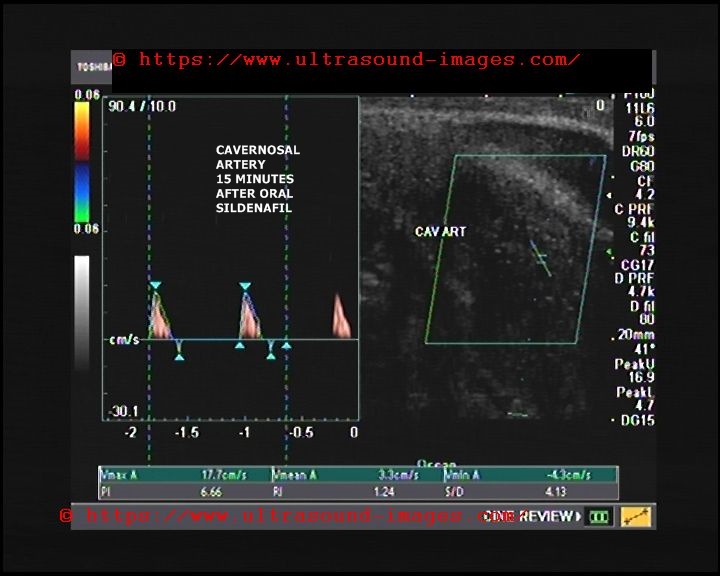
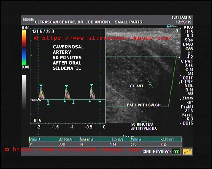
This 37 year old male patient had erectile dysfunction for which oral sildenafil (Manforce) 50 mg was administered and serial penile Doppler ultrasound scan was done.
following findings observed:
- multiple calcific plaques along tunica albuginea of corpora cavernosa seen
- lack of response to oral sildenafil as cavernosal artery diameter did not increase significantly even after 1 hour (< 75 % increase)
- PSV of cavernosal artery increased from 17 cms./sec to only 21 cms./sec after 1 hour of oral sildenafil. (less than 25 cms./sec)
Thus it was concluded that the erectile dysfunction in this patient was arteriogenic and labelled as Peyronie's disease.
© 2026, Joe Antony, MD All images and content in this site are copyrighted. www.ultrasound-images.com When visiting this site, we may use cookies to improve the performance of this site. Use of this site means you accept the use of cookies. Read our privacy policy at this page: https://www.ultrasound-images.com/privacy-policy/