
Ultrasound images of diseases of the caecum
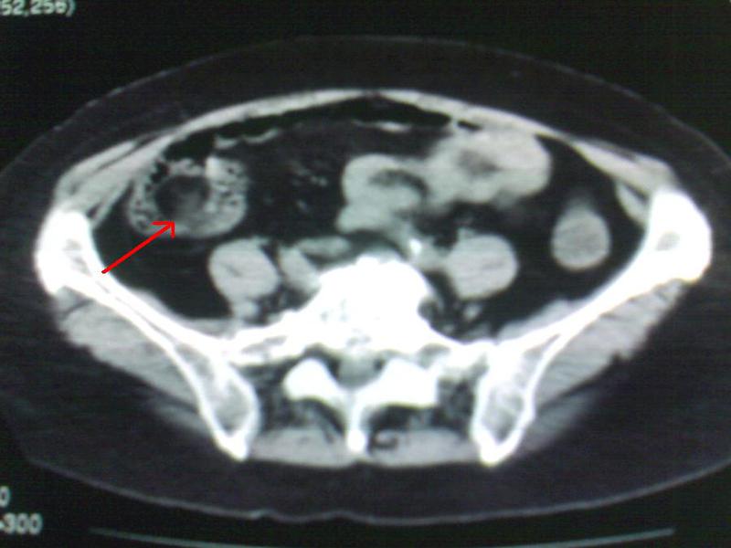

This 64 year old female patient underwent sonographic imaging of the abdomen. Ultrasound image (right) shows an echogenic, rounded mass lesion in the caecum. CT scan of the pelvis (Left) reveals a fat density mass in the caecum. These images suggest a diagnosis of lipoma of the caecum. Caecal lipomas are usually seen in elderly female patients and are usually symptomatic if their size is more than 3 cms. They can often cause intussusception. Images courtesy of Dr. Ravi Kadasne, UAE (Sonography done using a Toshiba Powervision color doppler machine).
Reference:http://www.jgld.ro/42005/393-396_11.pdf(free article and images)
© 2026, Joe Antony, MD All images and content in this site are copyrighted. www.ultrasound-images.com When visiting this site, we may use cookies to improve the performance of this site. Use of this site means you accept the use of cookies. Read our privacy policy at this page: https://www.ultrasound-images.com/privacy-policy/