
Ultrasound images of pediatric abdomen
Contents of this page
- Ectopic kidney or Renal ectopia
- Pelvic kidney in neonate
- Nephrocalcinosis in neonates
- Communicating hydrocele in neonate
- Bilateral communicating hydrocele in neonate
- Persistent vesico-urachal diverticulum in child
- Sonography of renal fungal balls in neonate
- Following are ultrasound images after 2 weeks
- Sonography of neonatal uterus
- Gall bladder calculi in neonate
- Cirrhosis of liver and hepatorenal syndrome in neonate
- Neuroblastoma in neonate
- Case-2: Left suprarenal (adrenal mass) in neonate
- Bilateral adrenal hematoma (hemorrhage)
- Congenital right diaphragmatic hernia in neonate
- Meconium peritonitis
- Necrotizing enterocolitis
- Unilateral UPJ (uretero-pelvic junction) or PUJ (pelvi-ureteric junction) obstruction in neonate
Ectopic kidney or Renal ectopia
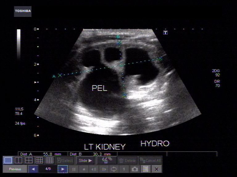
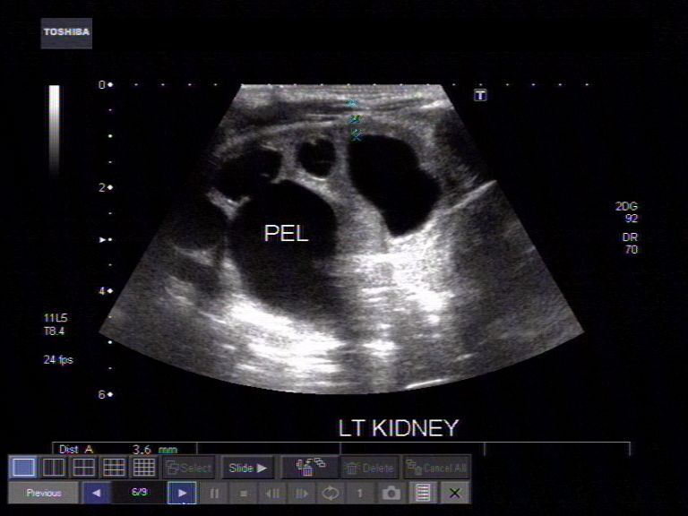
Pelvic kidney in neonate

My e-book on neonatal ultrasound for Amazon kindle is available at:
Atlas of neonatal ultrasound- cranium and abdomen
for other countries:
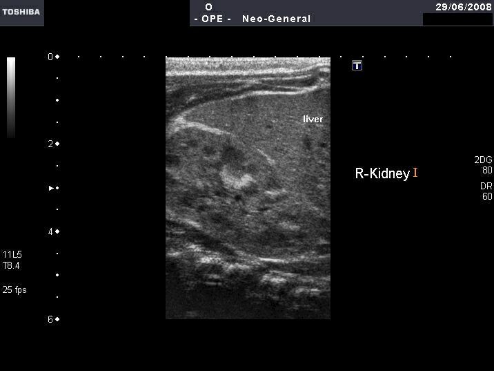
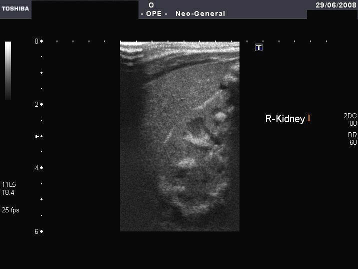
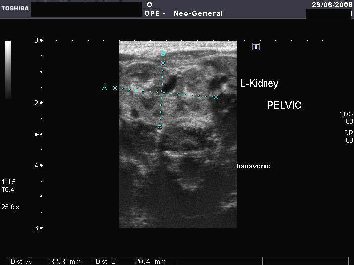
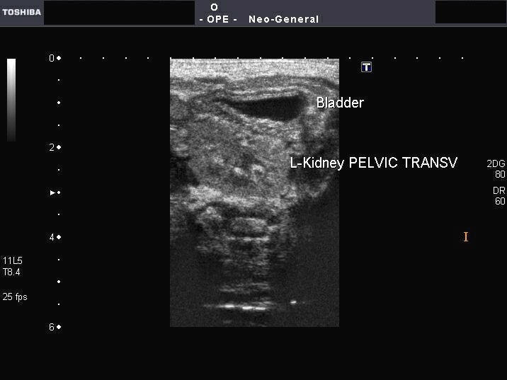
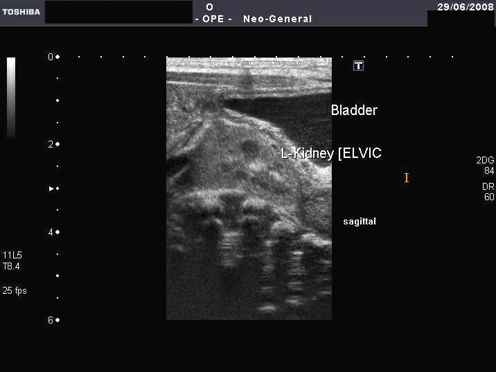
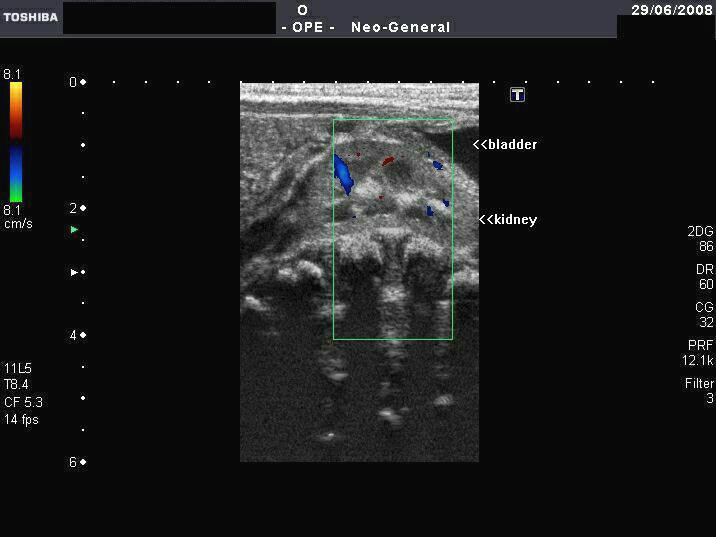
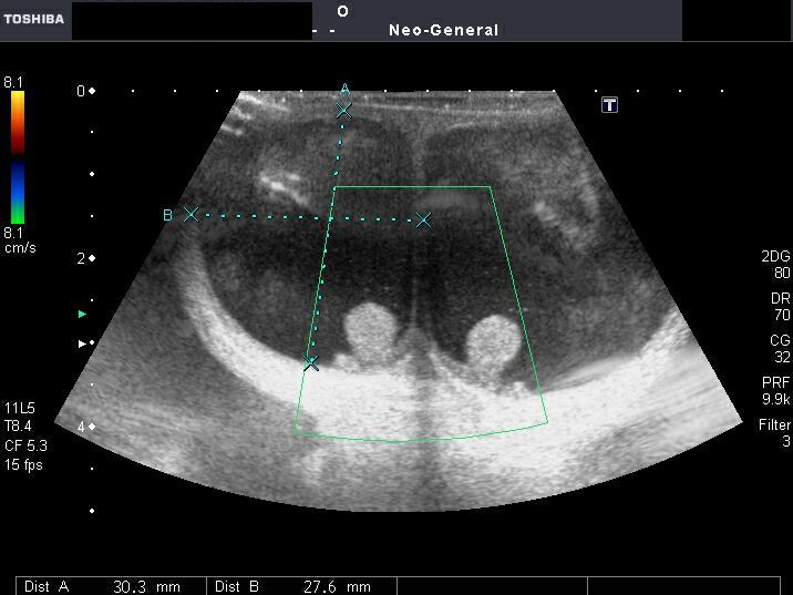
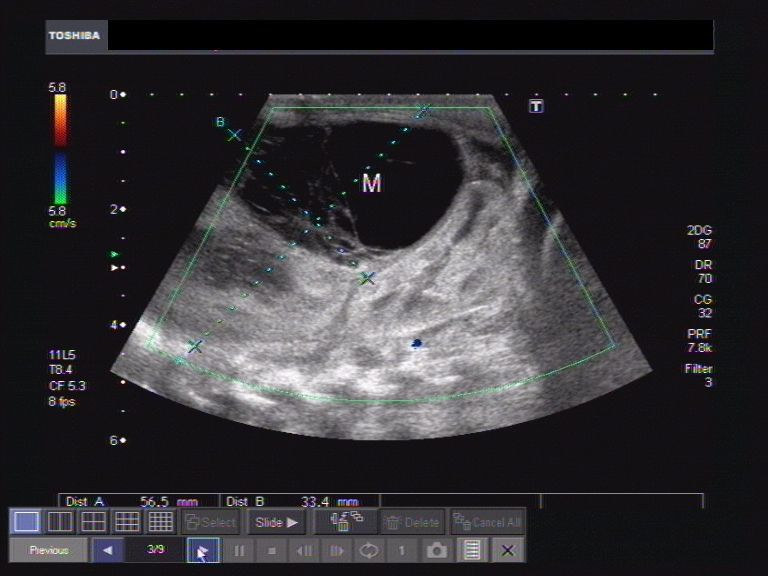
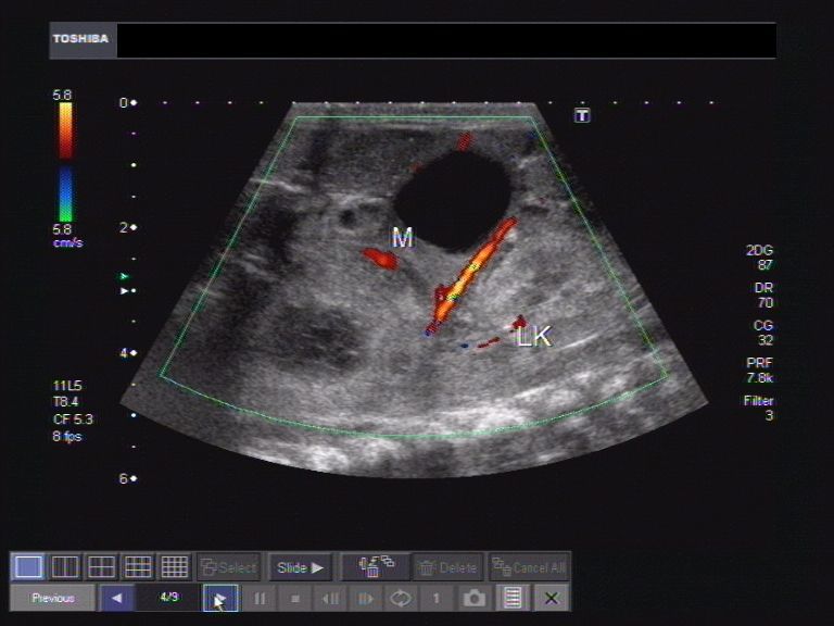
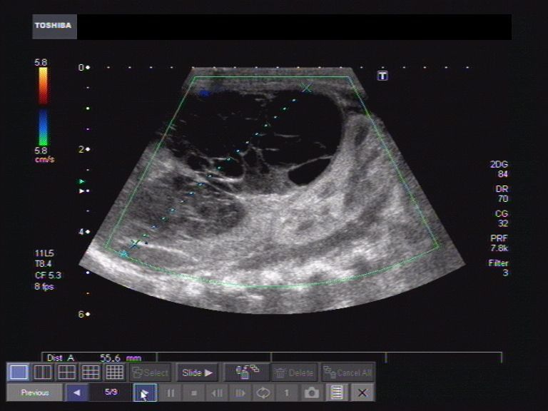
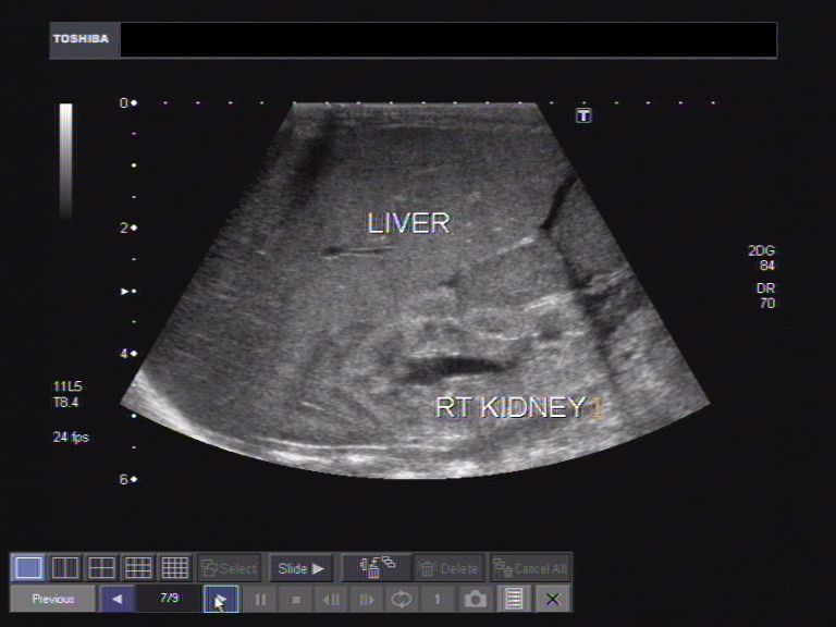
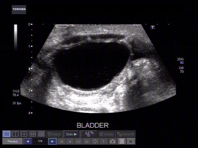
This female neonate presented with imperforate anus. Sonography reveals normally located right kidney. However the left kidney is not visualized in left renal fossa. Ultrasound images reveal a pelvic kidney that appears somewhat rounded, located posterior and superior to the bladder. The renal pyramids of both kidneys appear echogenic suggestive of early nephrocalcinosis. The uterus of this neonate was not visualized well. Color doppler imaging shows the vascularity of the pelvic kidney. Images taken using a Toshiba Xario machine.
Reference:http://www.thefetus.net/page.php?id=549(a good article on pelvic kidney, with free images).
Nephrocalcinosis in neonates
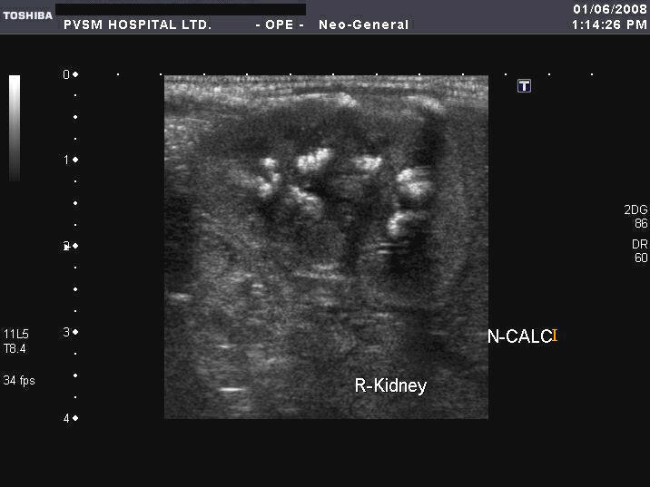

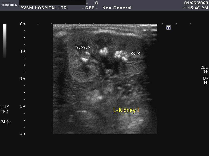
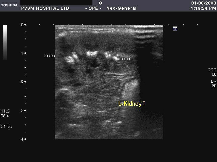
These ultrasound images of neonatal kidneys show- markedly echogenic renal pyramids, bilaterally, with chief involvement of the tips suggestive of medullary nephrocalcinosis. Such nephrocalcinosis in neonates usually follows long term parenteral therapy or due to use of certain drugs notably gentamicin and furosemide. It can also be caused by low fluid intake in the neonate and due to oxygen dependency. This condition, in neonates, usually resolves following correction of the etiological factors. Case courtesy of Joe Antony, MD, India. Images taken using a Toshiba Xario machine.
Also see: http://www.ultrasound-images.com/renal-calculi.htm (where I describe nephrocalcinosis in adults).
Communicating hydrocele in neonate
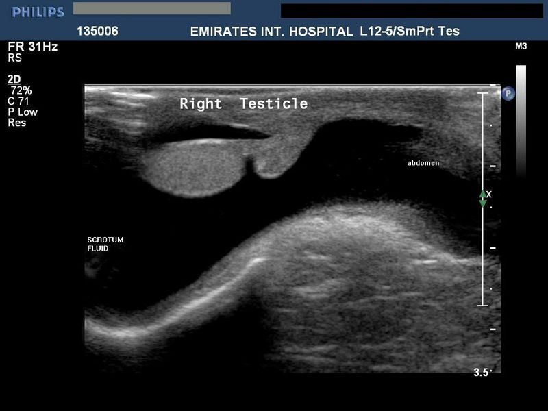
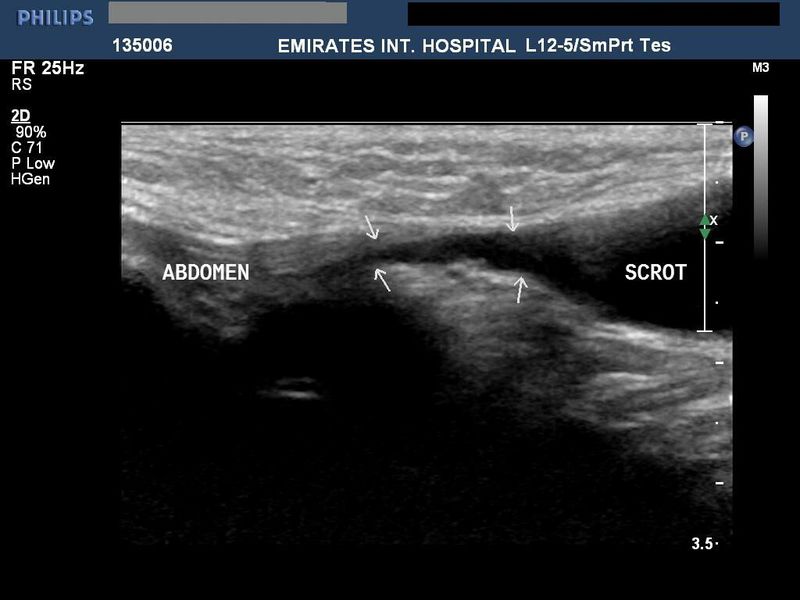
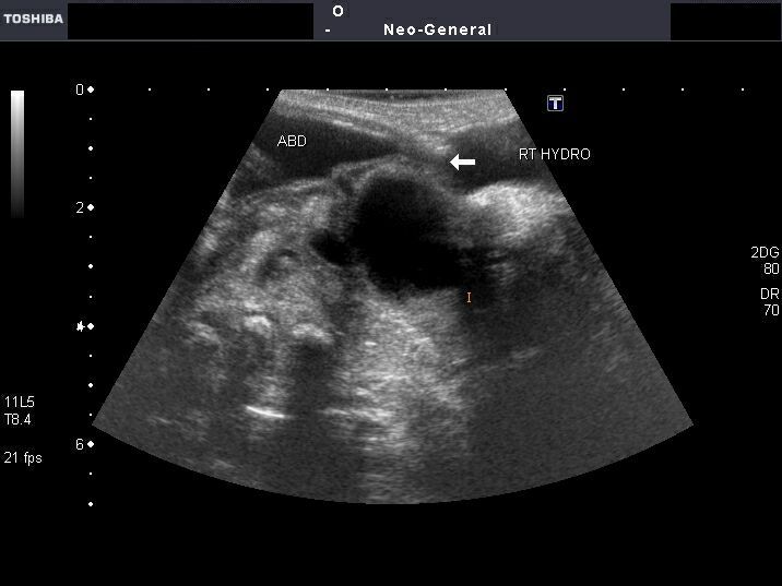
This neonate showed a swelling of the Rt. scrotum. Sonography of the pelvis and scrotum revealed a right sided hydrocele (seen as a hypoechoic collection surrounding the right testes). The hydrocele also appears to communicate (arrows) with the abdomen through a patent processus vaginalis. This patent communication may cause a hernia or hydrocele to persist in infants. These ultrasound images are diagnostic of communicating hydrocele. Images courtesy of Dr. Ravi Kadasne, Radiologist, UAE, who used a Philips IU 22 ultrasound machine, here.
Reference:http://www.emedicine.com/ped/topic1037.htm(free article).
Bilateral communicating hydrocele in neonate

The above ultrasound images show gross bilateral communicating hydrocele in a neonate, which was found to have Hydrops fetalis in utero. Note the presence of ascitic fluid with communication with the hydrocele (arrow in figure below).
Persistent vesico-urachal diverticulum in child
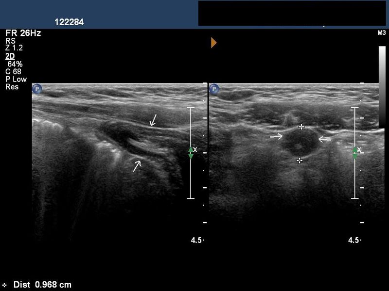
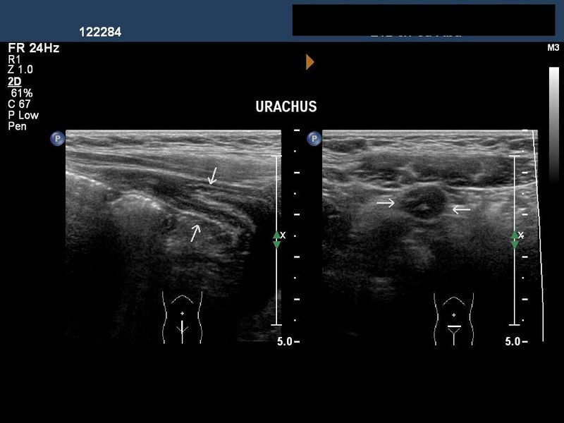
This 4 yr. old male child had severe pain in the lower abdomen. Sonography revealed a tubular hypoechoic structure leading to the urinary bladder from just below the umbilicus. These ultrasound images suggest a diagnosis of inflammation of the persistent vesico-urachal diverticulum. The urachus is a duct that leads from the urinary bladder to the umbilicus in the fetus. Peristence of the urachal duct can present as a sinus or diverticulum in children. Ultrasound images taken using a Philips IU 22 machine by Dr. Ravi Kadasne. UAE.
Reference:
1)http://www.emedicine.com/ped/topic1402.htm (free article)
2)http://www.nlm.nih.gov/medlineplus/ency/article/002993.htm
Sonography of renal fungal balls in neonate
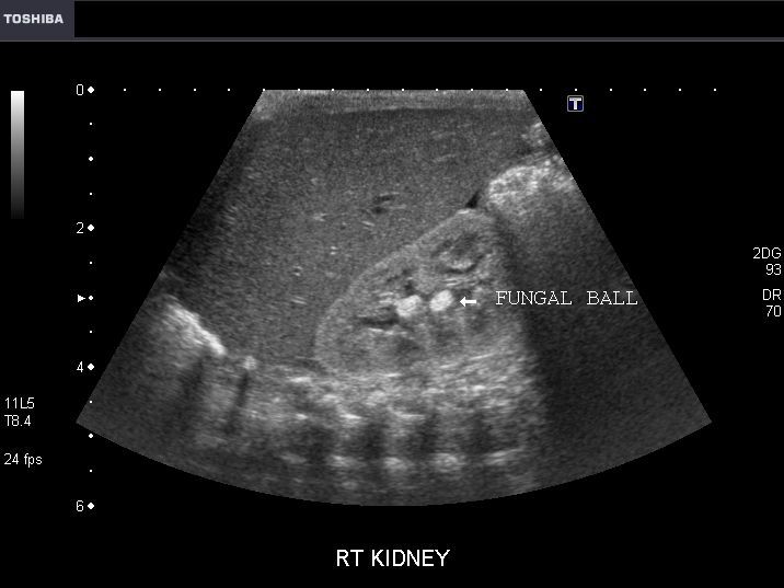

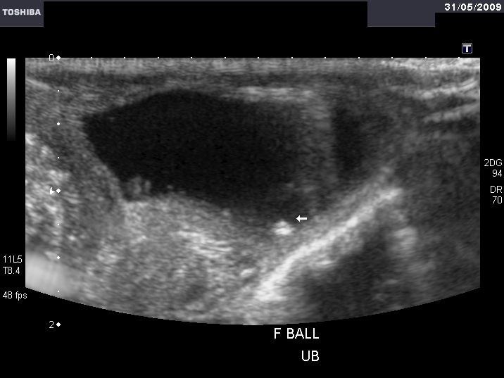
The above ultrasound images (LEFT) shows 2 echogenic, rounded, non-shadowing mass lesions in the renal pelvis of right kidney. The left kidney of the neonate shows pelvicalyceal dilation with a few echogenic foci in the calyces. The neonate was undergoing prolonged antibiotic therapy following sepsis after preterm delivery.
Following are ultrasound images after 2 weeks
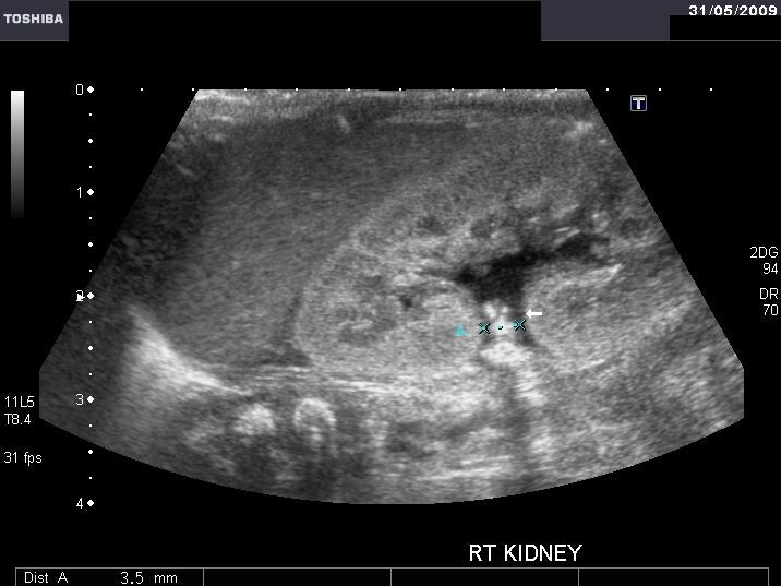
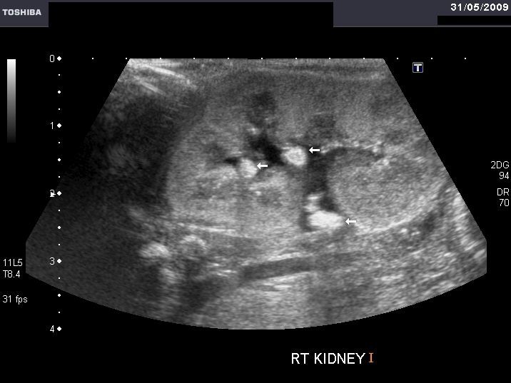
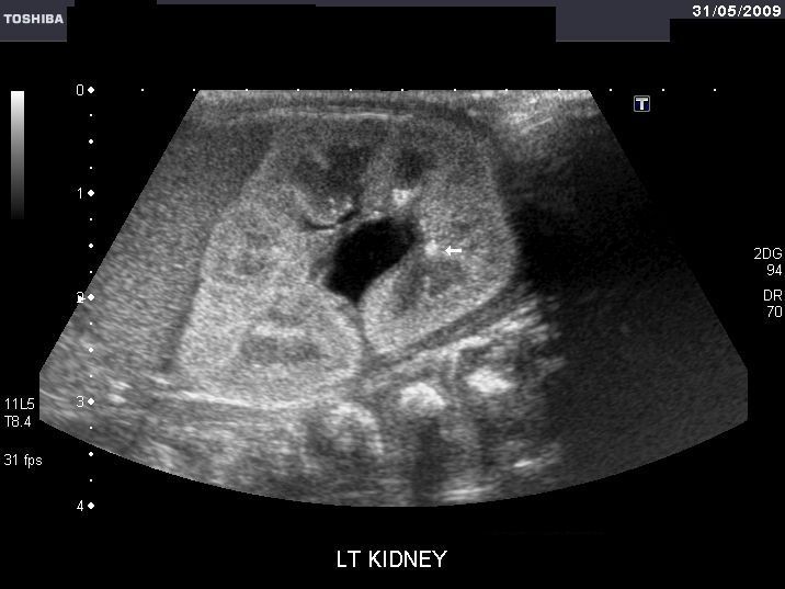
The follow up ultrasound images of the same neonate after 2 weeks show presence of the more rounded masses in the right kidney which also shows mild dilation of the renal pelvis. Smaller lesions are seen in the left kidney with pelvicalyceal dilation. The right kidney also shows rounded lesions in the pelvi-ureteric junction region. The urinary bladder also shows small rounded lesions floating within it. Such lesions are usually due to fungal balls or blood clots. Since blood clots in the renal collecting system usually dissolve after a few days, these ultrasound images suggest fungal balls in the kidneys and urinary bladder. The dilated pelvi-calyces of both kidneys suggest obstruction produced by the fungal balls in the collecting system.
Reference:http://radiology.rsnajnls.org/cgi/reprint/142/2/473.pdf
Sonography of neonatal uterus

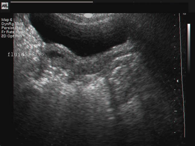
The above 2 ultrasound images show neonatal uterus in a female. The endometrial cavity (uterine cavity) shows small hypoechoic collections within it suggestive of fluid. A small amount of fluid may normally be seen in a perfectly normal female baby and should be of no clinical concern. Both these sonographic images are courtesy of Mr. Shlomo Gobi, Israel.
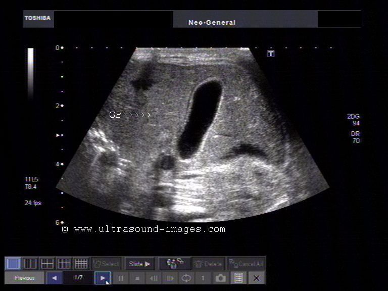
Gall bladder calculi in neonate
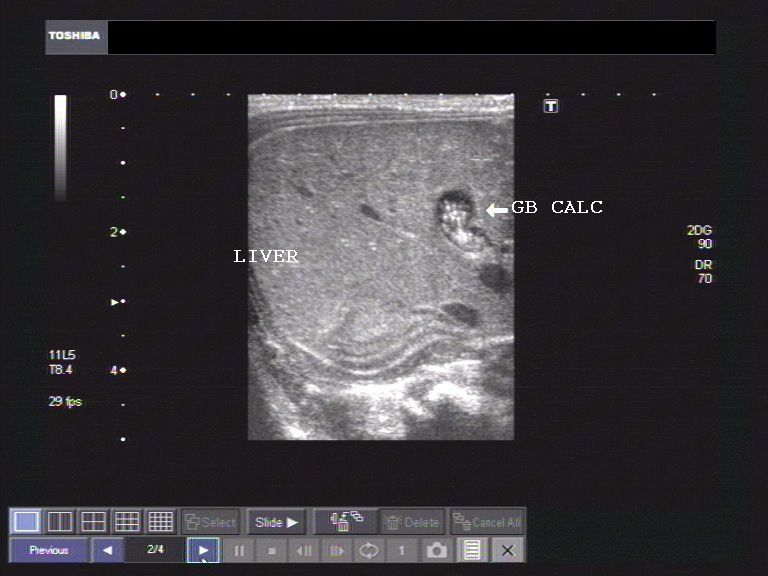
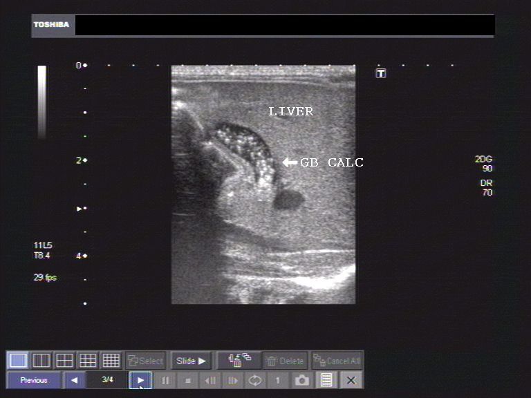
The above ultrasound images show a long section through a neonatal gall bladder (GB). Multiple echogenic lesions are seen in the lumen of the neonate's gall bladder suggestive of numerous gall stones/GB calculi. Presence of gall bladder calculi in the last trimester of fetus and immediately after birth is a known occurrence. However, this type of gallstones in the neonate are of little clinical significance, as follow up sonography usually shows resolution of the condition. Ultrasound images are by Joe Antony, MD, using a Toshiba Xario ultrasound system.
Reference:http://emedicine.medscape.com/article/927522-overview
Cirrhosis of liver and hepatorenal syndrome in neonate
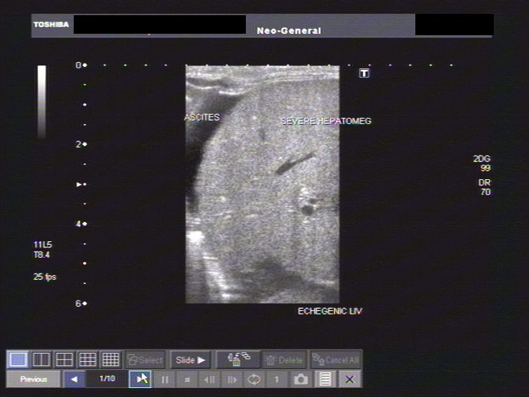


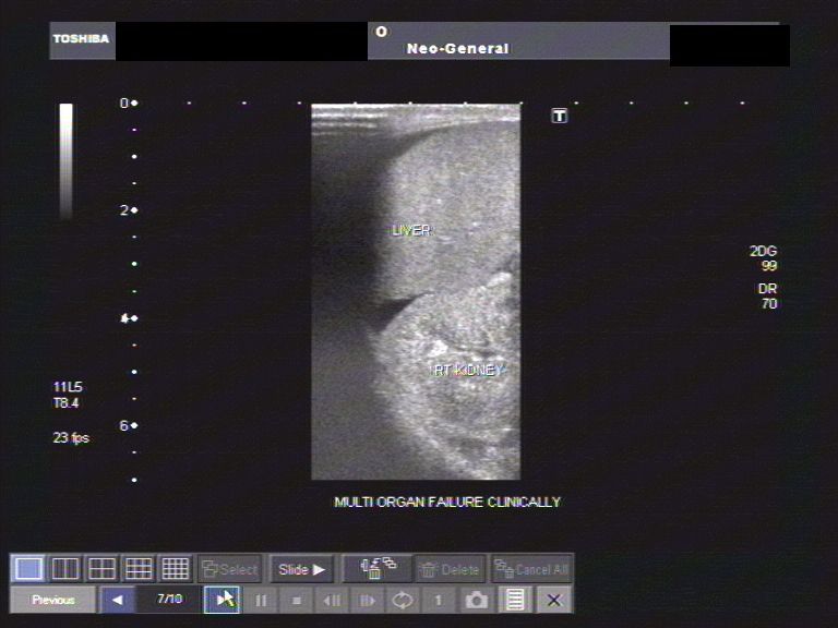
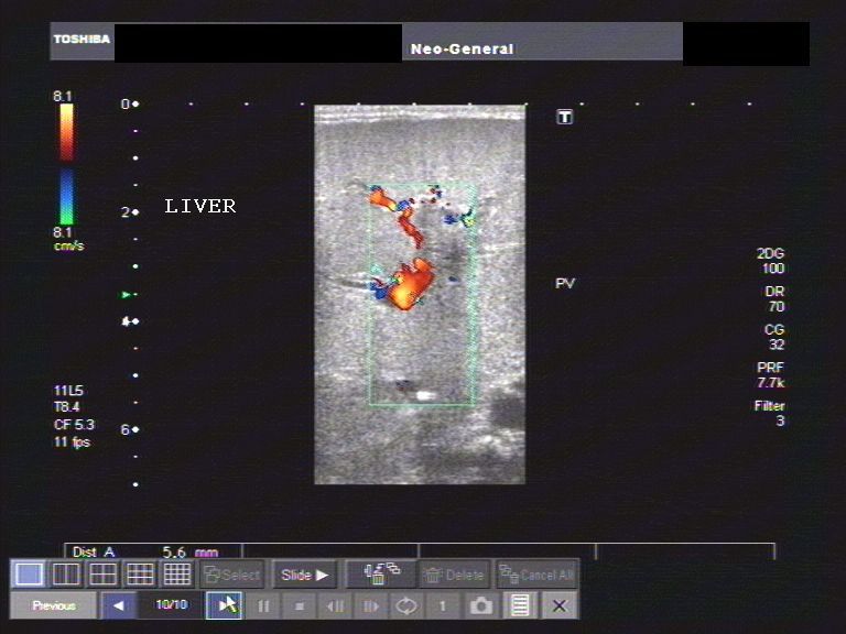
This female neonate presented with abdominal distension and oliguria. Sonography of the neonatal abdomen showed evidence of moderate ascites and echogenic, nodular liver. The liver showed moderate hepatomegaly with nodularity best seen outlined by the fluid in peritoneal cavity. The right kidney was also markedly echogenic as was the left kidney (not shown here). Color Doppler ultrasound image also shows mildly dilated portal vein (6mm. at porta hepatis). These ultrasound findings suggest advanced cirrhosis of the liver with associated renal failure in this neonate (hepato-renal failure). The cause of the cirrhosis was not determined and this condition was labeled idiopathic cirrhosis with multi-organ failure.
Reference:
http://www.jultrasoundmed.org/cgi/reprint/19/4/285.pdf
http://pediatrics.aappublications.org/cgi/content/abstract/33/5/721
Neuroblastoma in neonate
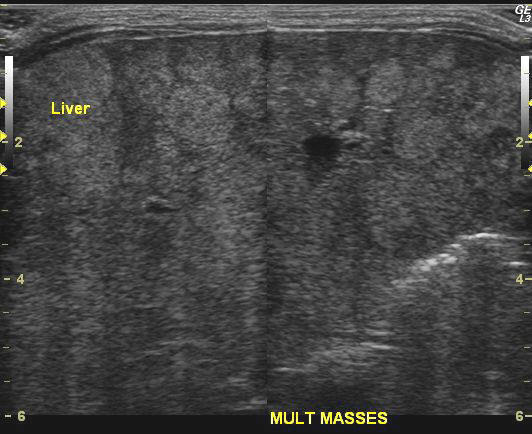
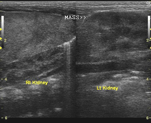
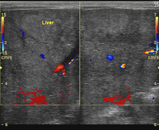
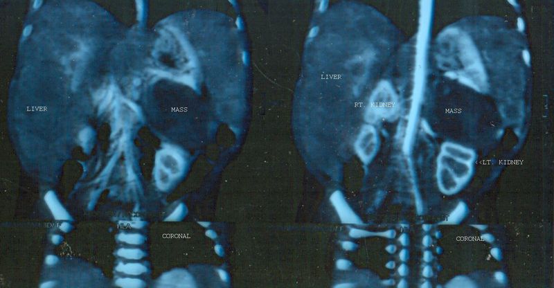
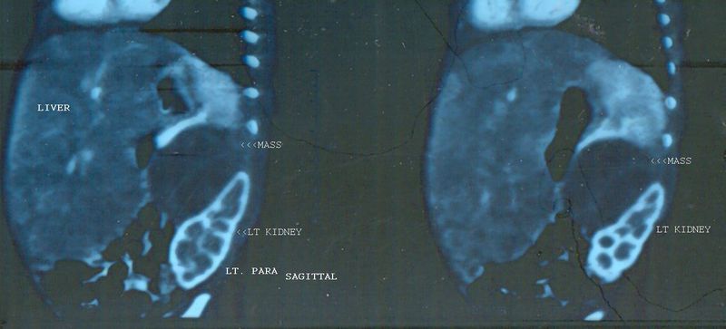
The above ultrasound and color Doppler images show multiple echogenic, poorly vascular masses throughout the liver in this neonate. The left kidney shows a poorly defined mass in the upper pole. The right kidney appears normal. A provisional diagnosis of neuroblastoma was made on these sonographic findings. However, it was felt that CT scan would be of better diagnostic importance. CT scan images of this neonate are shown below (coronal and sagittal reformatted images). The CAT scan images of this neonate confirmed the presence of a left suprarenal mass, which was hypodense and poorly enhancing. The left kidney was also found to be displaced laterally and downwards. These findings confirmed a left adrenal neuroblastoma with metastases to the liver. The liver shows massive hepatomegaly.
Reference: http://emedicine.medscape.com/article/411694-imaging
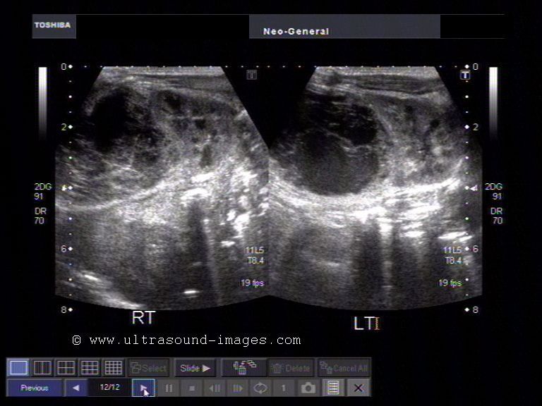
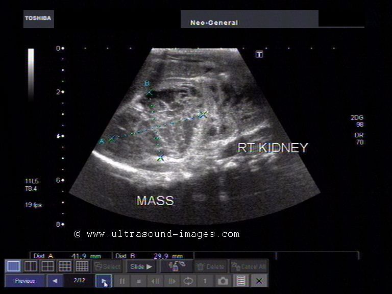
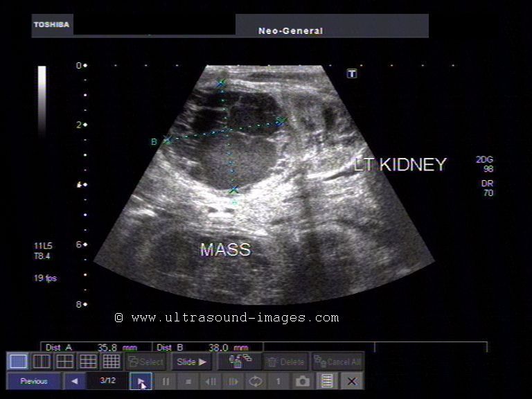
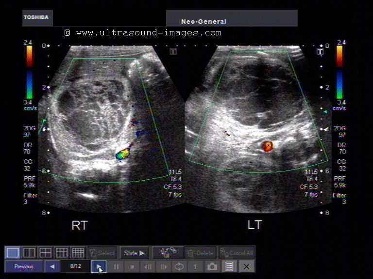
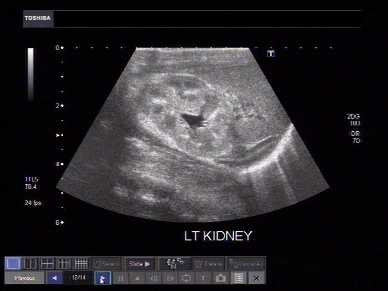
Case-2: Left suprarenal (adrenal mass) in neonate


This neonate was anemic but delivered at full term. However, the neonate's abdomen showed a palpable mass in the left flank with palpably enlarged liver. Ultrasound images of the left adrenal region showed a huge mass of 5.5 cms. in diameter with both solid and cystic regions within it. Color Doppler imaging showed some vessels within it. The infant died within days after the imaging study. The few vessels within the mass suggest a left adrenal neuroblastoma (as in the previous case above). However, the marked anemia and the complex nature suggest co-existing left suprarenal hemorrhage within the adrenal mass. The left kidney is clearly pushed downwards by the mass with the renal margins well defined. The right kidney appears normal. There is associated hepatomegaly.
Reference:
http://www.rsroc.org.tw/db/ejournal/article/V24/N3/A3/A3.htm
http://emedicine.medscape.com/article/376445-imaging
Bilateral adrenal hematoma (hemorrhage)
Both adrenal: transverse section
Rt. kidney and suprarenal gland: long section
Left kidney and suprarenal gland- Long section
Both adrenals: transverse section

Color Doppler image: both adrenals
This female neonate presented with palpable mass in the abdomen suspected to be enlarged spleen. Ultrasound images of the suprarenal regions showed 3 x 4 cms. sized masses in both sides. Both the adrenal masses displace the kidneys downwards and distort the upper poles of the kidneys. The suprarenal masses on both sides are complex, but mainly cystic with anechoic and echogenic fluid within multiple septae. Calcification is not seen. Color Doppler shows absence of flow within the masses. CT scan was also done which confirmed these findings. These ultrasound images suggest bilateral adrenal/ suprarenal hematoma (hemorrhage). The only other possibility in such cases of neonatal bilateral adrenal masses are neuroblastoma. However, neuroblastomas are usually unilateral and show presence of considerable vascularity.
References: Ultrasound imaging of adrenal hemorrhage
Sonography of bilateral adrenal masses in neonates
Congenital right diaphragmatic hernia in neonate


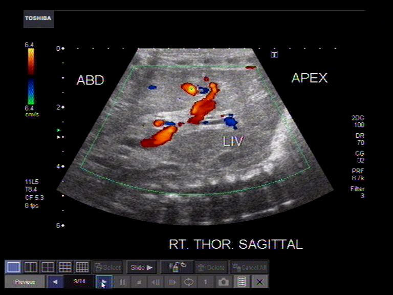

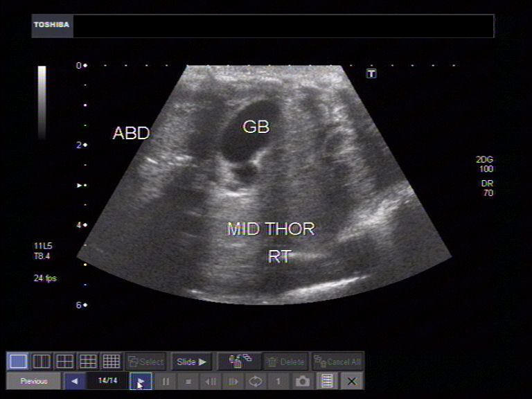
This female neonate showed opacification of the right hemithorax on X-ray chest. Ultrasound images show herniation of the liver almost entirely into the right hemithorax, with the gall bladder (GB) seen in the mid thorax on the right side. The color Doppler image shows normal vascular pattern typical of liver, in the right hemithorax. In addition, multiple tubular structures are seen in the right hemithorax, suggestive of bowel. These ultrasound and color Doppler images are suggestive of right diaphragmatic hernia, and a very large one at that. Transverse section color Doppler ultrasound image shows the heart to the left of the herniated liver. (apex= apex of right hemi-thorax). This neonate has very likely pulmonary hypoplasia in addition, as suggested by the massive nature of right diaphragmatic hernia. Both the kidneys appear grossly normal, though, very often such neonates would have hypertrophy of the kidneys.
References:
http://emedicine.medscape.com/article/407519-imaging
http://www.sonoworld.com/fetus/page.aspx?id=1581
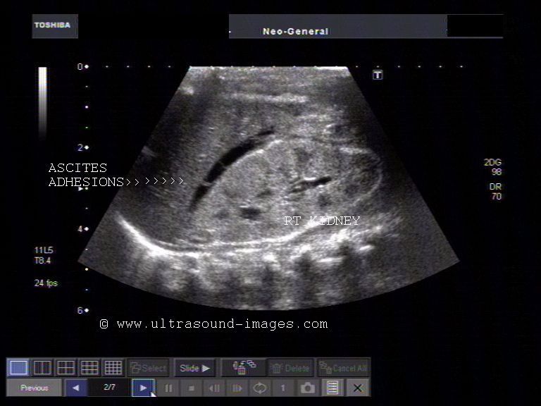
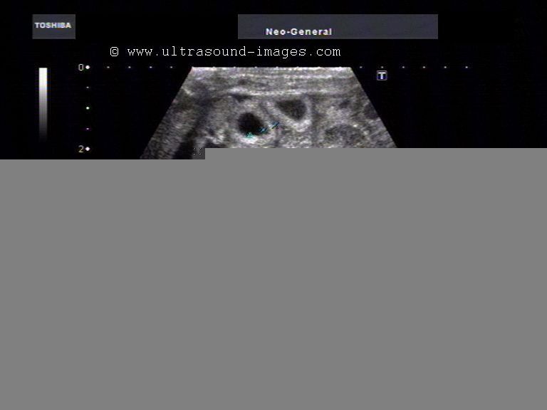
Meconium peritonitis
Gall bladder shows edematous walls
Fluid in Morison's pouch
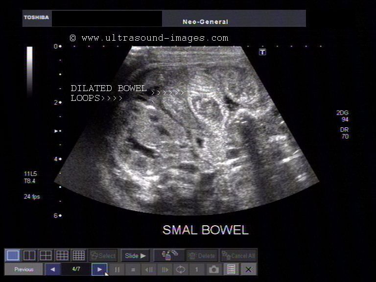
Dilated loops of small intestine with edematous walls
Concertina pattern of dilated jejunal loops
Dilated small intestine loops filled with meconium plugs
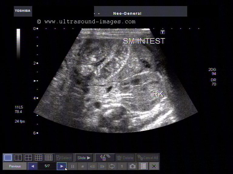
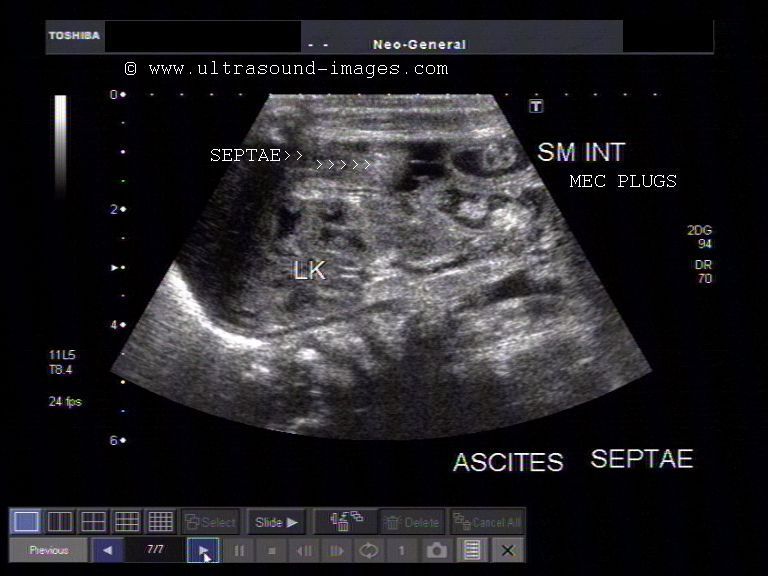
Ultrasound images of this 5 day old neonate show ascites with multiple septae or adhesions within the fluid between the loops of small intestine and also between the liver and right kidney (Morison's pouch). Though the quantity of ascites is not excessive, the presence of adhesions within the fluid indicates the presence of inflammation. The loops of small intestine are seen distended with the jejunal loops showing the typical signs of intestinal obstruction- the "concertina pattern". There is also evidence of multiple meconium plugs within the distended small intestine which are seen as echogenic lumps within the small bowel. These meconium plugs may be the cause of the intestinal obstruction and resultant leak of intestinal contents (leakage of meconium) from a small rupture of the small bowel. The other feature seen in this sonography is the edema of the bowel wall. These ultrasound images show typical sonographic features of meconium peritionitis. There are reported to be 4 types of meconium peritonitis: 1) fibroadhesive 2) cystic 3) generalized and 4) healed. The case described above shows ultrasound features of the fibroadhesive type of meconium peritonitis. Among the causative factors responsible for meconium peritonitis are: meconium ileus or intestinal obstruction due to meconium plugs, small or large bowel atresia and cystic fibrosis of the pancreas.
References:
http://sonoworld.com/fetus/page.aspx?id=253 (meconium peritonitis in fetus- images and article)
http://www.bahrainmedicalbulletin.com/june_2008/Meconium_peritonitis.pdf(free article)
http://www.learningradiology.com/archives2008/COW%20320-Meconium%20Ileus/mecileuscorrect.htm
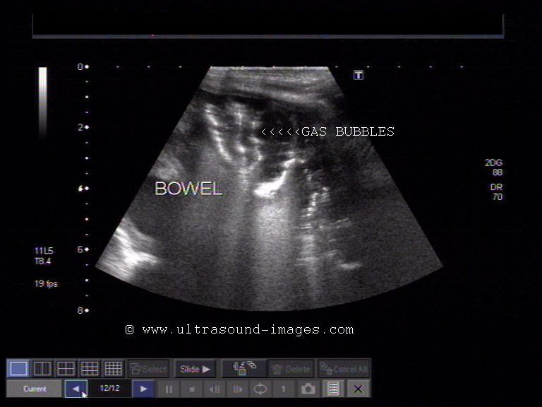
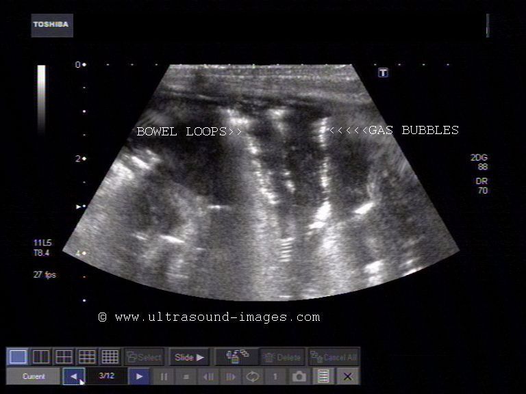
Necrotizing enterocolitis
Echogenic walls of intestine loops
Tram track sign
More images of gas bubbles in walls of intestine
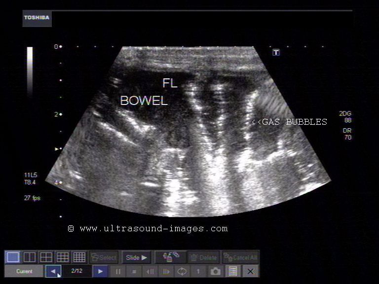
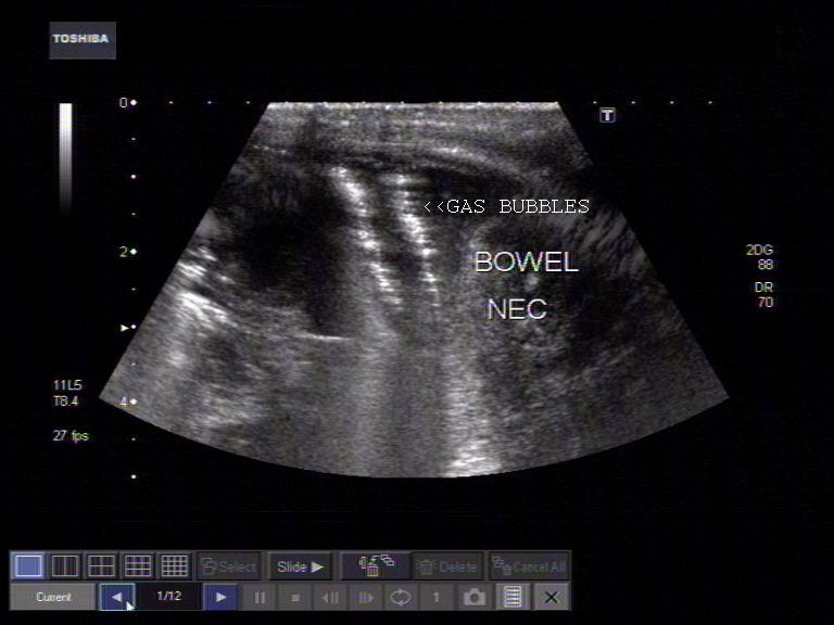
Dilated small bowel loops with gas bubbles in wall of intestine
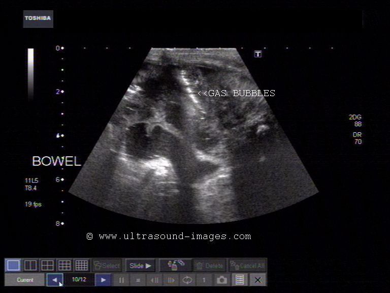

This neonate showed multiple dilated small bowel loops on x-ray, following abdominal surgery. Close sonographic examination of the loops of small and large intestine show fluid distended intestine loops with multiple echogenic foci within the thickened walls of the bowel- these represent air bubbles within the bowel wall. The parallel echogenic lines - the so called tram track sign is diagnostic of necrotizing enterocolitis. The presence of air bubbles within the intestinal walls is called "pneumatosis intestinalis" or pneumatosis of the small intestine. These ultrasound images of necrotising enterocolitis can also be confused with normal bowel gas. However, in this case (normal bowel gas), the gas is present within the bowel lumen, a normal appearance.
References:
Ultrasound study of necrotizing enterocolitis (radiographics article- free)
Sonography of pneumatosis of the small intestine
http://www.ncbi.nlm.nih.gov/pubmed/10398791- the circle sign of necrotizing enterocolitis
Unilateral UPJ (uretero-pelvic junction) or PUJ (pelvi-ureteric junction) obstruction in neonate
Normal right kidney

Hydronephrosis of left kidney
Marked thinning of the left renal cortex
Normal bladder and ureters
This neonate shows marked hydronephrosis of the left kidney with ballooning of the renal pelvis on that side. The left kidney was palpably enlarged on clinical examination. Ultrasound images show absence of hydroureter on either side with normal distension of the urinary bladder. Obviously the cause of the left hydronephrosis is left UPJ (ureteropelvic junction) /PUJ (pelvi-ureteric junction) obstruction. The commonest cause of congenital UPJ obstruction in neonates is failure of canalization of the ureteral bud on the affected side. Other causes are extrinsic obstruction or compression due to aberrant renal vessels, or due to bands or kinks in the ureter.
References: Article on UPJ obstruction (e- medicine)
© 2026, Joe Antony, MD All images and content in this site are copyrighted. www.ultrasound-images.com When visiting this site, we may use cookies to improve the performance of this site. Use of this site means you accept the use of cookies. Read our privacy policy at this page: https://www.ultrasound-images.com/privacy-policy/