
3D ultrasound images of fetus:
This is a collection of 3D and 4D (3-Dimensional) fetal images, of normal face as well as other normal parts.
Contents of this page
- 3D fetus in profile-1
- 3D fetal face in profile-2
- fetal face in 3d ultrasound- 25 weeks
- fetal face with open eyes
- fetal sole of foot-3D ultrasound
- fetal fist-3D ultrasound
- fetal face in coronal view-3D ultrasound
- 3D ultrasound image fetal lips
- 3D ultrasound image fetus in fetoscopic view
- 3D and 4D ultrasound scan- the basics
- fetal ears in 3D ultrasound
- fetal yawn- 3D ultrasound
- fetal face in 3D- another good one
- 3D ultrasound images of fetal choroid plexus
- 3D doppler ultrasound images of fetal circle of willis
- fetal-anus-3D-ultrasound
- fetal-sole-of-foot-3D-ultrasound
- triplets-triamniotic-trichorionic
- 4D-ultrasound-video-12-weeks-fetus
- 3D-fetal-ultrasound-images-week-by-week
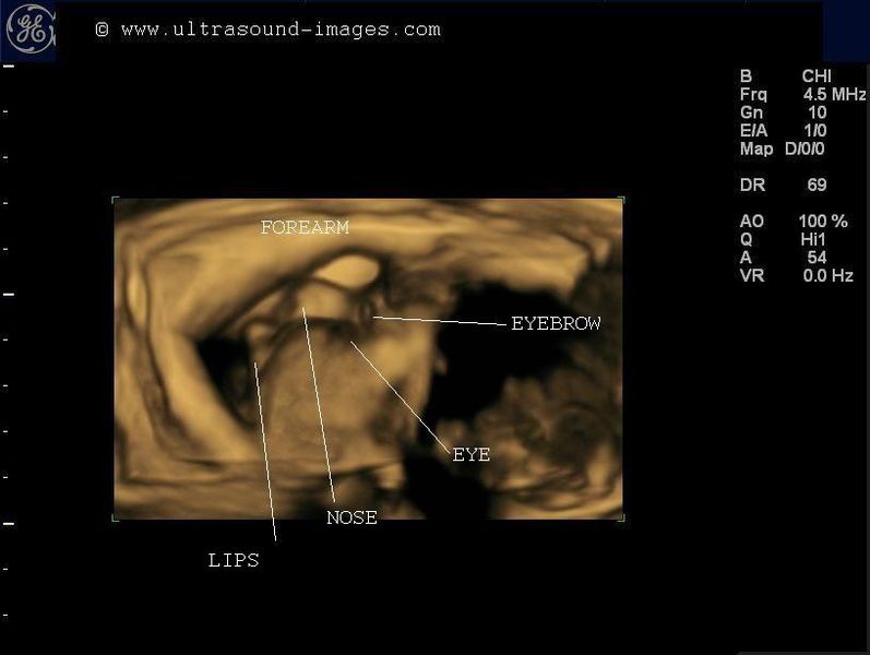
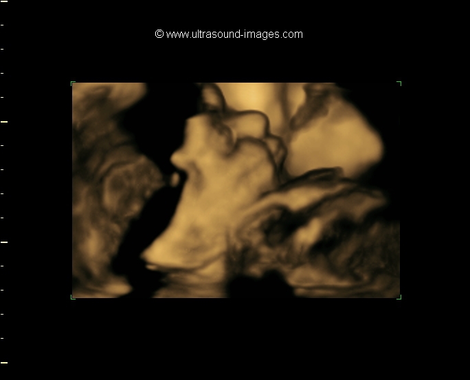
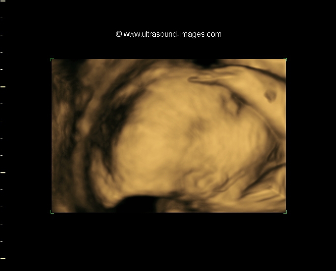
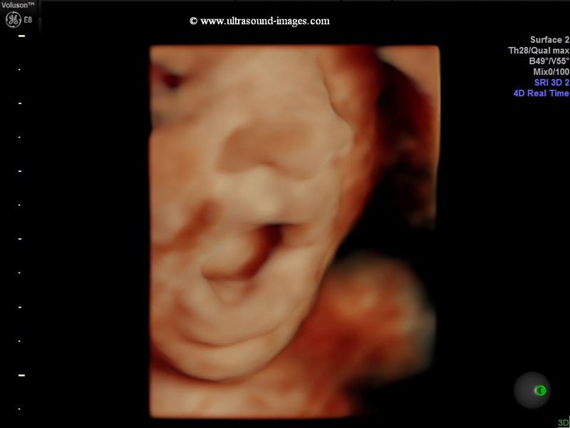
3D fetus in profile-1

Download my E book for Amazon Kindle (download free Kindle reader for i Phone or Android)
Assorted ultrasound cases- by Joe Antony, MD
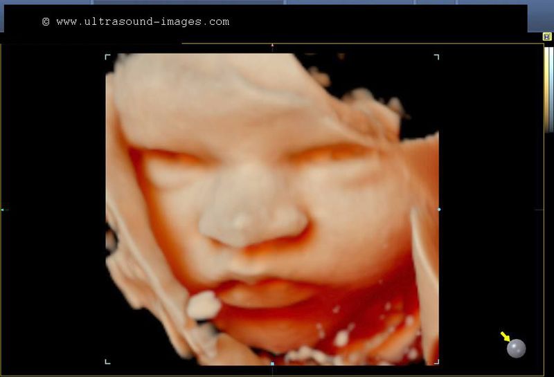
This 3D ultrasound image of a fetus shows the nose, lips and shut eyes in profile. In fact the excellent resolution of the 3D fetal face images actually show the eyebrow and look carefully- even the eyelashes.
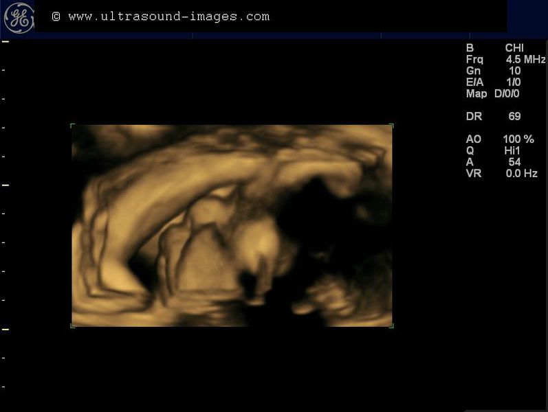
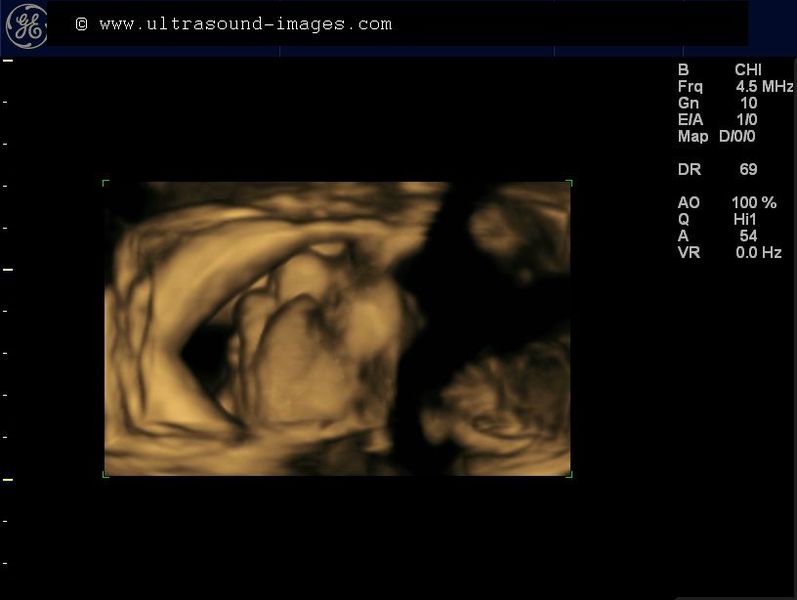
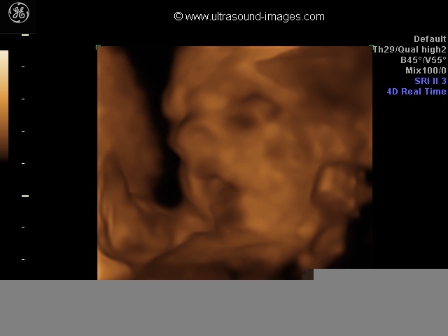
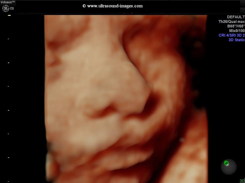
3D fetal face in profile-2

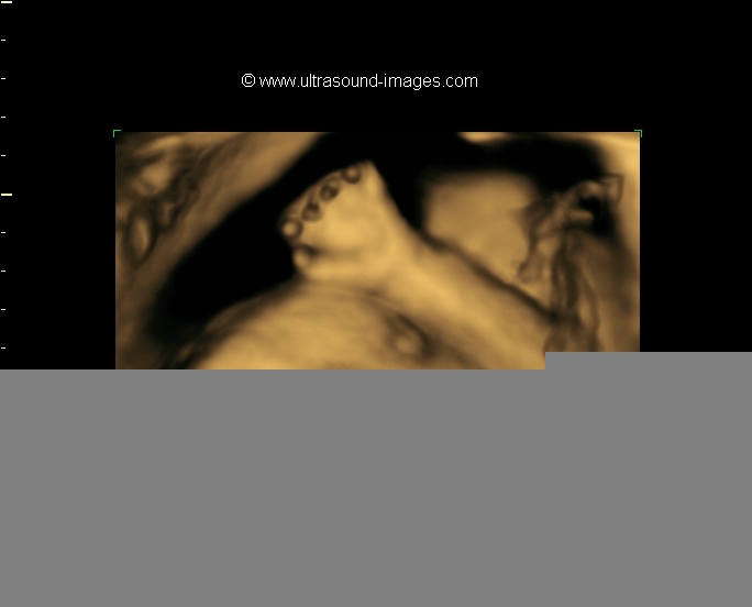
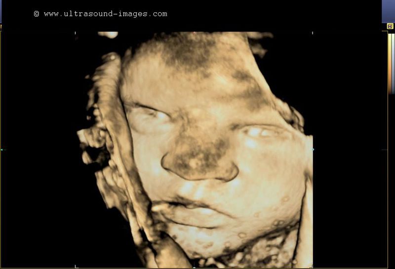
These 3D fetal ultrasound images of the face (32 weeks) are stunning in detail. Observe the cheek and other facial features. Note the pinna of the fetal ear shown in striking clarity.
The fetal fingers are also seen well.
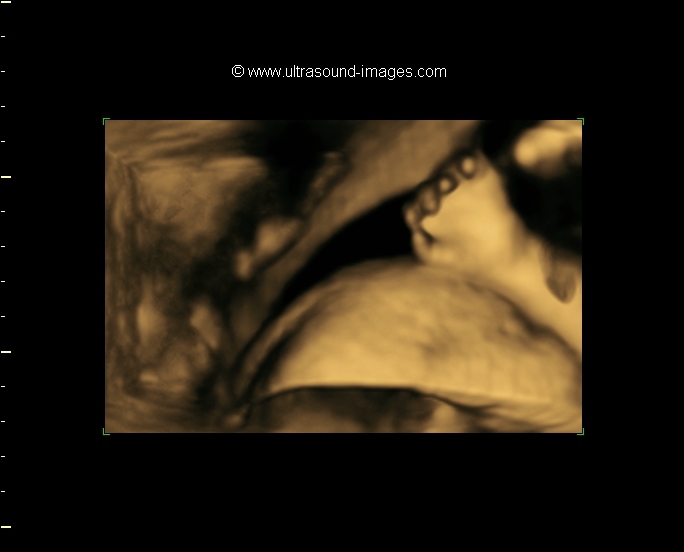
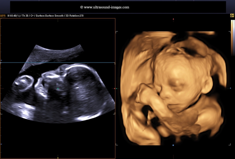
fetal face in 3d ultrasound- 25 weeks

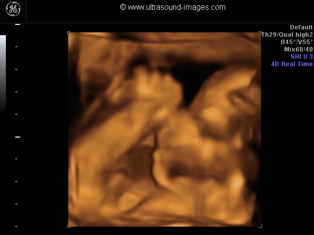
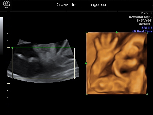
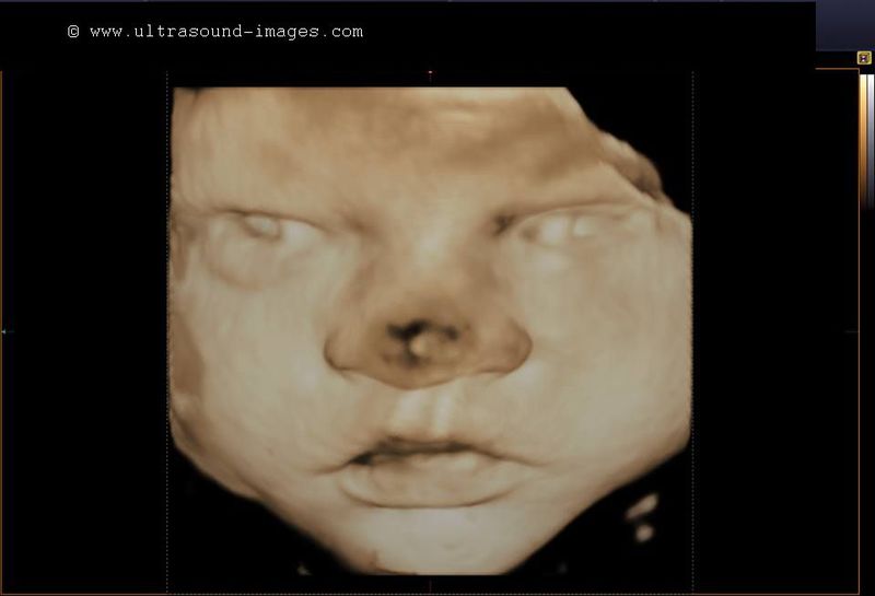
This was a 25 week old fetus with vigourous Fetal movements which could only be captured using 4D ultrasound. The machine used was Voluson 730 system from GE. With the default settings at surface rendering, we could capture 3-D ultrasound images of the hands and Fetal face from multiple angles.
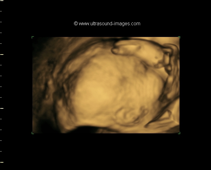
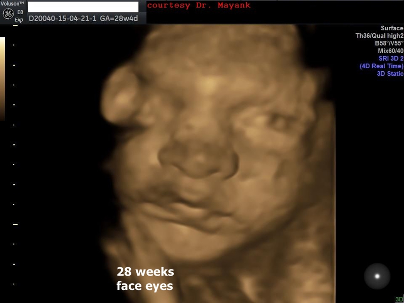
fetal face with open eyes
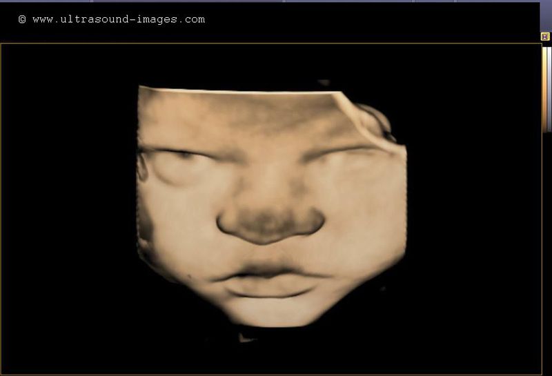
It is extremely rare for the fetus to open its eyes and to catch it during sonography. But these 3-D ultrasound images by Dr. Arun Mahajan, MD, India, show just that. The fetal eyes are imaged with the lens and possibly the iris visualized in detail in these 3-D ultrasound pictures.
fetal sole of foot-3D ultrasound
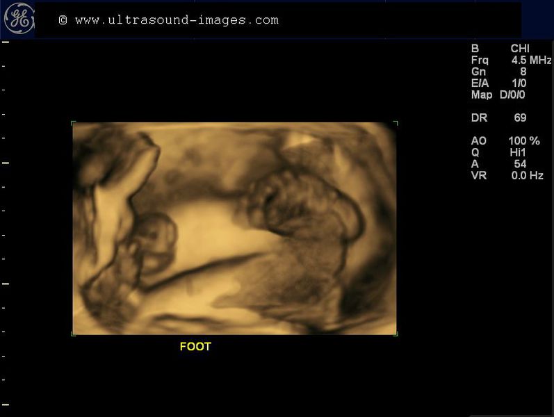
This 3D ultrasound image captures the fetal foot, with the sole of the foot facing the ultrasound beam. The toes and sole are seen well in this image. The fetal face is seen in profile to the left of the 3D ultrasound image.
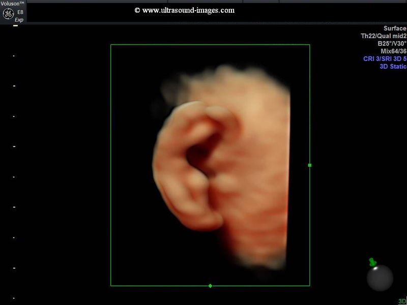
fetal fist-3D ultrasound

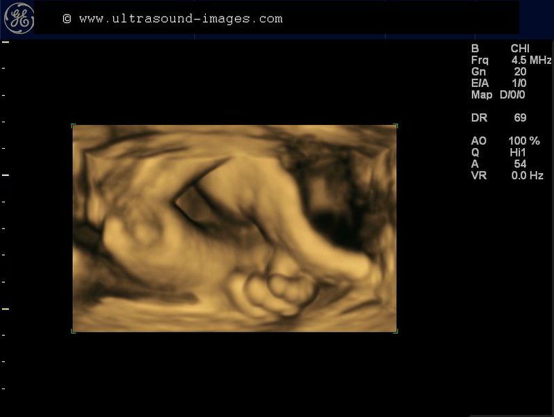
This fetus is captured in 3D ultrasound clenching its fist near the face. Observe the detail of the fetal fingers. Wish I could image the finger-nails also!
fetal face in coronal view-3D ultrasound
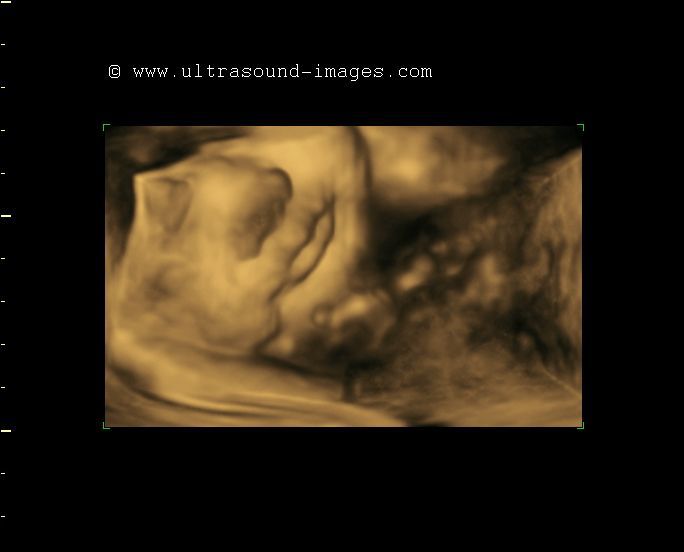
This is a good view of the fetal face in face on view or coronal section, with the lips and nose displayed well in this 3D ultrasound image.
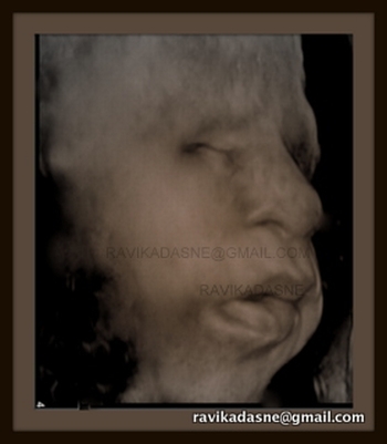
3D ultrasound image fetal lips
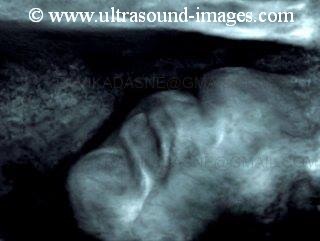
This 3D ultrasound image focuses and captures the lower part of the fetal face focussing on the fetal lips. This image is courtesy of Dr. Ravi Kadasne, MD, UAE.
3D ultrasound image fetus in fetoscopic view

This fetus was captured in 3D using a special technique, the so-called fetoscopic view, to produce true to life surface rendering and color, in these 3D ultrasound images. Images are courtesy of Dr. Gunjan Puri, MD, India. The ultrasound system used is the Accuvix A 30 from Samsung/ Medison.
3D and 4D ultrasound scan- the basics
3 D and 4 D ultrasound scan- advantages and limitations:
Advantages in pregnancy and fetal imaging:
3D scanning means seeing the still image of the baby in uterus in 3dimensions just like seeing a statue (3D scan image) versus a picture or photograph(2D ultrasound image). 4D scanning means seeing the 3-D ultrasound image in real time- that is, with baby's or mother's movements.
1. Realistic three-dimensional views of the entire foetus or baby during early pregnancy from 6 weeks to 25 weeks. The entire baby can be seen with visualization of the legs, hands, face, head and body.
These images can be given in a CD or DVD.
2. From about 30 weeks to 36 weeks it is possible to see the fetal face very clearly (almost) just like how it would look after delivery. In addition one can see the baby's spine, feet and certain other parts like the hands.
3. Thus one can detect various anomalies like cleft lip and club feet with 3-D and 4-D ultrasound.
4. One can detect with greater precision and detail, anomalies of the baby’s spine like spina bifida and meningocele.
5. Even the patient can easily view these normal and abnormal parts of the baby and appreciate the problems as well as the beauty of the child.
In gynecology :
During ultrasound of the non-pregnant uterus and ovaries it is possible to see anomalies of uterus like septate or bicornuate uterus etc. One can better appreciate tumors like fibroid and malignant masses in greater detail.
Limitations and problems of 3-D and 4-D ultrasound scanning:
1.In fat/ obese patients or in pregnancy with less amniotic fluid the resolution (and hence quality) of the images is very poor.
2. If the baby moves too much or too fast then the resolution is poor.
3. The baby's face may not be seen if the baby lies in a difficult position such as prone position or if the baby's face is apposed to the uterine walls.
4. In the last 1 month of pregnancy it may be difficult to see the baby in 3 dimensions.
5. Lastly, 4 d scan can only be used to compliment ordinary 2-D or B- mode ultrasound scan..it has the same problems and limitations as ordinary 2-D scan. As always it cannot detect many anomalies and defects if conditions for imaging are not
optimal.
Dr. Joe Antony, MD.
fetal ears in 3D ultrasound

These 3D ultrasound images of the fetal ear (pinna) are among the best I have seen so far. Taken by Dr. Mayank Chowdhury, MD, using one of the best 3D ultrasound systems in the market, the GE Voluson E-8, Expert. Ultrasound study of the fetal external ear is often helpful in ruling out syndromes associated with anomalies of the ear.
fetal yawn- 3D ultrasound
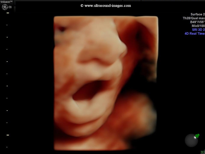
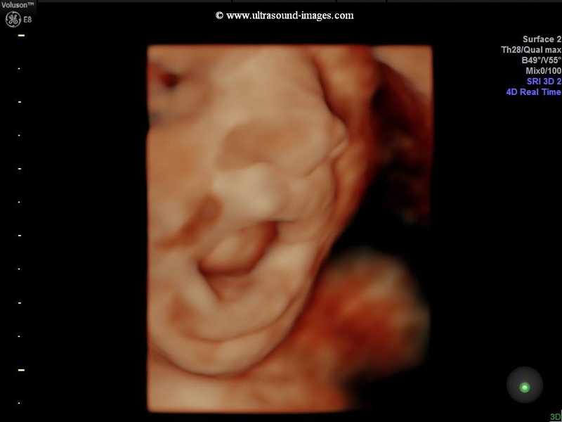
An excellent capture in 3D/ 4D sonography of different stages of fetal yawn. This is a late 2nd trimester fetus (25 weeks). 3D sonographic images courtesy of Dr. Mayank Chowdhury , MD.
fetal face in 3D- another good one
This is yet another great 3D ultrasound image of the fetal face (image courtesy of Ravi Kadasne, MD). The fetal nose, eyes and lips are visualized in fine detail.
3D ultrasound images of fetal choroid plexus
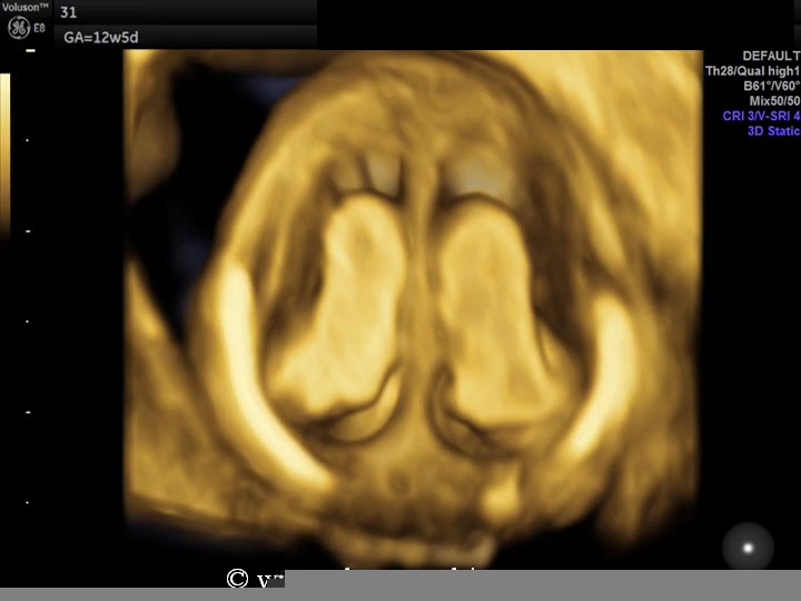

The progress of current cutting edge 3D and 4D ultrasound imaging technology means that fetal internal structures including the fetal brain and its inner anatomy can be displayed in spectacular detail. These 3d ultrasound images (courtesy of Mayank Chowdhury, MD) reveal the choroid plexuses of the fetal brain at 12 weeks. The choroid plexus is seen from multiple viewing angles in volume sections. The ultrasound system used here is the Voluson E8 from GE.
3D doppler ultrasound images of fetal circle of willis
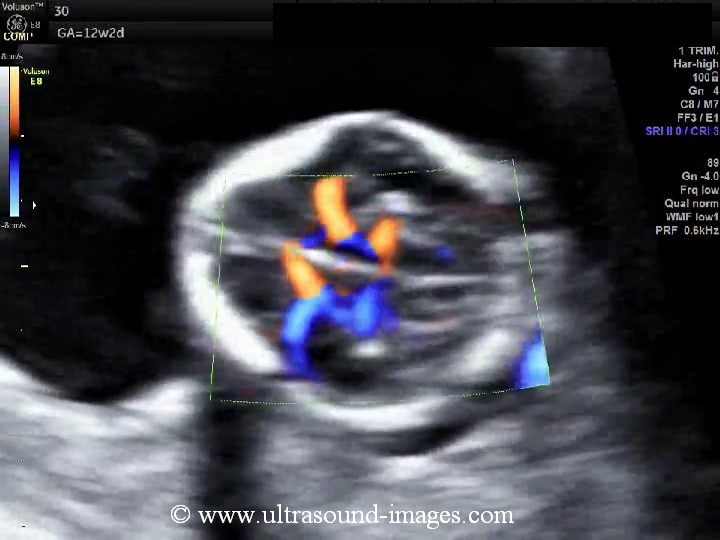
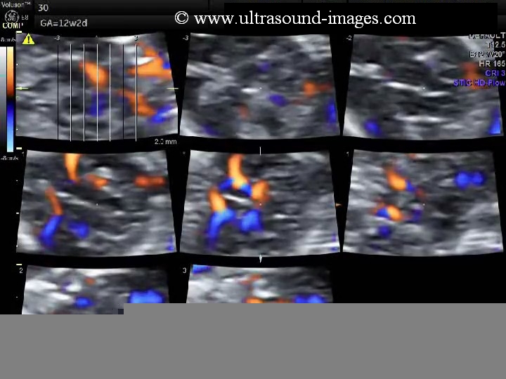
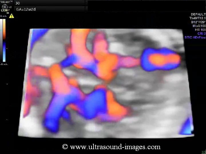
3d Color Doppler imaging of the circle of Willis in a fetus at 12 weeks. These 3d doppler images leave nothing to the imagination with the anterior, middle and posterior cerebral arteries within the circle of Willis visualized. (Images are courtesy of Mayank Chowdhury, MD)
fetal-anus-3D-ultrasound
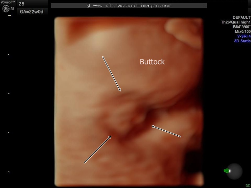
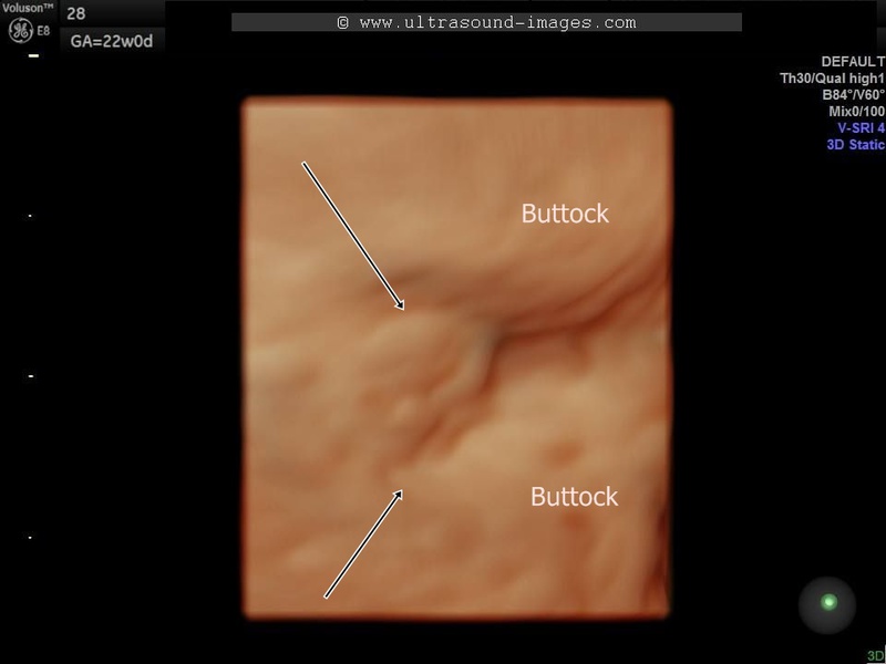
3D ultrasound images (fetus at 22 weeks gestational age) showing a surface rendering of the fetal buttocks and anus. Arrows point to the fetal anus whilst the fetal gluteal folds are seen on either side. These 3D surface rendering images are courtesy of Mayank Chowdhury, MD.
fetal-sole-of-foot-3D-ultrasound

This is yet another 3D ultrasound image showing the foot, but this time it shows the sole of the foot or in medical parlance, the plantar surface of the foot in stunning detail. Each toe is visualized. Image is courtesy of Mayank Chowdhury, MD.
triplets-triamniotic-trichorionic
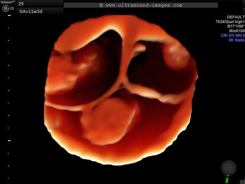
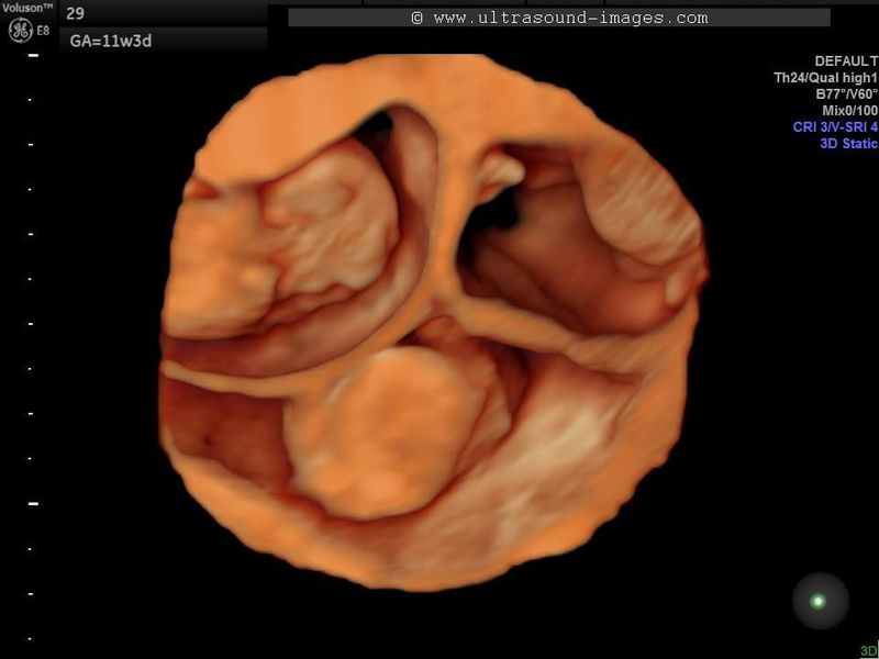
3D ultrasound images of triplets gives an eery appearance to this rare medical phenomenon. Images are courtesy of Mayank Chowdhury, MD. Each of the fetuses at 11 weeks has its own gestation sac, amnion and chorionic plate.
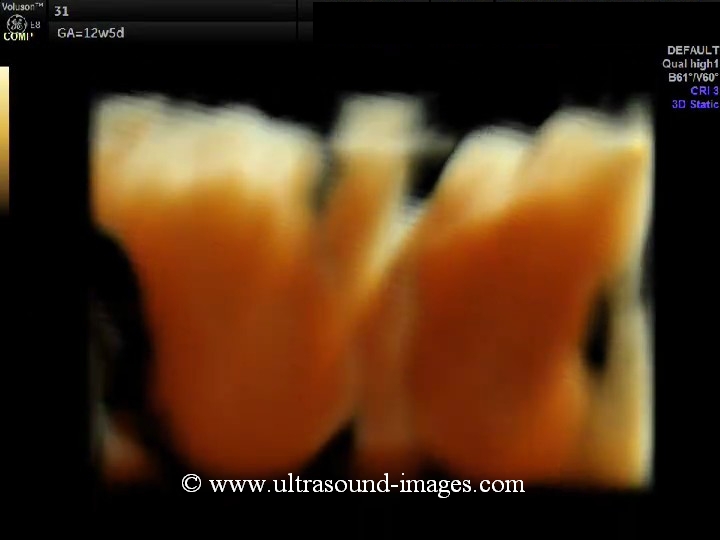
4D-ultrasound-video-12-weeks-fetus
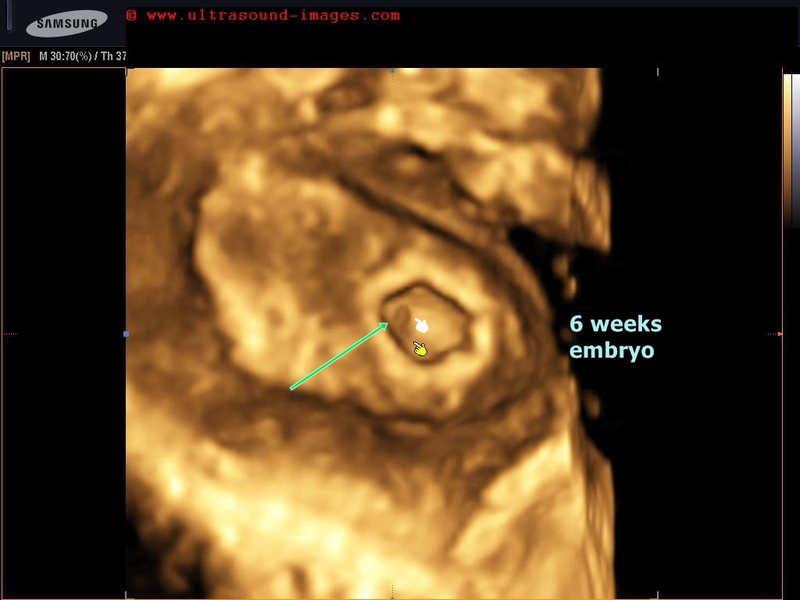
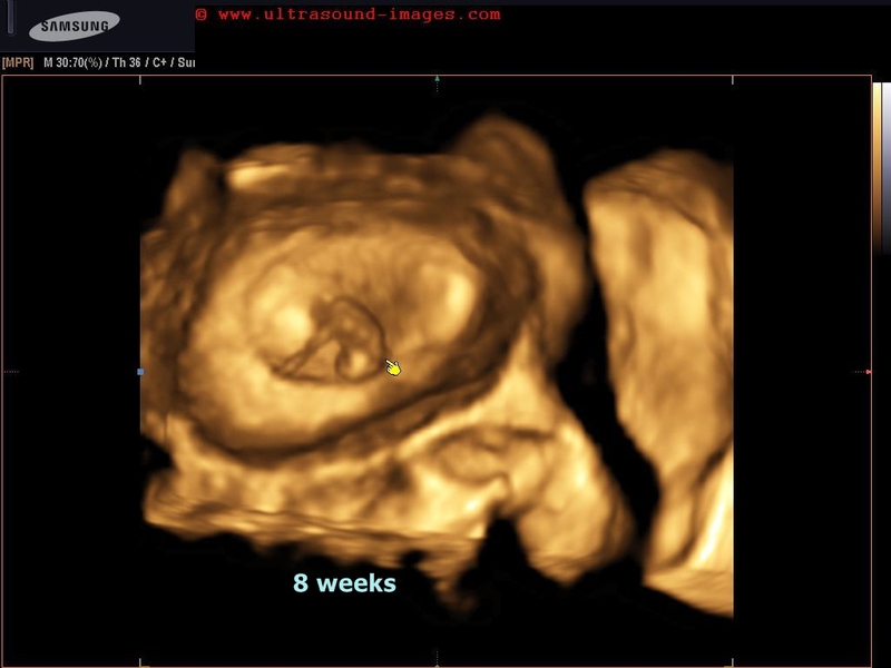
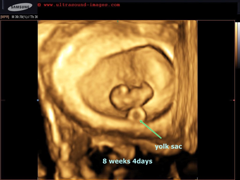
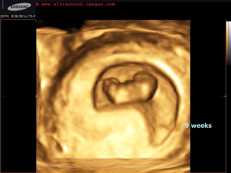
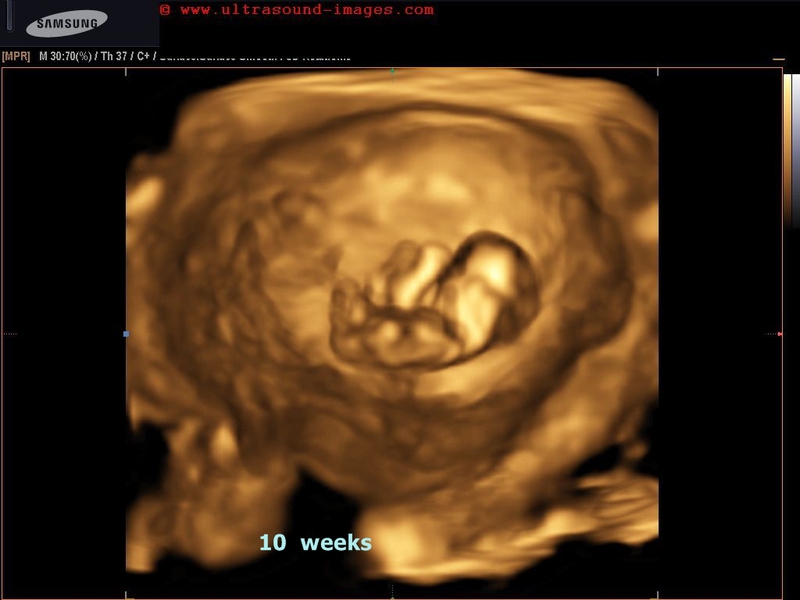
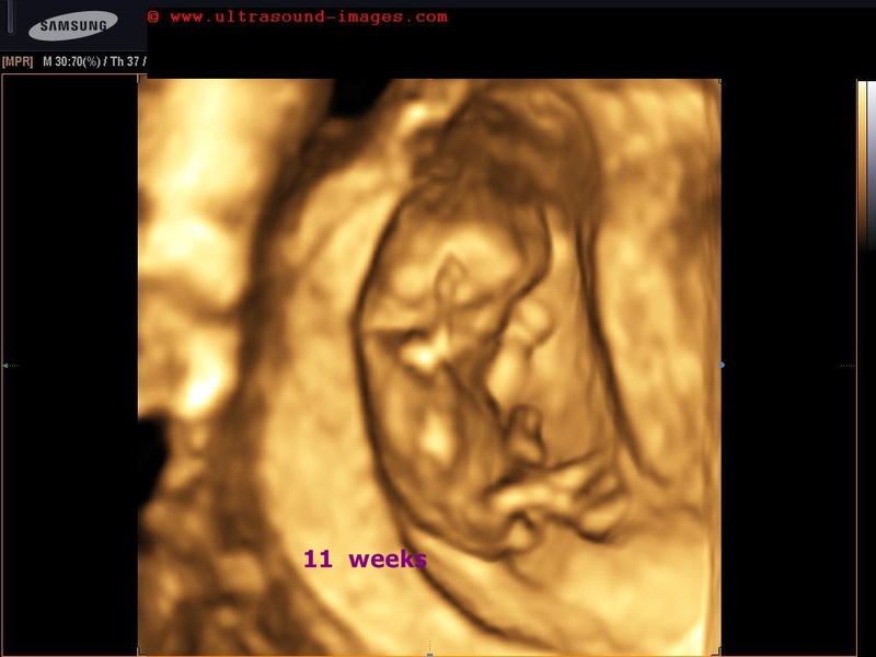
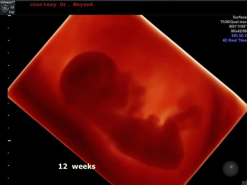
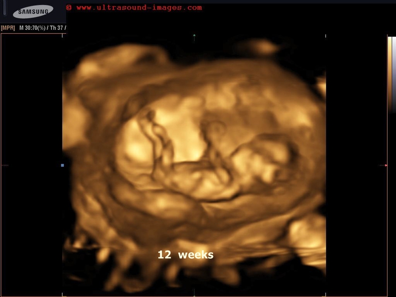
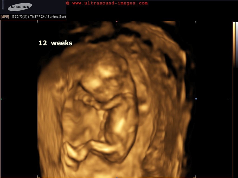
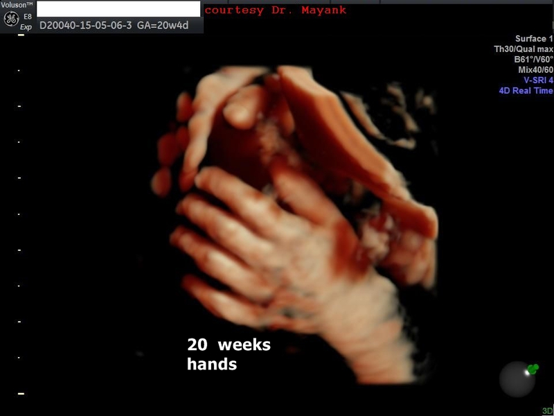
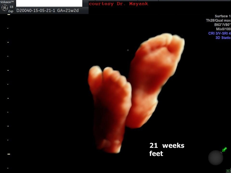
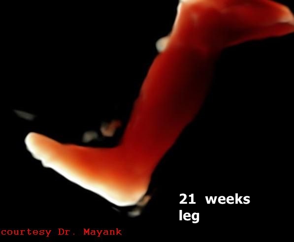
3D-fetal-ultrasound-images-week-by-week
1st and 2nd trimester fetus-
some of the images below are courtesy of Dr. Mayank Chowdhury.


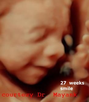
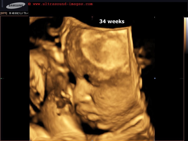
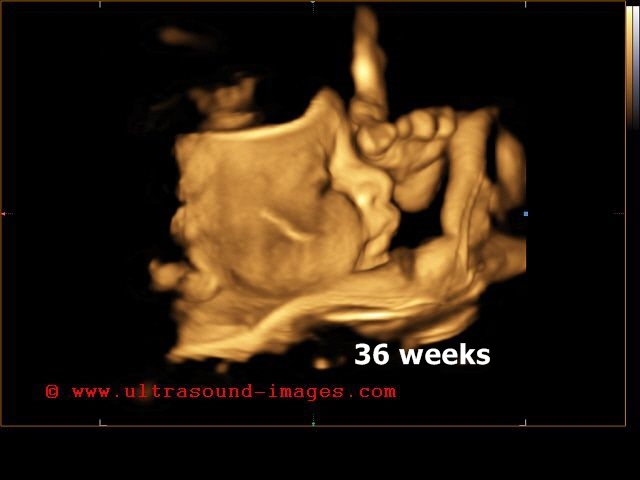
3D ultrasound 3rd trimester fetus

© 2026, Joe Antony, MD All images and content in this site are copyrighted. www.ultrasound-images.com When visiting this site, we may use cookies to improve the performance of this site. Use of this site means you accept the use of cookies. Read our privacy policy at this page: https://www.ultrasound-images.com/privacy-policy/