
Ultrasound images of diseases of the kidneys
Contents of this page
- Normal variants
- SONOGRAPHY OF RENAL PATHOLOGY
- Autosomal Dominant Polycystic Kidney Disease (ADPCK)
- CYSTIC LESIONS OF THE KIDNEYS
- Cortical cysts or simple renal cyst
- Calyceal diverticulum or calyceal cyst
- Milk of Calcium cyst
- Complex cysts of the kidney (complex renal cysts)
- Hemorrhagic cyst of kidney
- Pseudoaneurysm in Transplanted kidney
- Ectopic/ Pelvic kidney
- Ectopic /thoracic kidney
- Horseshoe kidney
- Renal cell carcinoma
- Case-2 (Renal cell carcinoma)
- Chronic renal failure (Medical renal disease)
- Severe renal failure with arterio-venous fistula (hemodialysis)
- Renal sinus lipomatosis
- Renal trauma- subcapsular hematoma
- Renal angiomyolipoma -(AML) of right kidney
- Exophytic renal cysts- Exophytic cortical cysts of the kidney
- Duplex kidney with complete duplication of ureters
- Xanthogranulomatous pyelonephritis of right kidney
- cross fused ectopic kidneys with duplex ureters
- trifid renal pelvis
- nutcracker phenomenon
- pyelonephritis-UTI-renal-edema
Normal variants
Fetal lobulation of kidneys

Download my E book for Amazon Kindle (download free Kindle reader for i Phone or Android)
Assorted ultrasound cases- by Joe Antony, MD
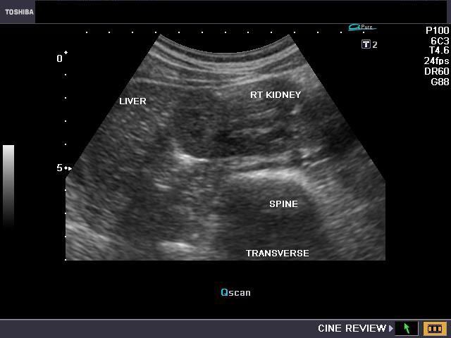
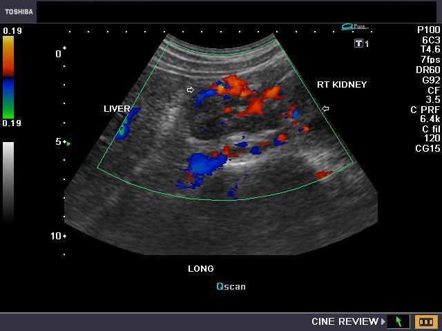
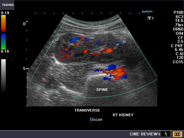
This patient, an adult female underwent routine sonography of the abdomen. Ultrasound images of the kidneys reveal multiple indentations (short arrows) of the renal cortex, with no evidence of fibrotic tissue (would be seen as hyperechoic bands). There is no evidence of thinning of the renal cortex or deformity of the pelvicalyces (ruling out chronic pyelonephritis). These ultrasound images show typical appearance of renal fetal lobulation. Fetal lobulation is a common finding and is a normal variant of the kidneys. It is caused by the persistence, into adult life, of the lobulation of the kidneys seen normally during fetal stage.
Hypertrophied column of Bertin or Junctional parenchyma

The left kidney in this male child shows a prominent column of renal cortex in the middle third of the kidney. This column of tissue is seen encroaching on the renal sinus, with the renal contour (outer margins of the left kidney) appearing normal. Color Doppler images of the left kidney show normal vascular pattern in the column. The echogenicity and echotexture of the column of tissue appears similar to the adjacent renal cortex. These ultrasound images and the sonographic findings are typical of what is called hypertrophied column of Bertin. The current terminology for this benign normal variant is junctional parenchyma. This new term for a long understood normal variant, is used as the renal tissue in the column of Bertin does not show any evidence of hypertrophy. The tissue in the junctional parenchyma/ hypertrophied column of Bertin is absolutely normal renal parenchyma and contains renal cortex, pyramids and septa or columns of Bertin. the renal tissue in the column is continuous and merges with the adjacent cortical tissue of the kidney. The column of Bertin is most commonly seen involving the left kidney as in the case above.
References: Sonography of junctional parenchyma (abstract)
Ultrasound imaging of hypertrophied column of Bertin
SONOGRAPHY OF RENAL PATHOLOGY
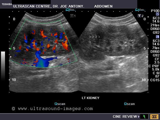
Autosomal Dominant Polycystic Kidney Disease (ADPCK)
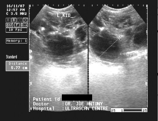
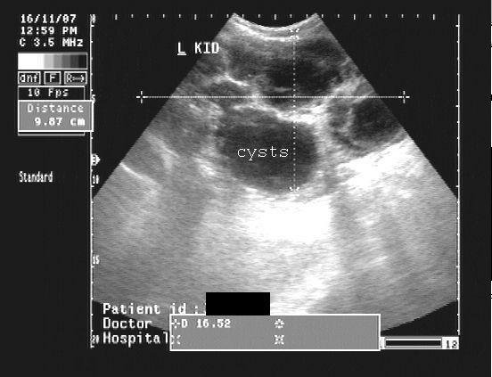
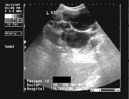
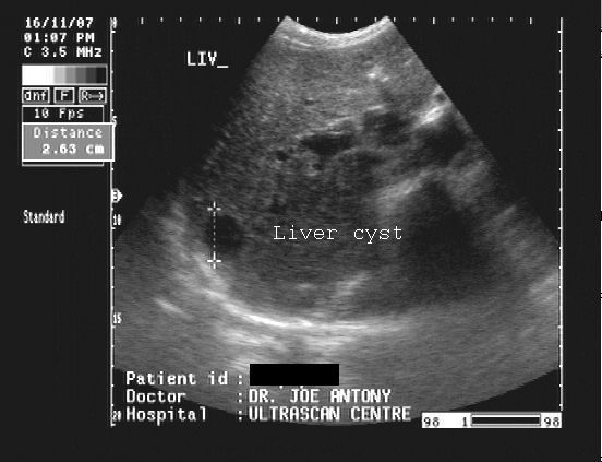
Sonography of this 55 yr. old male patient, who presented with hematuria, reveal bilateral grossly enlarged kidneys (14 to 16 cms. in length, 8 to 10 cms. width). The kidneys are replaced by multiple large cysts (more than 2 in each kidney) of sizes varying from 2 to 5 cms. (Criteria for diagnosis of ADPCK: a) more than 2 renal cysts in one/ both kidneys, if age less than 30 yrs. b) 2 or more cysts in each kidney, if aged from 30 to 59 yrs. c) 4 cysts in each kidney if aged more than 60 yrs.) There is very little normal renal parenchyma seen in these images. Note that the cysts are non-communicating ruling out hydronephrosis. The right kidney shows an echogenic focus, in one cyst, which may be a calculus or calcification. A solitary cyst is seen in the right lobe of liver. These ultrasound images are diagnostic of Autosomal Dominant Polycystic Kidney (ADPCK) disease.
References:
1) http://en.wikipedia.org/wiki/Polycystic_kidney_disease (free article)
2) http://www.emedicine.com/med/topic1862.htm
See this link:
autosomal dominant polycystic kidneys with multiple calculi
this 58-year-old Male patient underwent routine sonography for non-specific complaints including mildly elevated serum creatinine and blood urea. Sonography shows multiple small cysts ranging from 3 mm up to 12 mm in size in both kidneys. Both kidneys are literally studded with these minute cystic lesions (more than 20 in each kidney) with many of them showing the presence of milk of calcium. In addition, there are also multiple minute calculi ranging from 2 to 4 mm in size. This is an unusual presentation of autosomal dominant polycystic kidney disease ( see the ultrasound images above) and could represent the initial stages of the disease. Both kidneys also show moderate enlargement with sizes of 11 x 6 cm.
CYSTIC LESIONS OF THE KIDNEYS
Cortical cysts or simple renal cyst
Case-1
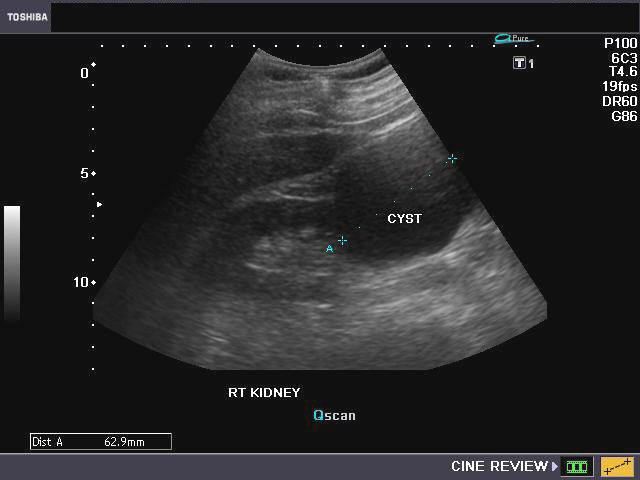
The above ultrasound images show solitary cortical cysts of the kidney. These are seen as single, well defined cystic lesions mainly involving the renal cortex and have thin walls. There is also acoustic enhancement posteriorly. The Power Doppler image (top row- right) shows blood vessels curving around the cyst. Cortical cysts are usually a benign condition requiring follow up to look for changes such as infection, rupture or increase in size.
Reference: http://emedicine.medscape.com/article/453831-diagnosis
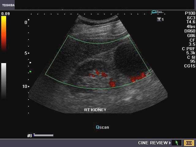
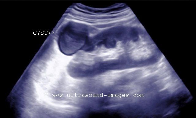
Calyceal diverticulum or calyceal cyst
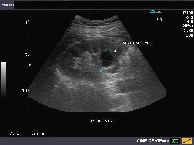
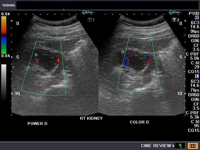
The above B-mode ultrasound image (left) and Color Doppler image (right) show a cystic lesion involving the collecting system (middle calyx) of the right kidney. This suggests a calyceal diverticulum /calyceal cyst of the right kidney. The walls of the cystic lesion are thin but irregular and possibly communicate with the renal pelvis. Again, in this case too, the cyst appears to displace blood vessels which surround the lesion.
Reference: http://www.radswiki.net/main/index.php?title=Calyceal_diverticulum
Milk of Calcium cyst

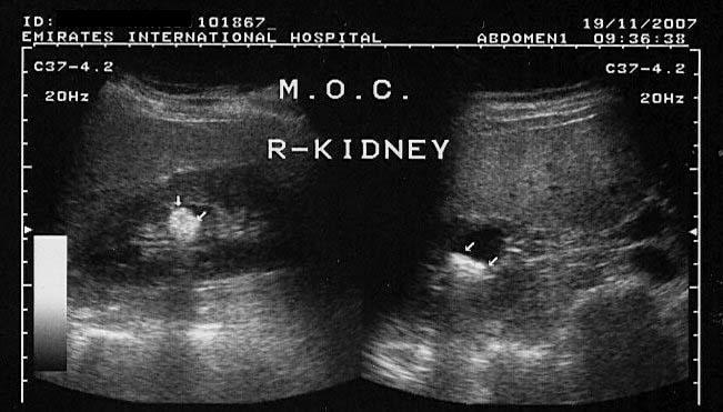

The above ultrasound images of the right kidney show echogenic debris layering at the dependent part of a small cyst. There is change in the fluid-debris interphase with change in position. There is minimal acoustic shadowing due to a relatively large amount of calcium debris. These are typical findings of Milk of Calcium within a renal cyst. Images courtesy of Dr. Ravi Kadasne, UAE.
Reference:
1) http://www.jultrasoundmed.org/cgi/content/abstract/11/5/195
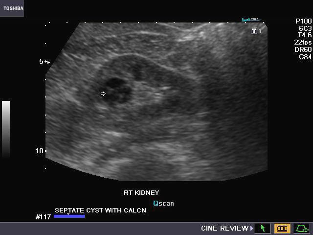
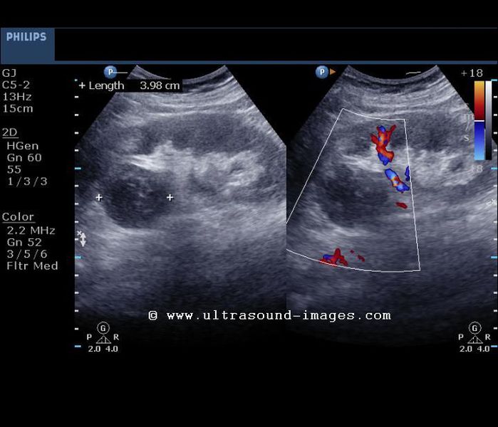
Complex cysts of the kidney (complex renal cysts)
Case-1
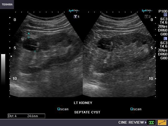
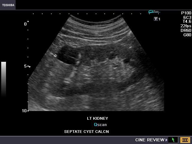
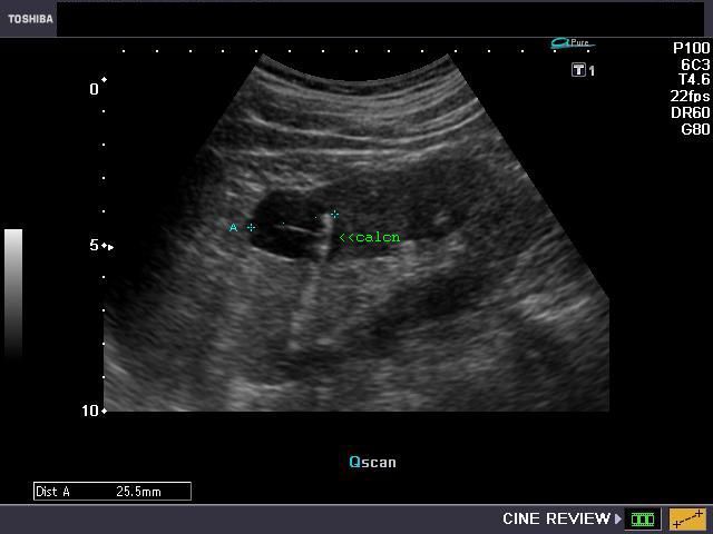

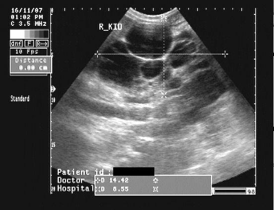
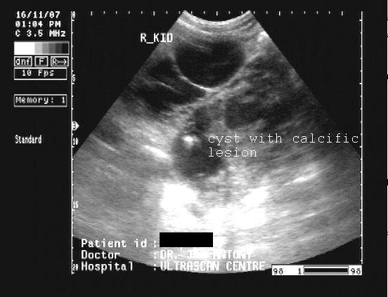
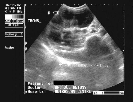
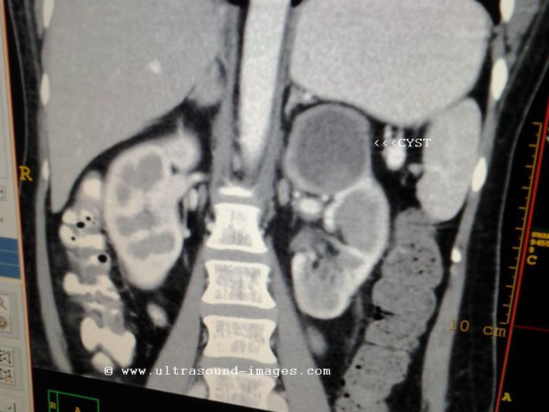
The above ultrasound images show a cyst of 2.5 cms. in the left kidney. What differentiates this cystic mass from the previously discussed cases of renal cysts is the presence of multiple fine septae within the cyst. In addition, there are 2 prominent calcific lesions in the cyst wall. These ultrasound images and findings are the hallmark of what is called complex renal cysts. The color Doppler image also shows the non vascular nature of the cyst walls and septae. The Bosniak classification describes the various grades of cysts from category-1 (simple renal cysts without septae or calcifications, category-2 (mildly complex but benign cysts of the kidney), category-3 (indeterminate cysts that need surgery in most of the cases), category 4 (cystic neoplasia or potentially malignant cysts). There is, in addition, category 2-F, cases that need follow up ultrasound/ CT imaging. This classification is a modification of the original Bosniak categorization of renal cysts. In the case above, ultrasound alone might be unable to clearly categorize the complex cystic mass, and CT imaging is advisable.
References:
http://emedicine.medscape.com/article/453831-diagnosis (free article)
Case-2

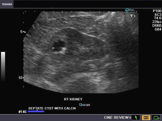
The above ultrasound images demonstrate a similar cyst in the right kidney of this middle aged male patient. Complex renal cysts of this nature are usually an incidental finding and need to be followed up. Though ultrasound imaging of complex cysts do give a lot of information and detail about renal cysts, Bosniak categorization of complex renal cysts is better done with CT scan imaging and where needed, MR imaging is also indicated.
Hemorrhagic cyst of kidney

Left kidney- upper pole cortical cyst with echogenic contents suggesting hemorrhage. Normal vascularity is present around the cyst. No vessels are seen within the cyst.
Long section ultrasound image of the left kidney

Echogenic matter is seen within the renal cortical cyst suggesting either hemorrhage or infective change.
CT scan images showing the hemorrhagic cyst of the upper pole of the left kidney

The CT scan images shows hyperdense matter within the cyst with enhancement of the edges of the lesion, without enhancement of the contents (post contrast study). Hemorrhage is a known complication of cortical or simple renal cysts.
References: 1) Imaging of hemorrhagic cyst of kidney
Pseudoaneurysm in Transplanted kidney
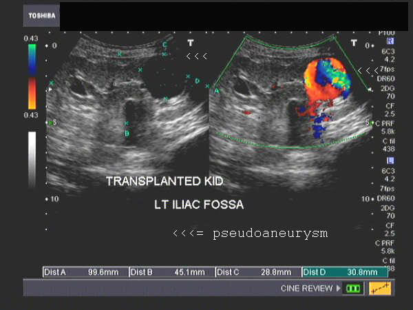

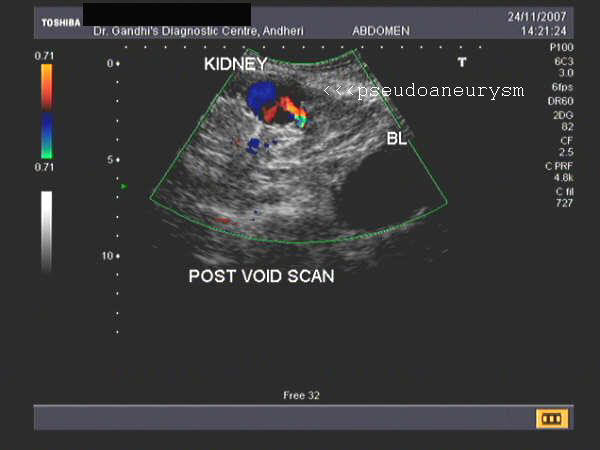
Sonographic imaging was done on this patient who underwent renal transplant some time ago. The transplanted kidney is seen in the left iliac fossa in these ultrasound images. The first image shows a cystic lesion (anechoic) in the lower pole of transplanted kidney. The 2nd image also shows mild dilation of the renal pelvis and upper ureter in this kidney. Color doppler images reveal swirling flow pattern within the cystic lesion. There is also evidence of a communication of the lesion with a segmental artery. Real time imaging also revealed to and fro flow between the lesion and the artery. Diagnosis: pseudoaneurysm in transplant kidney. Images courtesy of Dr. Jaydeep Gandhi, Mumbai, India.
Reference:http://www.sonoworld.com/Sonoworld/Cases/Cases.aspx?CaseID=363(free article)
Ectopic/ Pelvic kidney
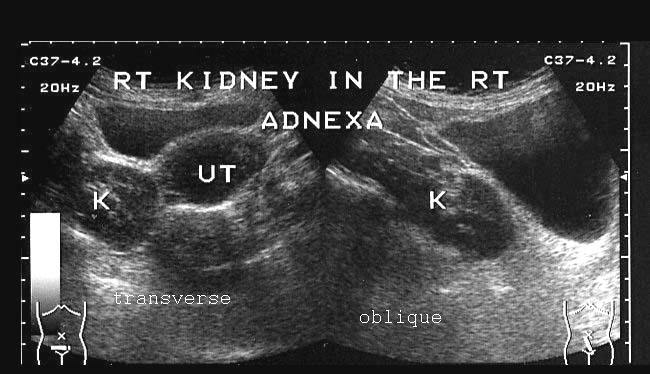
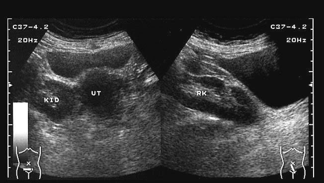
This female patient underwent routine abdominal ultrasound imaging. These ultrasound images show the right kidney just to the right of the uterus in the true pelvis. There was no kidney visualized in the right renal fossa. These sonographic images are diagnostic of ectopic kidney (in this case, a pelvic kidney). It is unusual to see the pelvic kidney so low down in the pelvis. Images courtesy of Dr. Ravi Kadasne, UAE.
Reference: http://www.pubmedcentral.nih.gov/articlerender.fcgi?artid=1398389(free article)
Case-2: Bilateral ectopic (pelvic) kidneys

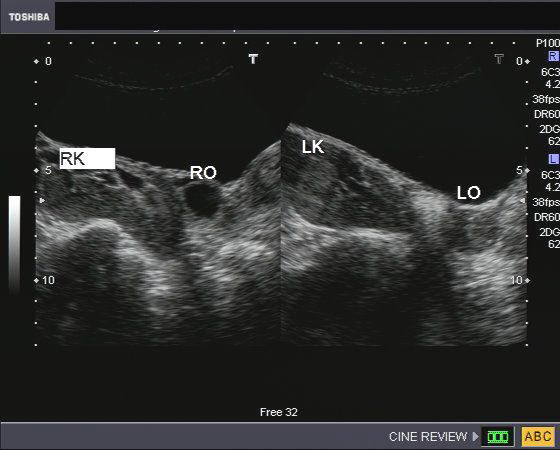
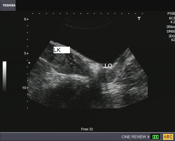
In this female patient, ultrasound images show an incidental finding, which is rather rare. Both kidneys appear to be ectopic and located in the pelvis, close to the ovaries (RTK= right kidney; LTK= left kidney; RO= right ovary; LO= left ovary). Such cases of bilateral pelvic kidneys can cause serious difficulty in normal delivery/ childbirth. In contrast, normal childbirth is possible in unilateral pelvic kidney. The renal arteries in such cases usually originate from the iliac arteries or lower part of aorta.
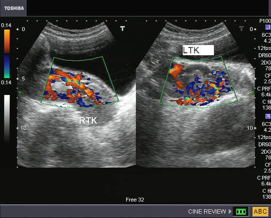
Another ultrasound image (right) shows the bilateral pelvic kidneys adjacent to the ovaries and posterior to the urinary bladder. Color Doppler ultrasound image on right shows normal vascularity of the pelvic kidneys. Such ectopic/ pelvic kidneys are usually complicated by infection and uretero-pelvic junction (pelvi-ureteric junction) obstruction. Patients also have higher incidence of renal calculi. Ultrasound images of bilateral pelvic kidneys is courtesy of Dr. Jaydeep Gandhi, MD, India.
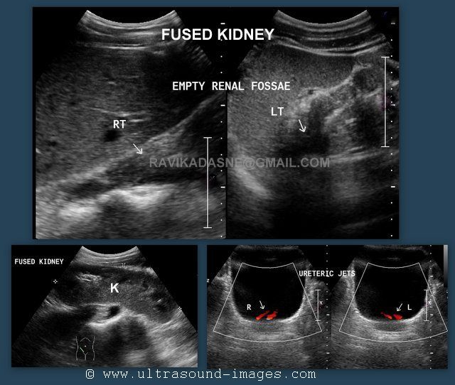
case-3: Bilateral ectopic fused kidneys (ectopia with fusion- S-shaped fusion of kidneys)
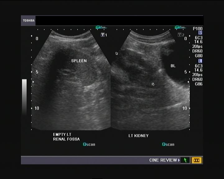
This 20 yr. male patient had lower abdominal pain. Ultrasound imaging of the kidneys revealed - empty left renal fossa with a normal spleen. Possibly (see ultrasound images below), we were dealing with left ectopic kidney. The left kidney was seen posterior to the dome of the urinary bladder. However, the ectopic left kidney showed an abnormal rotation, and appears oriented along the transverse plane (horiziontally).
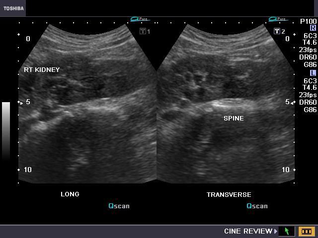
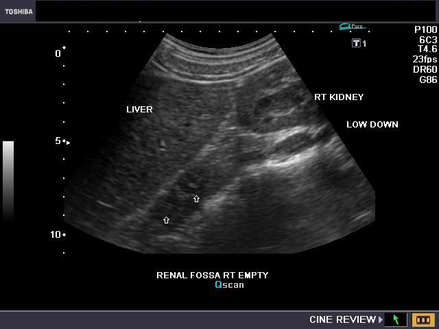
On sonography of the right kidney, we similarly saw an empty right renal fossa with the right kidney seen much lower down (a lower lumbar kidney). However, the right kidney also appeared malrotated and was also oriented along the transverse plane, anterior o the spine.
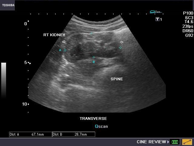
The above ultrasound images of the right kidney suggest a more ominous anomaly of the right and left ectopic kidneys (bilateral ectopic kidneys).
Observe the link of renal tissue connecting both horizontally oriented kidneys seen along the sagittal plane in the midline. It suggests a partial or complete fusion of both ectopic kidneys. The color Doppler ultrasound images confirm a normal vascularity of this fused ectopic kidney complex. These ultrasound images confirm what appears to be a S-shaped fusion of bilateral ectopic kidneys. S-shaped fusion of bilateral ectopic kidneys is a very rare anomaly with very little literature available online or in print. These types of renal fusion anomalies are associated with renal infection, calculi (stone) disease and hydronephrosis (obstructive changes) in the involved kidneys. None of these are however, seen in this patient.
One can view ultrasound and Color Doppler videos of this case at:
http://ultrasound-videos.blogspot.com/2010/11/unusual-ectopic-fused-kidneys.html
References: Pictorial review of renal ectopia and fusion anomalies
Ectopic /thoracic kidney
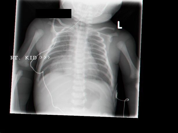
This young child underwent routine ultrasound scan of the abdomen. Ultrasound images reveal absent right kidney in right renal fossa. However, scan upwards revealed the right kidney to be located high just within the right hemithorax, close to the heart. The right kidney is ectopic (see X-ray image), and has slipped through the foramen of Bochdalek into the right hemithorax (thoracic ectopic kidney). On plain X-ray of the chest, this is seen as a mass in right paracardiac region (arrows). Ultrasound images are courtesy of Mr. Shlomo Gobi, Israel.
Reference: http://www.nejm.org/doi/full/10.1056/NEJMicm0807840
Horseshoe kidney
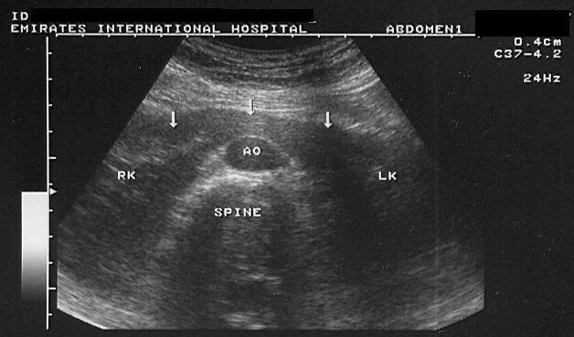
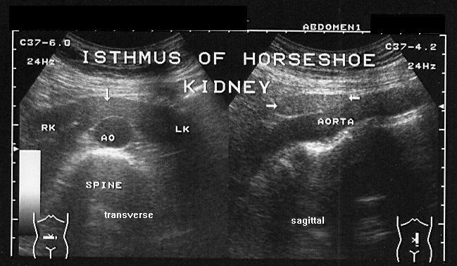

Routine abdominal sonography of the abdomen in this patient revealed: a) kidneys located at a lower level than normal b) the lower poles of both kidneys pointing medially, c) a bridge of renal tissue or isthmus connecting the two kidneys. This isthmus is seen passing anterior to the abdominal aorta. Often, the lower poles of the kidneys are difficult to visualize in such cases. These ultrasound images are diagnostic of horseshoe kidney. Images taken by Dr. Ravi Kadasne, UAE, using a Toshiba Powervision ultrasound machine.
The above color doppler images show a large isthmus lying anterior to the abdominal aorta.
Images courtesy of Dr. G. V. Ramaiah, AP, India.
References: E-medicine article on Horseshoe kidney (free article and images- rated comprehensive)
maternal horseshoe kidney in pregnancy
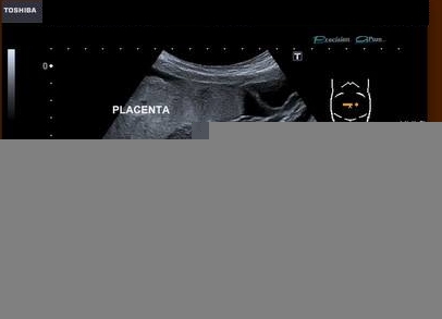
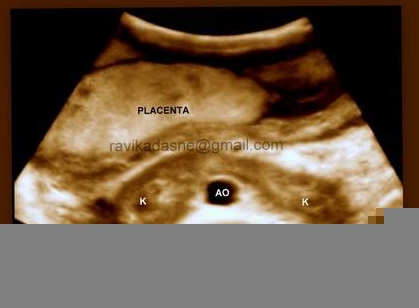
This 3rd trimester pregnancy was imaged for fetal anomalies. An incidental finding of maternal horseshoe kidneys was observed. Transverse section ultrasound and 3D ultrasound images through the upper abdomen reveal a bridge of connecting renal tissue passing anterior to the maternal abdominal aorta, connecting both maternal kidneys. This is diagnostic of maternal horseshoe kidneys. The placenta is seen to be fundic and anterior. These ultrasound images of maternal horseshoe kidneys in a pregnant lady are courtesy of Dr. Ravi Kadasne, MD.
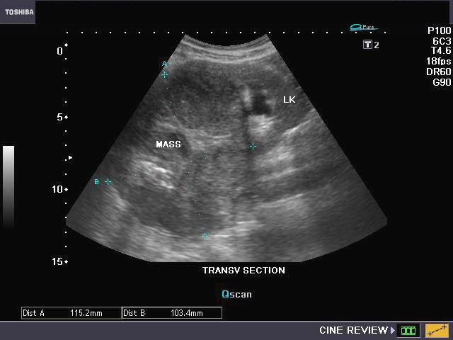
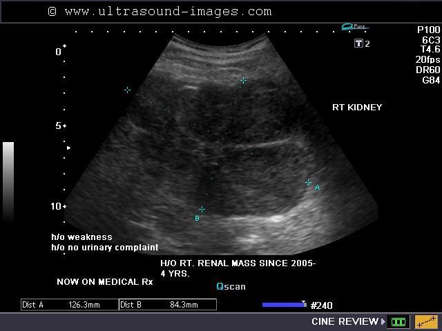
Renal cell carcinoma
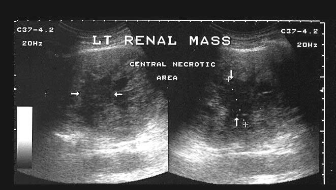
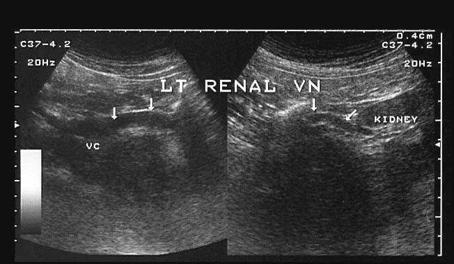
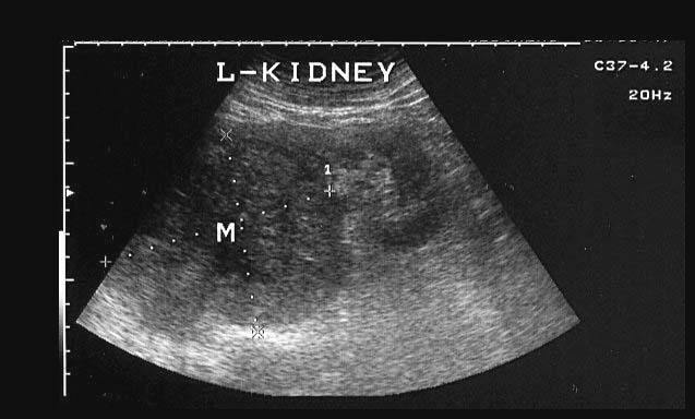
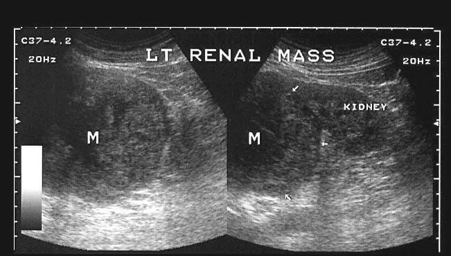

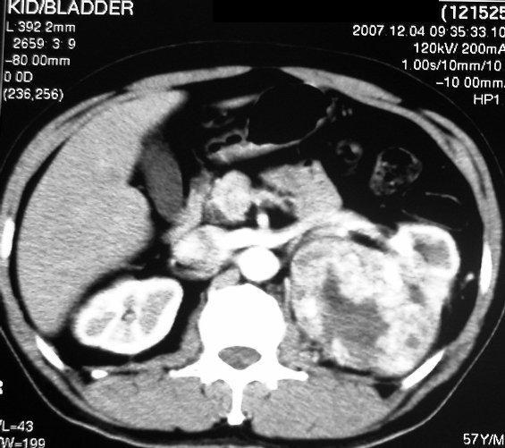
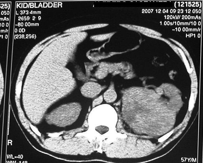
This 57 yr. old male patient presented with abdominal pain and mass in abdomen. Sonography reveals a large, hypoechoic, inhomogenous mass (10 x 8 cms.) at the upper pole of the left kidney. The central portion is markedly hypoechoic suggestive of necrosis. The left renal vein appears normal. The right kidney shows a small cortical cyst and mild pelvic dilation. The urinary bladder was normal (not shown here). CT scan images confirm the large left renal mass with enhancement of the tumour and necrotic central area. These ultrasound images are suggestive of renal cell carcinoma. Ultrasound images taken with Toshiba Power Vision machine, courtesy of Dr. Ravi Kadasne, UAE.
Reference: http://www.emedicine.com/med/topic2002.htm (free article)
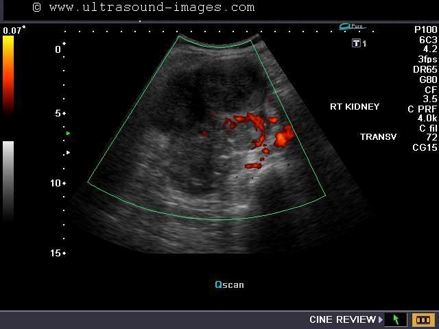
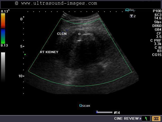
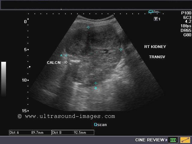
Case-2 (Renal cell carcinoma)
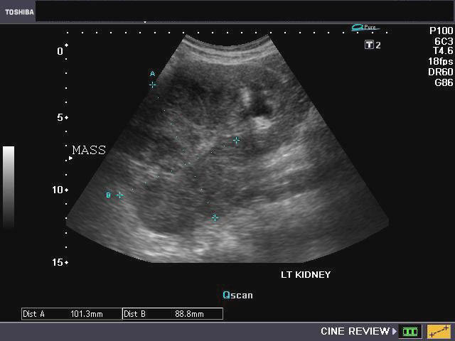
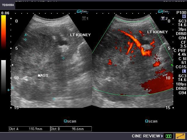
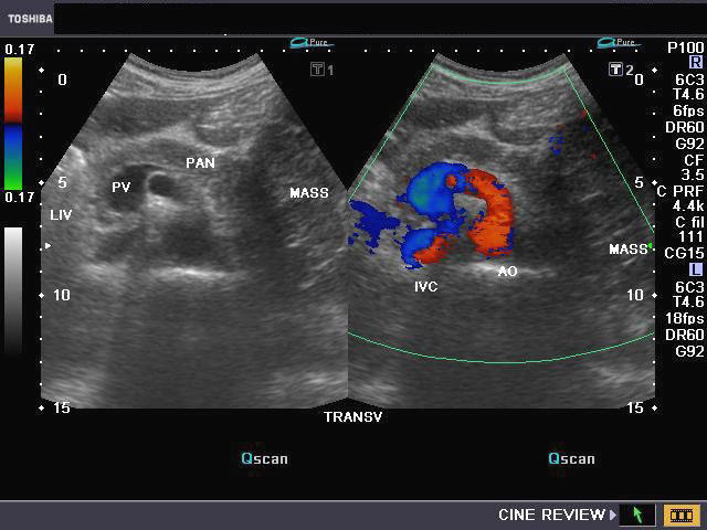
Another case of renal cell carcinoma. This elderly male patient presented with hematuria. Sonography of the left kidney shows a large, solid mass (11 x 8 cms.) involving the upper half of the left kidney. The affected kidney shows mild obstructive changes. Power Doppler image (bottom left) shows multiple vessels entering the mass. Transverse section of the liver and pancreas (ultrasound image on bottom right) shows the mass to be in close relation to the tail of pancreas. (Liv= Liver, Pan = pancreas, PV= Portal vein, AO = aorta, IVC= Inferior Vena Cava).
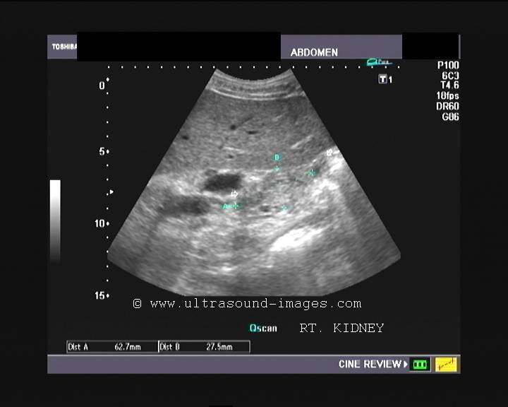
Chronic renal failure (Medical renal disease)
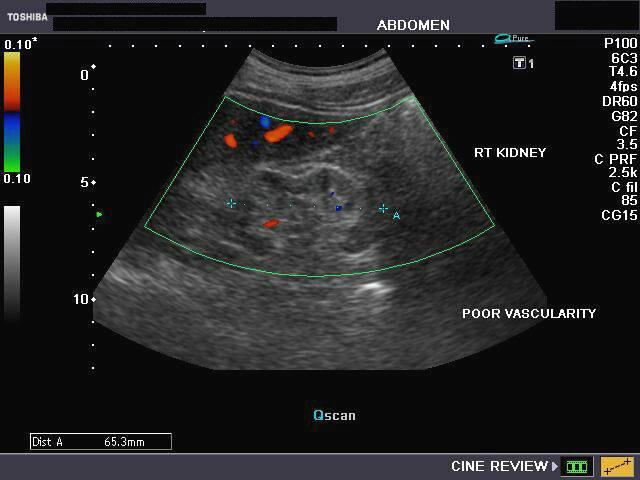
This elderly male patient presented with symptoms of medical renal disease. Sonography of the kidneys revealed: 1) bilateral echogenic (hyperechoic renal cortex) kidneys 2) both kidneys appear small in size (atrophic) 3) reduced thickness (thinning) of renal cortex (10mm.) 4) reduction in cortico-medullary differentiation 5) Color doppler imaging of the kidneys revealed reduced flow in both kidneys. These ultrasound images are diagnostic of chronic medical renal disease (or chronic renal failure). All ultrasound images above (taken using Toshiba Nemio-XG Color Doppler imaging system, by Joe Antony, MD, India.
Reference:http://emedicine.medscape.com/article/238798-diagnosis (free article).
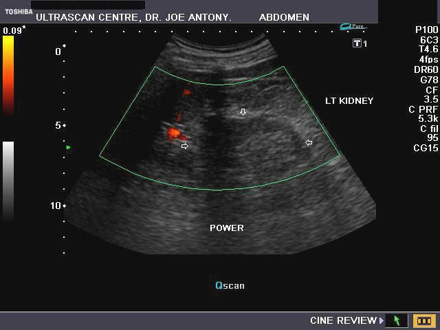
Severe renal failure with arterio-venous fistula (hemodialysis)
Ultrasound image- Left kidney- Chronic renal failure
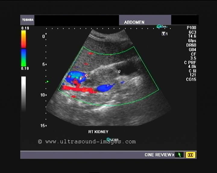
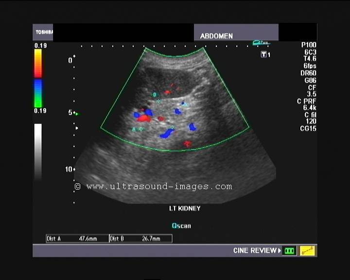
This 30 year old female patient was undergoing hemodialysis for long standing renal failure (chronic medical renal disease). The kidneys are poorly visualized and are very echogenic with almost no normal renal tissue or morphology seen suggesting that this lady has severe renal failure. The upper pole of both kidneys show prominent vessels on color Doppler imaging, suggesting AV fistula in both kidneys following renal biopsy some time ago.
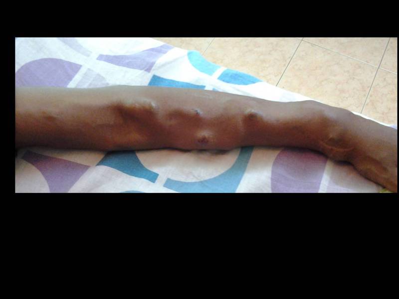
Images of the iatrogenic A-V fistulae in the arm for vascular access for hemodialysis

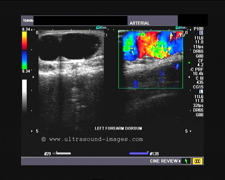
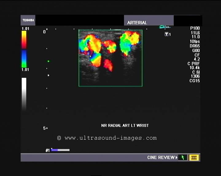
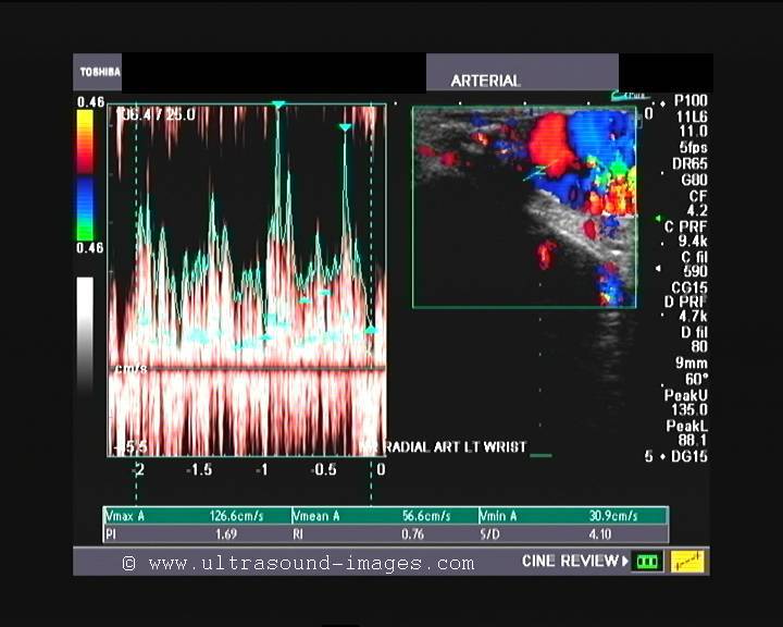
To facilitate the hemodialysis, this lady underwent a surgery to create an artificial AV fistulae in the arm. The ultrasound and color Doppler images show multiple dilated vessels in the arm which represent the AV fistula. Here the radial artery is connected with the cephalic vein with markedly turbulent high velocity flow in the fistulous veins.
Image of the left arm showing multiple AV fistulae in the left arm
References:
AV fistula as vascular access for hemodialysis
Article in hemodialysis- wikipedia
Renal sinus lipomatosis

The above ultrasound images show echogenic tissue in the lower part of the left kidney within the renal sinus. The first diagnosis that seems likely is angiomyolipoma of the left kidney. But observe closely the other image- (lower row) - both kidneys appear affected and it is the renal sinus that is extensively involved. The color Doppler image of the left kidney shows poor vascularity in the lesion. These ultrasound images are clearly suggestive of fatty tissue within the renal sinus- the condition being called renal sinus lipomatosis.
Reference: Ultrasound video clips of renal sinus lipomatosis
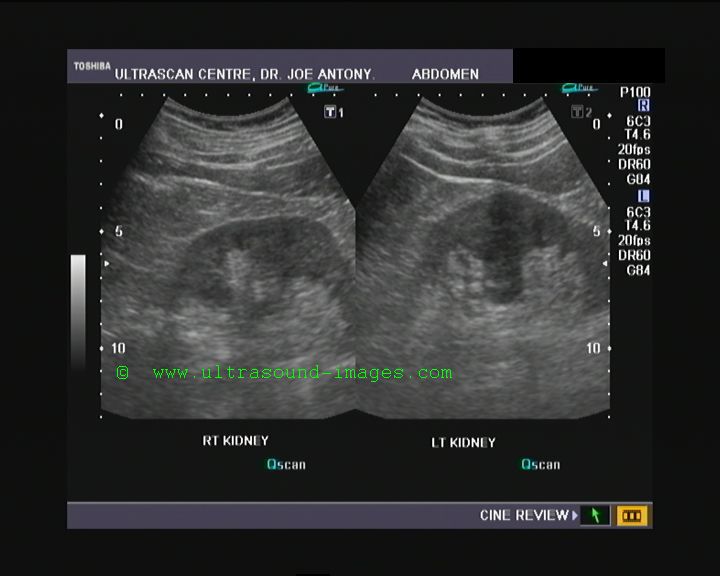
Renal trauma- subcapsular hematoma
Subcapsular hematoma- Rt. kidney
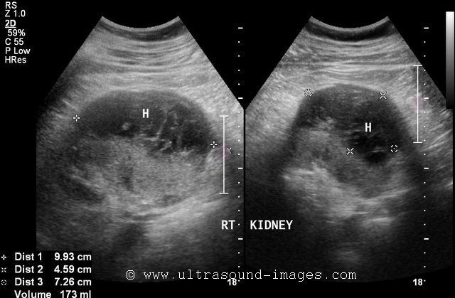
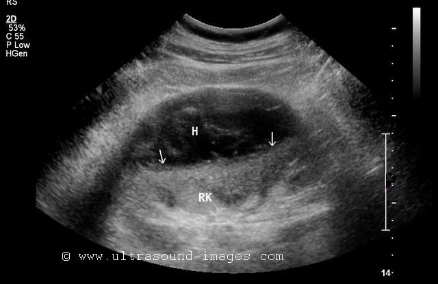
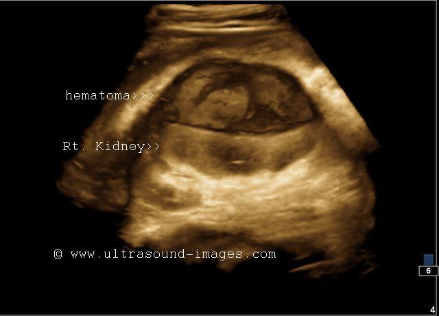
The ultrasound images above show a large subcapsular hematoma of the right kidney in a case of blunt abdominal trauma. Sonography shows a 9 x 4.5 cms. sized markedly hypoechoic collection in the subcapsular part of the right kidney in an oval shape. The collection compresses upon the intact part of the right kidney, (arrows), without changing the reniform contour of the kidney. Smaller subcapsular (intracapsular) hematomas appear crescent shaped. The renal hematoma shows sharp outer margins suggesting that it is limited by the renal capsule. The pelvicalyces appear intact. Such renal injuries may be classified as grade-1 renal trauma. However, CT evaluation is advisable to detect the extent of renal laceration if any. Color Doppler imaging would help in determining the viability of the kidney when large renal hematomas of this nature cause compression and resultant ischemia to the organ. Ultrasound images are courtesy of Dr. Ravi Kadasne, M.D., UAE.
References:
http://emedicine.medscape.com/article/379085-overview#a22
Article on sonography of renal subcapsular hematoma (free images article- sonoworld)
Renal angiomyolipoma -(AML) of right kidney
Long section of right kidney- echogenic mass and renal calculus


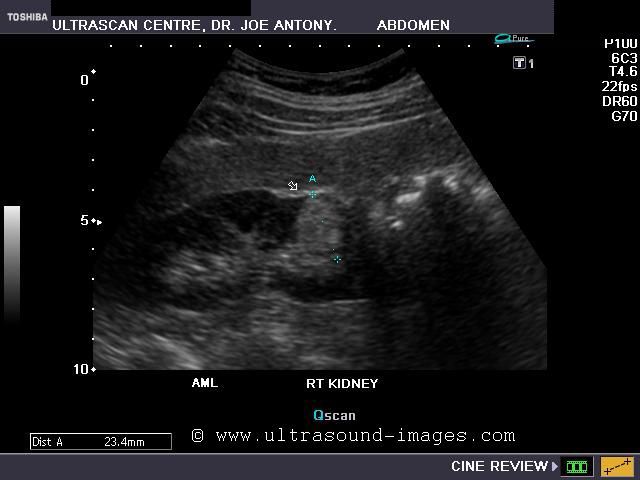
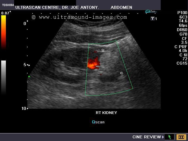
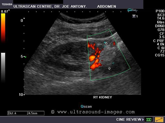
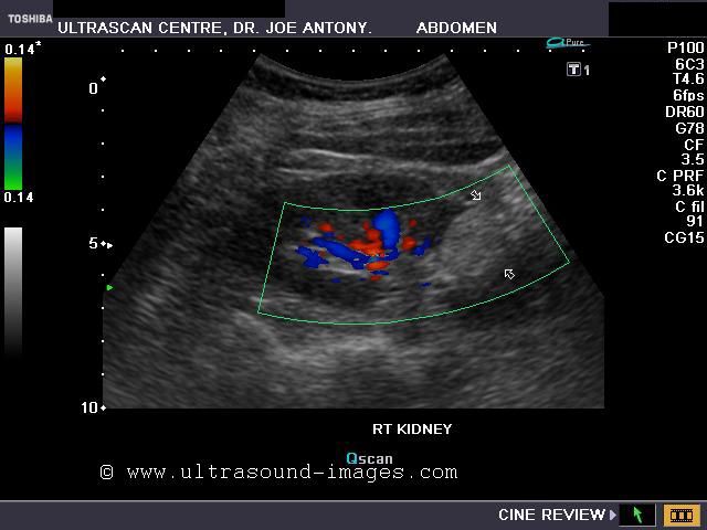
This 50 year old female patient complained of pain and discomfort in the right flank. This lady also complained of hematuria. Sonography of the abdomen, showed a moderately large (2.5 cms.), hyperechoic mass which was well defined and non calcific involving the lower pole renal cortex and extending up to the renal sinus. In addition, a small calculus was seen in the middle calyx of the left kidney. The color Doppler and Power Doppler images show poor vascularity of the renal tumor. Such markedly hyperechoic, well defined masses are the typical features of angiomyolipoma of the kidney. Renal angiomyolipomas are usually incidental findings and usually involve the right kidney. These are usually small, less than 2 cms., asymptomatic and do not significantly grow in size. They consist of small thickened vessels, fatty tissue and smooth muscle. It is the amount of fatty/ adipose tissue within the mass that makes the tumor echogenic. Usually, AML is a benign tumor; however, larger tumors may cause hematuria and may turn malignant. Larger tumors (like the case above), may develop small aneurysms which can rupture and cause severe hemorrhage. D/d: The main differential diagnosis for AML is a small renal cell carcinoma, which can also be echogenic. However, RCC (renal cell carcinoma) may show an hypoechoic rim (halo) and may have small cystic spaces within the tumor. Besides AML usually produces posterior acoustic shadowing. CT imaging is usually helpful in determining the presence of fat within the tumor in AML.
References
http://www.ajronline.org/content/141/3/575.full.pdf(free article)
http://casereports.bmj.com/content/2010/bcr.01.2010.2624.full.pdf (free article)
http://radiology.rsna.org/content/225/1/78.full.pdf+html(free article)
Exophytic renal cysts- Exophytic cortical cysts of the kidney


3D ultrasound images of exophytic renal cysts 2D B-mode ultrasound image
This kidney shows a large cortical cyst which has expanded outside the confines of the kidney surface. Such renal cysts are called exophytic cysts of the kidney. Note that this is simply a renal cortical cyst; the term exophytic is used to denote the fact the cyst has bulged outside the outer margins of the renal surface, as opposed to renal cortical cysts that remain within the renal margins. The danger of rupture of such renal cysts (exophytic) is greater. The images above also show a fluid-debris level due to particulate matter within the cyst. Images above are courtesy of Dr. Ravi Kadasne, MD, UAE.
References:
http://radiology.rsna.org/content/216/3/792.full
http://radiology.rsna.org/content/243/1/158.full
Duplex kidney with complete duplication of ureters
Rt. kidney shows duplication of pelvicalyces: Ultrasound images
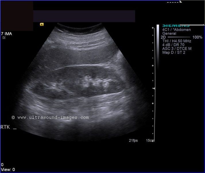

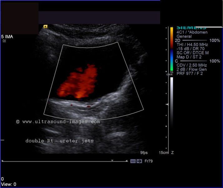

This patient has an incidental finding on ultrasonography- the right kidney shows duplication of the renal pelvis. (The kidney appears larger due to this anomaly). In addition the ureter also appears completely duplicated with one ureter for each moiety of the duplex right kidney. The color Doppler ultrasound image shows a resultant double right ureteric jet emanating from duplication of the right ureteric openings. Thus this is a duplex right kidney with complete duplication of the collecting system (double right ureters). Such anomalous variants of the kidney are prone to vesico-ureteric reflux and resultant urinary tract infections as well as stone formation. Usually these patients are asymptomatic and the diagnosis is usually made on sonography or other modes of imaging. Ultrasound images of duplex kidney are courtesy of Mr. Shlomo Gobi, sonographer, Israel.
References:
http://emedicine.medscape.com/article/378075-overview
Duplication of collecting system
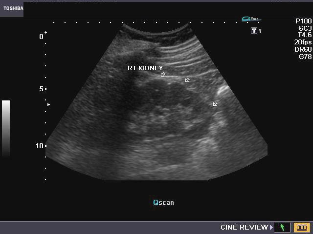
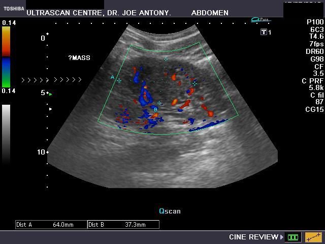
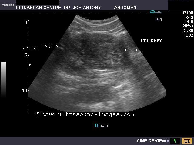
Xanthogranulomatous pyelonephritis of right kidney
Xanthogranulomatous pyelonephritis (XPN) diagnosed as right renal mass
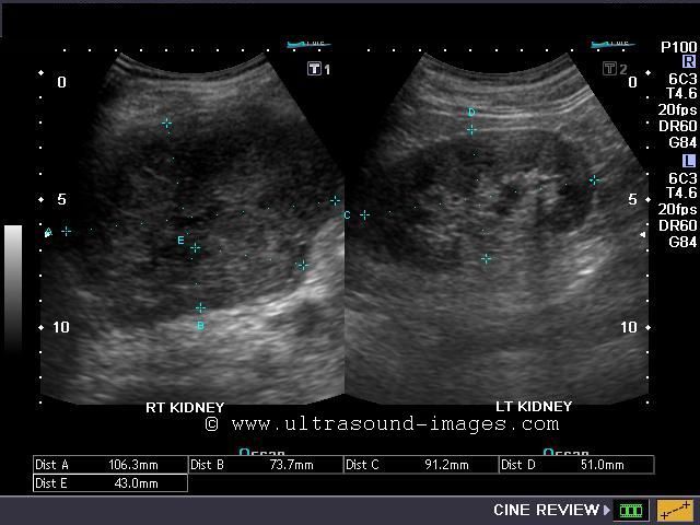
This elderly male patient had history of few episodes of urinary tract infection. He was clinically suspected to have a right renal mass
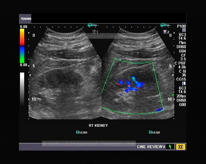
Power Doppler image of right kidney
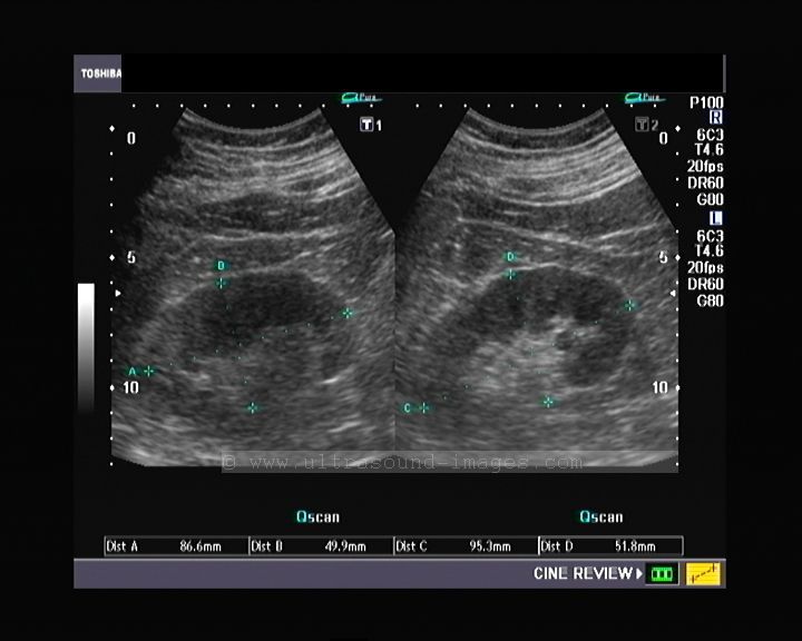
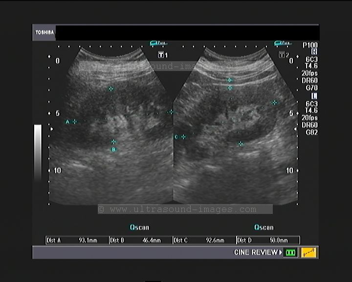
The ultrasound images of the right kidney show the following features: the kidney is enlarged with multiple hypoechoic and inhomogenous, rounded masses within it. There are two calculi within the middle third of the right kidney. There is no evidence of perinephric fluid collections. The right kidney also appears considerably enlarged compared to its fellow. The presence of these ultrasound findings in a patient of renal or urinary tract infection is a clear evidence in favor of xanthogranulomatous pyelonephritis of this kidney. This is a relatively rare form of renal infection resulting in the destruction of the kidney from long standing chronic urinary obstruction with renal calculi. The color Doppler/ Power Doppler ultrasound images further confirm that the hypoechoic masses studding the right kidney are poorly vascular or non vascular unlike renal cell carcinoma or other renal malignancies. The hypoechoic masses in the kidney represent renal parenchyma, both cortical and medullary, that has been destroyed by the disease process and associated hydronphrosis, and replaced with lipid laden macrophages. These masses appear as lobulated yellow areas on pathological examination of the affected kidney. The ultrasound images above show a form of XPN (xanthogranulomatous pyelonephritis) of the diffuse variety. The other form of XPN is the focal type affecting a small part of the kidney. This kidney (case above) is practically non functional due to diffuse XPN. The differentiation of XPN from other renal masses helps to plan the management and surgical approach for the patient.
References
http://www.jultrasoundmed.org/content/23/3/409.full.pdf+html
http://radiographics.rsna.org/content/11/3/485.full.pdf+html
cross fused ectopic kidneys with duplex ureters
This male patient shows absence of both right and left kidneys in their normal positions- that is the renal fossae. Both kidneys appear as a single mass in the right iliac fossa- a clear case of cross fused ectopia of the kidneys. Such a kidney is called a pancake kidney also. These ultrasound images show a typical case of cross fused ectopia of the kidneys with an added anomaly also- there is bilateral duplication of the ureters. The urinary bladder shows 2 ureteric jets- suggesting duplication of the collecting system and ureters, which are seen to open on either side of the urinary bladder.
These ultrasound images of the cross fused renal ectopia with bilateral duplication of the collecting system are courtesy of Dr Ravi Kadasne, MD.
References:
http://www.mypacs.net/cases/CROSSED-FUSED-ECTOPIA-1091607.html
Cross fused ectopia- emedicine article
http://www.pediatricurologybook.com/kidney_fusion.html
trifid renal pelvis
In this elderly patient, ultrasound images of the right kidney shows three separate divisions of the renal pelvis- suggesting a trifid renal pelvis. The left kidney appeared to be normal. A trifid renal pelvis is a very rare renal anomaly, and very few cases have been documented in medical literature.
References: Trifid renal pelvis- case study
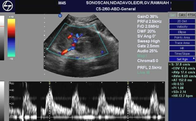
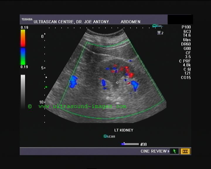
nutcracker phenomenon
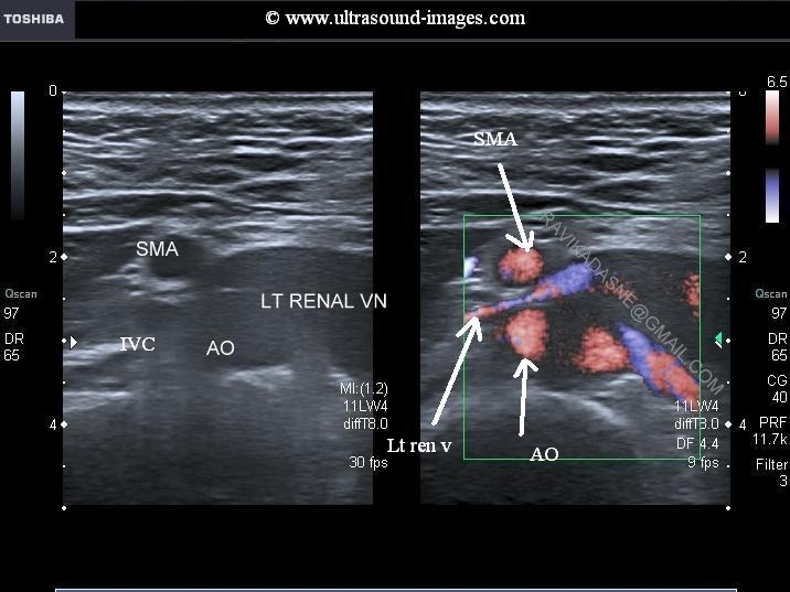
Nutcracker phenomenon is the name given to compression of the left renal vein between the superior mesenteric artery anteriorly and the abdominal aorta posteriorly. this results in dilation of the distal part of the left renal vein due to partial obstruction of venous blood flow as a result of compression between these two structures. These ultrasound and colour Doppler images show a typical case of nutcracker phenomenon. if the Nutcracker phenomenon is associated with symptoms due to this condition, such as hematuria, pelvic varices, varicocele and abdominal pain, this condition is called nutcracker syndrome.These ultrasound images of Nutcracker phenomenon are courtesy of Dr. Ravi Kadasne, MD, UAE. The machine used here is the Toshiba APlio system.
References: http://www.ncbi.nlm.nih.gov/pmc/articles/PMC2878259/
pyelonephritis-UTI-renal-edema
This patient has a history of UTI or urinary tract infection. Both kidneys show renal parenchymal edema evident as early loss of cortico-medullary differentiation with swelling of the renal parenchyma suggesting renal inflammation or pyelonephritis. In additon, observe the hypoechoic nature of the renal tissue. In late cases of pyelonephritis there may be focal abscesses and scarring of the renal cortex. Focal hypoechoic areas may also be seen in acute pyelonephritis.
References:http://www.jultrasoundmed.org/content/20/1/5.full.pdf
© 2026, Joe Antony, MD All images and content in this site are copyrighted. www.ultrasound-images.com When visiting this site, we may use cookies to improve the performance of this site. Use of this site means you accept the use of cookies. Read our privacy policy at this page: https://www.ultrasound-images.com/privacy-policy/