
cholecystectomy
Contents of this page
Ultrasound images of post-cholecystectomy abdomen
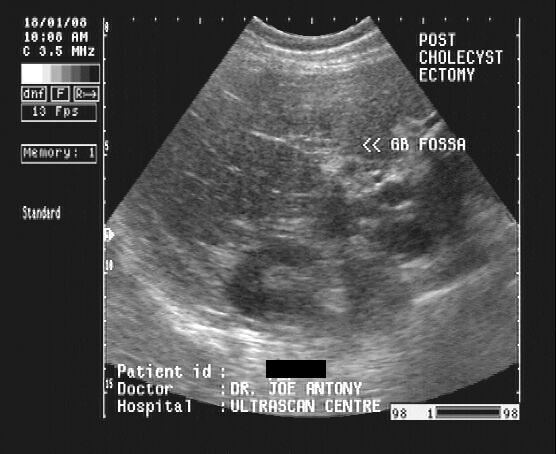
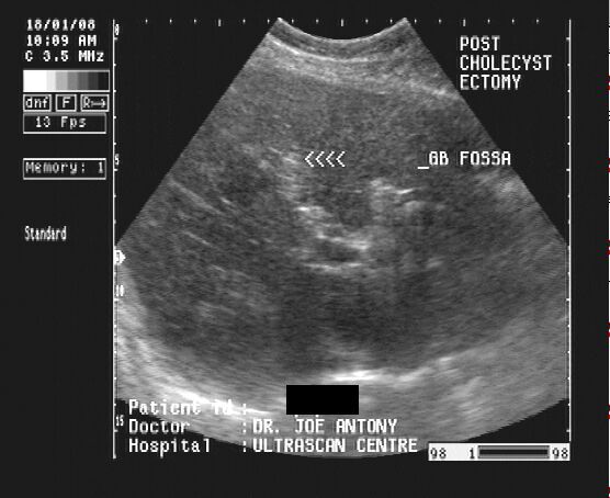

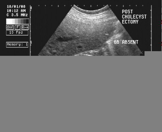
These are ultrasound images of middle aged female patient who underwent surgical removal of the gall bladder. The liver is visualized with the gall bladder absent in the location of the gall bladder fossa. This area is replaced by dense fibrous tissue, seen as a linear echogenic lesion (arrowed). The lower right image shows a parasagittal section through the right kidney and liver. Images taken using a Pie scanner 100 Falco, by Dr. Joe Antony, India.
Case-2: Cholecystecomy

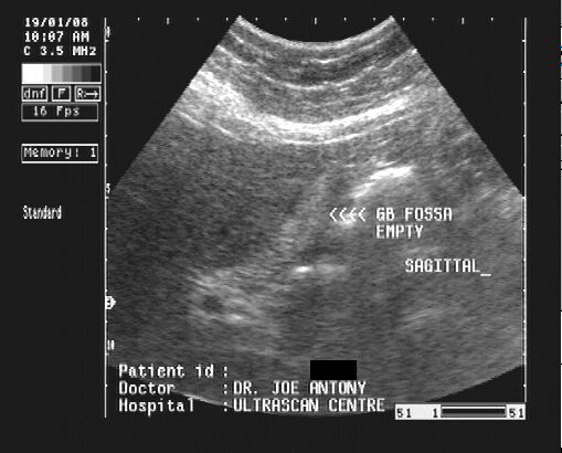
And here are some more images of another patient, post-cholecystectomy abdomen (50 yr. old female)
Reference: http://en.wikipedia.org/wiki/Cholecystectomy
© 2026, Joe Antony, MD All images and content in this site are copyrighted. www.ultrasound-images.com When visiting this site, we may use cookies to improve the performance of this site. Use of this site means you accept the use of cookies. Read our privacy policy at this page: https://www.ultrasound-images.com/privacy-policy/