
Ultrasound images of carcinoma of endometrium
Contents of this page
- Adenocarcinoma of endometrium
- Adenocarcinoma- presenting as Endometrial polyp
- Uterine-ultrasound-quiz-2
Adenocarcinoma of endometrium
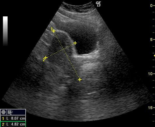


Transabdominal and transvaginal sonography of the uterus in this postmenopausal patient reveals: 1) Grossly thickened endometrium 2) fine cystic lesions within the endometrial mass 3) invasion of the myometrium by the mass 4) increased vascularity of the lesion on color doppler imaging. These ultrasound images suggest Stage 1B (FIGO) carcinoma of the endometrium. The patient underwent hysterectomy after histopathological study confirmed the presence of malignancy (adenocarcinoma).
Reference:
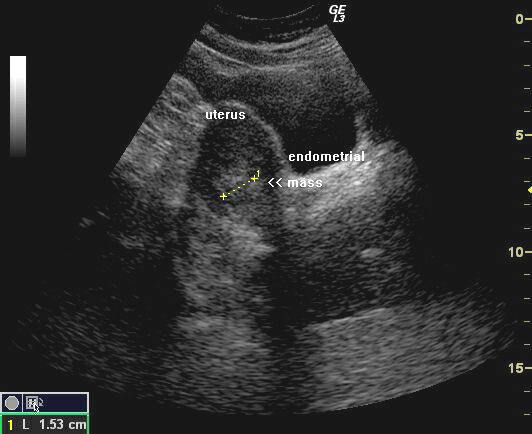
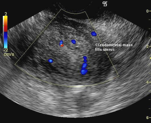
Adenocarcinoma- presenting as Endometrial polyp
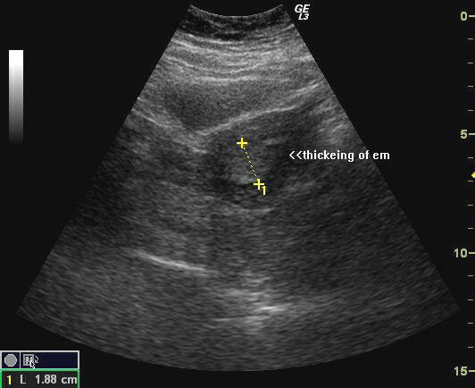
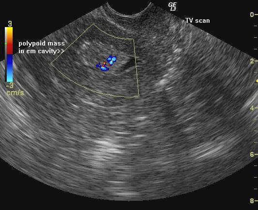
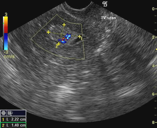
Ultrasound images of malignant polyp of uterus.
The above ultrasound images show 1) apparently marked thickening of the endometrium (19mm.) on transabdominal sonography. 2) on transvaginal imaging, there is a large polyp like mass (14 x 22 mm.) occupying the uterine cavity. 3) color doppler imaging shows feeding vessels supplying the polyp. Histopathology confirmed endometrial carcinoma (adneocarcinoma).Images by Joe Antony, MD, Cochin, India.
Uterine-ultrasound-quiz-2
© 2026, Joe Antony, MD All images and content in this site are copyrighted. www.ultrasound-images.com When visiting this site, we may use cookies to improve the performance of this site. Use of this site means you accept the use of cookies. Read our privacy policy at this page: https://www.ultrasound-images.com/privacy-policy/