
Sonography of lymph node diseases
Contents of this page
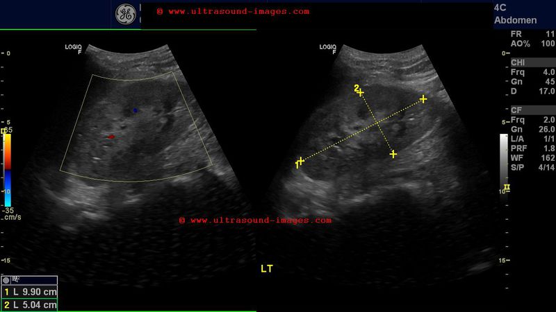
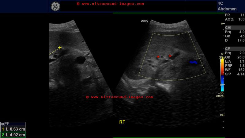
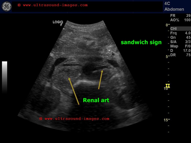
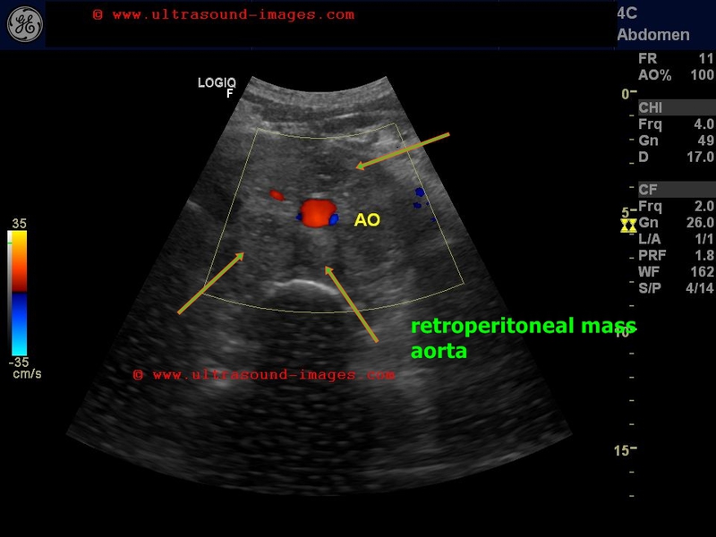
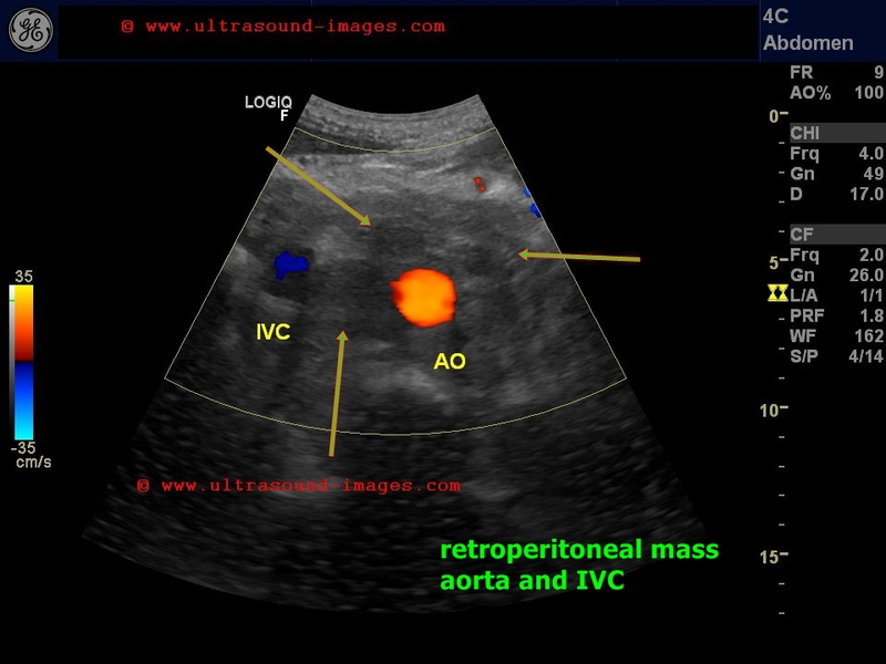
retroperitoneal-lymphoma
This elderly male patient, a known case of non Hodgkin's lymphoma has a large retroperitoneal mass on ultrasound imaging of the abdomen. Non Hodgkin's lymphoma is known to cause a lymph node conglomerate mass as in this case, to surround the aorta and inferior vena cava encasing these two major vessels. The arteries emerging from the aorta, namely the renal arteries and the celiac artery are also encased by the lymph node mass- the "sandwich sign". This condition is called retroperitoneal lymphoma.
References: Ultrasound imaging of retroperitoneal lymphoma


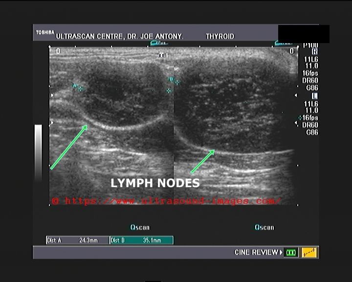
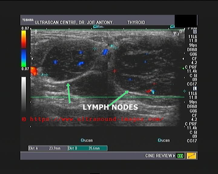
tuberculous-lymphadenitis
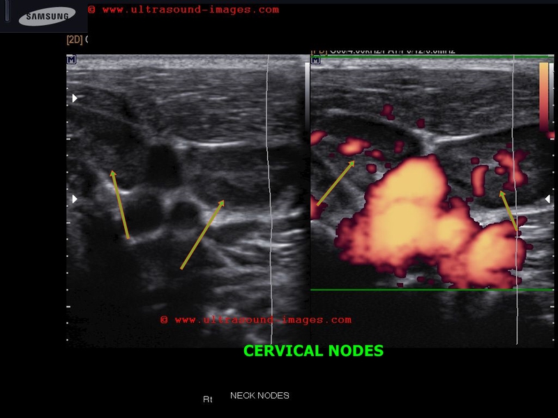
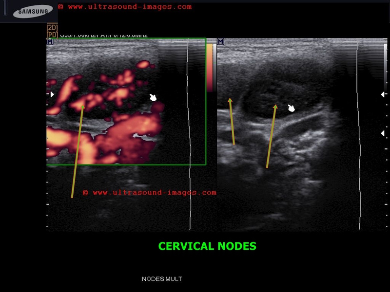
This child shows multiple cervical lymph nodes which show "reactive changes" or changes of lymphadenitis due to Kochs or tuberculous lymphadenitis.
Features seen here include:
- enlarged cervical nodes
- hypoechoic nature of the nodes
- prominent hilar vascularity
Tuberculous lymphadenitis is a common finding and a very common cause of neck swellings in the pediatric age group in developing countries.
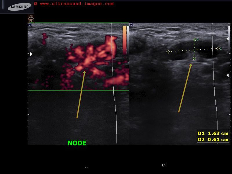
Enlarged nodes are also seen in metastases and lymphoma.
However, here the vascularity is primarily peripheral as opposed to hilar vascarity in inflammatory nodes.
Reference: AJR sonography of lymph nodes
Reactive vs malignant lymph nodes
axillary-lymphadenitis
This young adolescent female patient shows multiple reactive or inflamed axillary lymp nodes again showing features similar to the case above.
Lymphadenitis in this case is the result of mastitis of the ipsilateral breast.
The axillary nodes are hypoechoic and show central vascularity especially near the hilum suggesting the inflammatory nature.
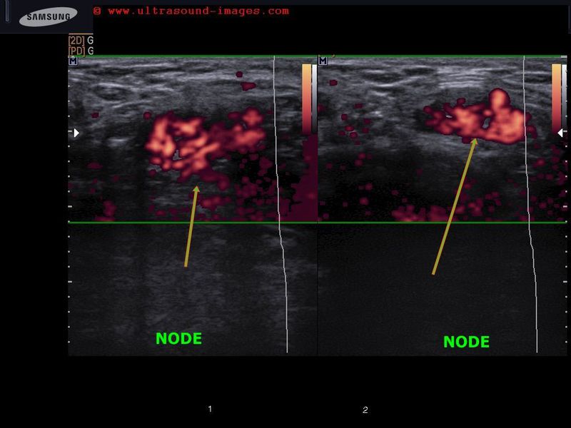
non-hodgkins-lymphoma
Known case of non Hodgkins lymphoma. Sonography of the neck shows two cevical nodes with typical features.
- hypoechoic and spherical enlarged lymph nodes
- poorly vascular -thus ruling out inflammatory causes
-non calcific nodes
- no central necrosis within the lymph nodes
see : sonography of lymphoma
© 2026, Joe Antony, MD All images and content in this site are copyrighted. www.ultrasound-images.com When visiting this site, we may use cookies to improve the performance of this site. Use of this site means you accept the use of cookies. Read our privacy policy at this page: https://www.ultrasound-images.com/privacy-policy/