Ultrasound images of diseases of the adrenals
Contents of this page
Normal adrenals (in neonate)
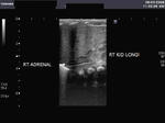
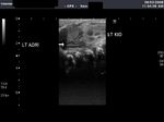
The normal adrenals are seen well in these ultrasound images in a neonate. They have a typical Y- shape and the central medulla is hyperechoic with a hypoechoic cortex.
Adrenal mass
Mass in suprarenal region
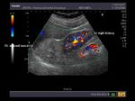
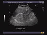
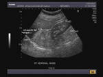
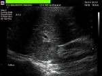
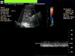
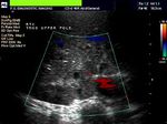
These sonographic images reveal a space occupying lesion above the right kidney in the region of the right adrenal gland . The mass is large, echogenic and poorly defined and pushes the kidney downwards. It shows an inhomogenous appearance, but no calcification is present. The retroperitoneal fat stripe is displaced anteriorly by the mass proving that it is of adrenal and not hepatic origin (see the arrowheads in third image). These ultrasound images of the right adrenal mass suggest likelihood of malignancy. Differential diagnosis includes: 1) adrenal adenoma 2) pheochromocytoma 3) metastasis. Large masses of the type seen here are highly suggestive of malignancy of the adrenal gland. However, the color doppler image shows scanty vascularity of the mass.
Ultrasound images courtesy of Dr. Vikas Arora, Ferozepur, India, using a Nemio 30 Ultrasound and color doppler machine.
Reference:
1) http://www.emedicine.com/RADIO/topic14.htm (free article on adrenal carcinoma ).
2) http://www.emedicine.com/RADIO/topic13.htm(free article on adenoma of adrenal)
Pheochromocytoma of adrenal gland
This patient underwent sonography of the abdomen. Images reveal a solid, rounded mass of 4 cms. arising from the right adrenal gland. Color doppler images reveal little flow within the mass. Histopathology studies confirmed the adrenal mass to be a pheochromocytoma. Ultrasound images taken by Shlomo Gobi, Israel, using a Philips/ ATL, HDI 3000 Ultrasound system.
Reference: http://www.emedicine.com/med/topic1816.htm(free article on Pheochromocytoma)

