
Normal sonographic anatomy of the fetal heart
Contents of this page
- The ductus arteriosus
- Ultrasound image of the Right Ventricular Outflow Tract (RVOT)
- Ultrasound images of the LVOT (Left Ventricular Outflow Tract)
- Some more images of LVOT and RVOT
- Four Chamber view
- Normal 3 vessel view in a 28 week fetus
The ductus arteriosus


Download my E book for Amazon Kindle (download free Kindle reader for i Phone or Android)
Assorted ultrasound cases- by Joe Antony, MD
Sonography of normal ductus.
The normal ductus arteriosus (arrowed) of a 2nd trimester fetus is seen well in this gif video. Sp= spine, rv= Right ventricle, Rt= right side of the fetus, Lt= left side of fetus, Ao= aorta .Ultrasound Video taken using a Pie Scanner 100 Falco, by Dr. Joe Antony. Ultrasound video edited by Dr. Anatoly, Ukraine.In these images (above) the ductus arteriosus is marked by an asterix.
Ultrasound image of the Right Ventricular Outflow Tract (RVOT)
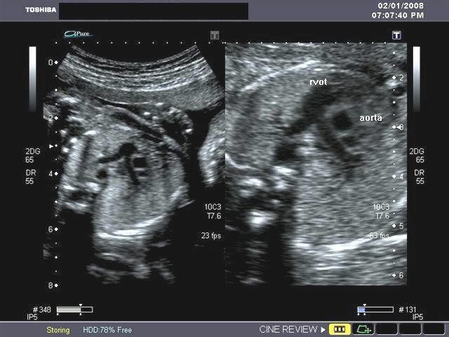
Ultrasound image shows the RVOT, the pulmonary trunk and bifurcation into the right and left pulmonary arteries. Image courtesy of Dr. Gunjan Puri, India.
Ultrasound images of the LVOT (Left Ventricular Outflow Tract)
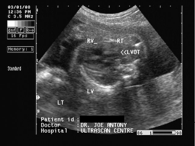
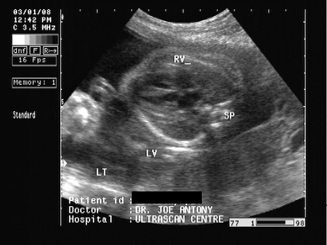
These 2 sonographic images of the fetal heart show the Left ventricular outflow tract as it leaves the fetal left ventricle . LV= left ventricle; RV= right ventricle.
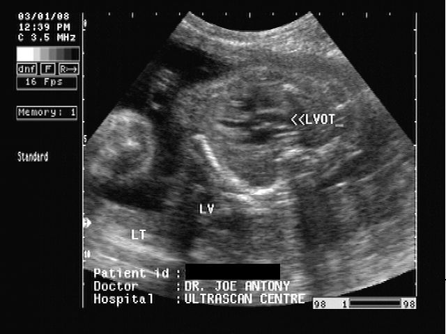
Some more images of LVOT and RVOT
The above ultrasound images of the fetal heart (fetal echocardiography, show the left ventricle (image on right) with the left ventricular outflow tract emerging from it to enter the aortic arch. The image on left shows the right ventricular outflow tract (RVOT) with the pulmonary trunk emerging from the right ventricle. This is seen to divide into the right and left pulmonary arteries. Ultrasound images courtesy of Gunjan Puri, MD, India./p>
Four Chamber view
This sonographic image shows the normal four chambers of the fetal heart.
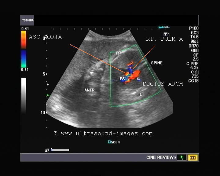
Normal 3 vessel view in a 28 week fetus
This 3 vessel view (ultrasound and color Doppler video) of the fetal heart in early 3rd trimester shows the Pulmonary artery- PA dividing into the right and left pulmonary arteries and continuing as the ductus arteriosus to meet the arch of aorta in a V shape (with the arch). The SVC is the superior vena cava and Ascending aorta (AO) is seen continuing as the Arch to meet the ductus arteriosus ( forming the characteristic V shape).
Fetal heart with labels showing each vessel

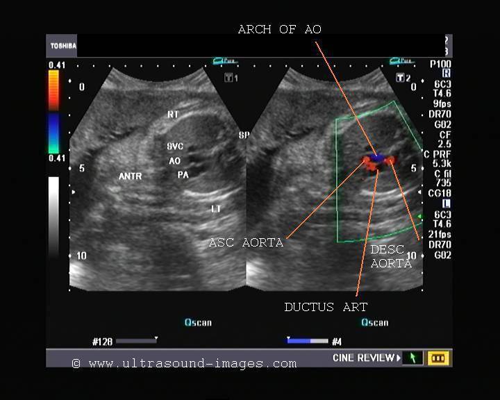
(ANTR= ANTERIOR, SP= FETAL SPINE. LT- LEFT OF FETUS, RT= RIGHT OF FETUS)
© 2026, Joe Antony, MD All images and content in this site are copyrighted. www.ultrasound-images.com When visiting this site, we may use cookies to improve the performance of this site. Use of this site means you accept the use of cookies. Read our privacy policy at this page: https://www.ultrasound-images.com/privacy-policy/