Amusing ultrasound pictures
Not too often, a sonologist comes across ultrasound images that make both doctor and patient smile. This is one page that brings such sonographic images to your computer monitor. Have a look and enjoy!
Contents of this page
- Fibroids playing Mickey mouse
- Pinna of fetal ear
- Static B-mode scanning machines
- The strange Bladder Cat
- And a bison too... in the pelvis
- A prostate fox
- schizencephaly-in-fetus
- Gall bladder turned swan
- fetal tarzan
- peacock-in-the-abdomen
- fetus calling mom
- Try-this-easy-quiz-on-uterus-below
Fibroids playing Mickey mouse
These are images of double fibroids that mimic the ears of a mouse with the face being the body of the uterus.
Images courtesy of Dr. Ravi Kadasne, UAE.
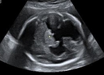
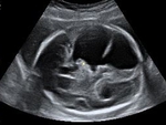
Pinna of fetal ear
The pinna of the ear is seen very nicely in these sonographic images of the fetal head. Images courtesy of Dr. Ravi Kadasne, UAE.
Static B-mode scanning machines
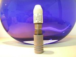
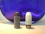
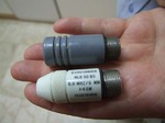
These are relics of a bygone era-- the ultrasound scan probes or scan heads of B-mode static scanning machines, probably of the early 1970s. These snaps by Mr. Shlomo Gobi, sonographer, from Israel, show that these probes were screwed onto the bulky, articulated arms of the early machines.
You can try this link to read more about the history of ultrasound machines:
http://www.ob-ultrasound.net/history1.html (free article and pictures)
The strange Bladder Cat
This interesting image of a cat face (in the urinary bladder) is the handiwork of sonography guru, Dr. Ravi Kadasne, a radiologist working in UAE. Fantastic imagination, Dr.Ravi!!
And a bison too... in the pelvis
also an owl
Images courtesy of Dr. Ravi Kadasne, UAE.
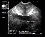
A prostate fox
Can you spot a fox in this TRUS image of the prostate?
schizencephaly-in-fetus
Ultrasound images showing schizencephaly. The cleft extends all the way from the ependyma of the lateral ventricle to the pia mater.
Gall bladder turned swan
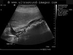
A nice long necked gall bladder turns into a work of art in the hands of Dr. Sanjiv Bhalla, MD. India.
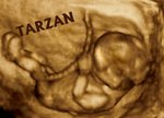
fetal tarzan
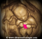
This 14 weeks old baby in the womb (fetus) caught playing Tarzan with the umbilical cord. This 3D ultrasound image is courtesy of Dr. Ravi Kadasne, MD, UAE.
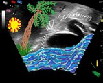
peacock-in-the-abdomen
Dr. Ravi Kadasne at work discovered this lively peacock in the abdomen. Enjoy Dr.Ravi's work of art!
Great piece.
fetus calling mom
Just another picture that makes us smile. This fetus seems to be calling its mom via a cell phone. Image and art courtesy of Dr. Ravi Kadasne, MD, UAE.
Try-this-easy-quiz-on-uterus-below
(For mobile devices, click this link):


