
Musculoskeletal
Contents of this page
- Tear-of-medial-cruciate-ligament
- Epidermoid cyst
- Case-2: Sebaceous cyst with leak
- Ultrasound image of transient synovitis of hip joint in a child
- Ultrasound image of olecranon bursitis
- Ultrasound images of inguinal hernia
- Sonography and Color Doppler imaging of Hemangioma in infant
- Hypertrophy of the Abductor Hallucis muscle of the foot
- Rib fracture
- Fracture radius with hematoma formation
- Intracranial mass/ Intracranial dermoid or epidermoid
- Baker cyst
- Rupture of the Achilles Tendon or Tendo achilles tear
- Bicipital tendonitis or Biceps tendonitis
- Glomus tumor (subungual location)
- Jersey finger or rupture of the tendon of the flexor digitorum profundus muscle
- Full thickness tear of supraspinatus tendon
- Myositis ossificans
- Suprapatellar effusion (suprapatellar hematoma)
- Tuberculous lymph node enlargement (tuberculous lymphadenitis)
- Divarication of recti
- 3D ultrasound image of foreign body
- inguinoscrotal hernia-3D ultrasound imaging
- xiphoid process- 3D ultrasound
- torn adductor longus muscle
- pilonidal cyst
- Achilles-tendinosis-or-tendinitis
- LSCS-scar-endometriosis
- epigastric-hernia
- trichilemmal-cyst-pilar-cyst
Tear-of-medial-cruciate-ligament
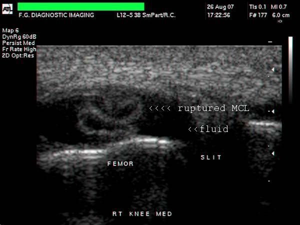
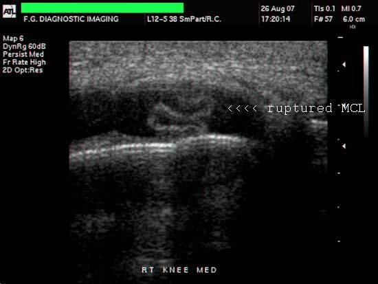
These are ultrasound images of the right knee joint following a motorcycle accident. There is a curvilinear echogenic structure within the medial part of the right knee joint cavity. Diagnosis: traumatic rupture of the distal insertion of the medial cruciate ligament, which now floats within the fluid distended (possible hemorrhagic) joint space. Images courtesy of Mr. Shlomo Gobi, Israel.
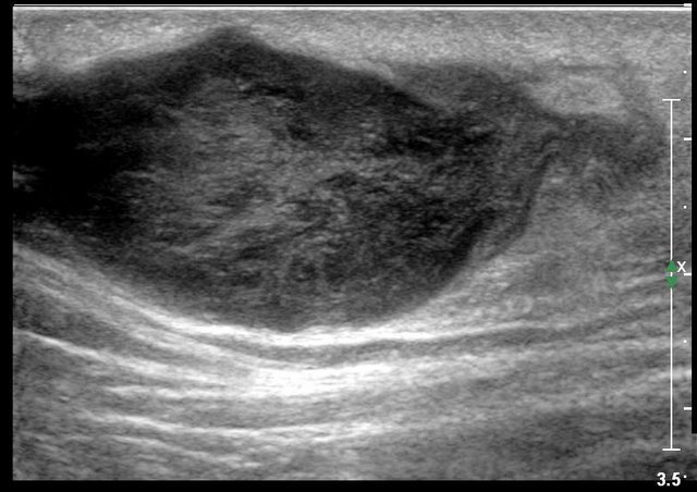
Epidermoid cyst
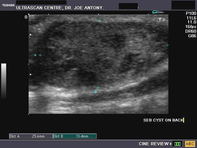
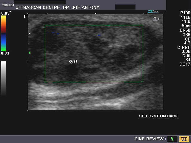
Sonography of a mass on the dorsal part of the chest was done to study a mass of 2.5 cms. Images reveal a hypoechoic mass of inhomogenous appearance measuring 2.5 x 1.3 cms. It is cutaneous in location and shows no vascularity within on color doppler imaging (image on right). No calcification is seen. These ultrasound images suggest a diagnosis of epidermoid cyst or what was often called a sebaceous cyst. Images by Dr. Joe Antony, Cochin, India, using Toshiba, Nemio XG color doppler machine.
Reference: E-medicine article on epidermoid cyst (free article).. rated excellent
Case-2: Sebaceous cyst with leak
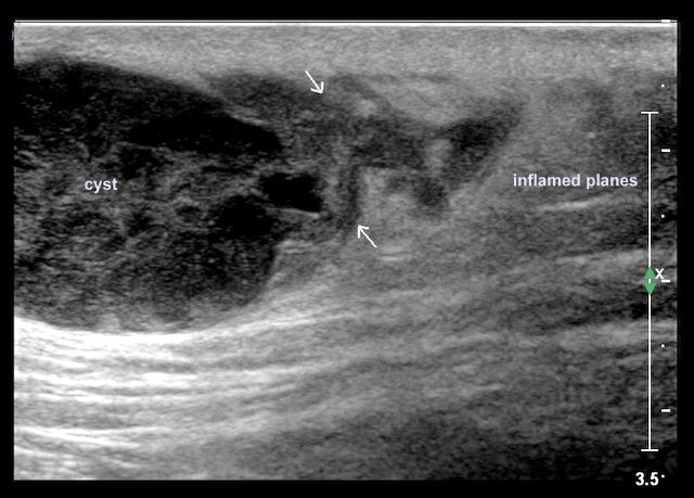
This patient has a large sebaceous cyst seen in the ultrasound images above as an inhomogeneous hypoechoic mass in the skin. But more importantly, the right of the image shows an extravasation of the keratinous material from the epidermoid cyst, into the surrounding subcutaneous tissue. Keratin being highly irritative to the subcutaneous tissue can provoke a strong inflammatory response and even result in abscess formation. Ultrasound images are courtesy of Dr. Ravi Kadasne, MD, UAE.
Ultrasound image of transient synovitis of hip joint in a child
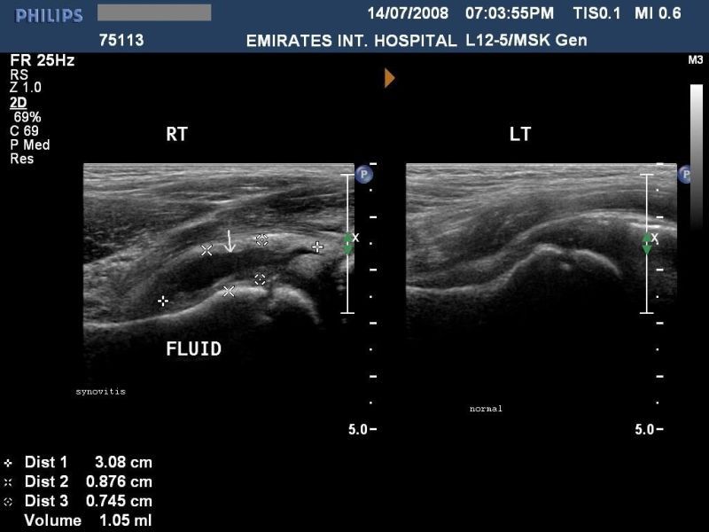
Sonography of the hip joints was done in this 5 yr. old child to c/o pain and swelling in the right hip. Ultrasound images show anechoic fluid collection around the right hip diagnostic of synovitis. The synovial fluid collection here measures 3 x 0.8 cms. The normal left hip joint is also shown for comparison. Ultrasound images courtesy of Dr. Ravi Kadasne, who used a Philips iU 22 machine.
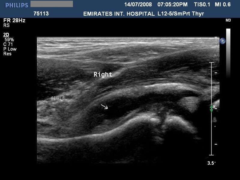
Ultrasound image of olecranon bursitis

This patient had pain and swelling of the left elbow. Sonography of the elbow shows a cystic lesion containing clear fluid over the tip of the elbow. The lesion is located between the skin and the underlying olecranon process of the left ulna. This ultrasound image is diagnostic of olecranon bursitis. Image courtesy of Dr. Ravi Kadasne, UAE. The machine used here is the Philips IU 22.
Reference:
1) http://www.jultrasoundmed.org/cgi/content/full/26/6/857 (free article and images).
2) http://www.emedicine.com/pmr/TOPIC91.HTM (free article)
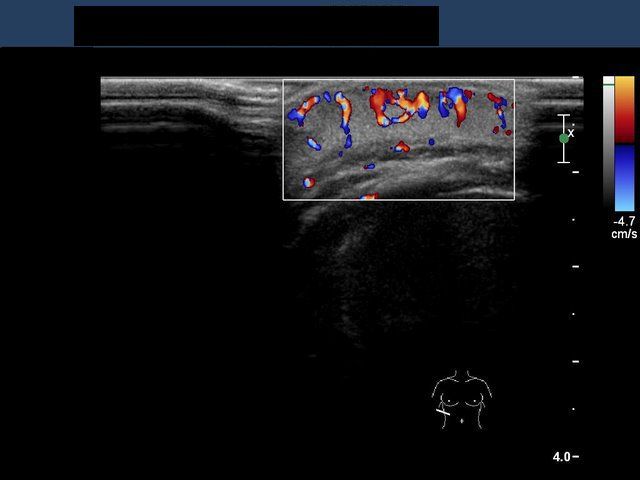
Ultrasound images of inguinal hernia
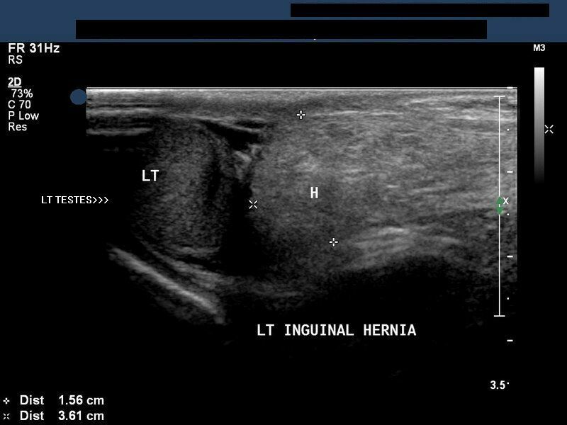
These ultrasound images show echogenic, linear/ tubular structures (intestine) within the left scrotal sac, just above the left testes, extending to the left inguinal canal s/o left inguinal hernia. (Images taken using a Philips iU 22 Ultrasound machine, by Dr. Ravi Kadasne, MD, UAE.
Reference:http://www.medscape.com/viewarticle/527887_8 (free article).
Sonography and Color Doppler imaging of Hemangioma in infant

An infant present with a large reddish patch in the right lumbar region. Ultrasound image (left) shows a hyperechoic lesion of about 2.3 cms. in the subcutaneous layer. Color Doppler image (right) shows the lesion to be markedly vascular. These ultrasound and color Doppler images are diagnostic of Hemangioma, possibly cavernous type. Images courtesy of Dr. Ravi Kadasne, UAE.
Hypertrophy of the Abductor Hallucis muscle of the foot
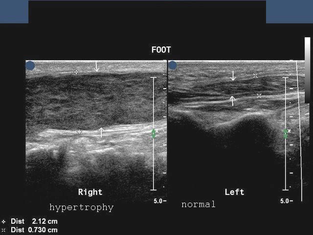
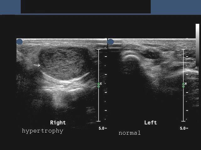
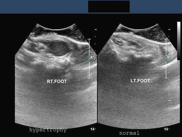
Ultrasound images showing hypertrophied muscle on medial part of plantar aspect of the foot.
This patient presented with persistent pain in the sole of the Rt. foot. Sonography of the plantar aspect of the right foot showed markedly thickened muscle along the medial aspect of the plantar region. The left foot was also imaged and showed the normal Abductor Hallucis muscle. It was concluded that the ultrasound images (above) showed a hypertrophy of the right abductor Hallucis muscle. The right abductor Hallucis showed a thickness of 2.1 cms. while the normal left side showed a thickness of about 0.7 cms. Ultrasound images are courtesy of Dr. Ravi Kadasne, MD, UAE.
Reference:http://www.ncbi.nlm.nih.gov/pmc/articles/PMC2565658/ (free article and images)
Rib fracture
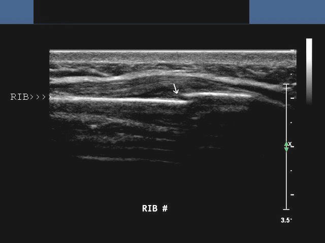
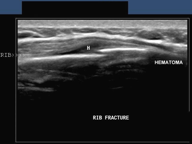
Ultrasound image of rib fracture.
This patient had a history of trauma to the chest. Sonography of the thorax (chest) showed a breach in continuity of the rib shown above (arrow). 3-D ultrasound image (image on right) shows fracture of the rib with small hematoma collecting anterior to the fracture site. The hematoma is seen as an anechoic collection. High resolution sonography/ ultrasound is now increasingly being used in imaging the skeletal system, especially when in a superficial location. Ultrasound images are taken using Philips IU22 system and are courtesy of Dr. Ravi Kadasne, MD, UAE.
Reference:http://www.ajronline.org/cgi/reprint/173/6/1603 (free article and images)
Fracture radius with hematoma formation
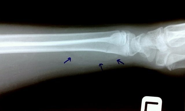
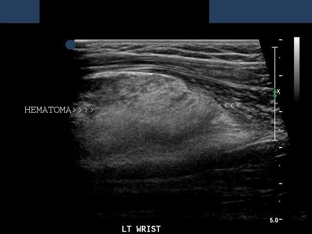
This X-ray of the wrist and forearm shows a fracture of the distal end of radius. Ultrasound image on right shows large hyperechoic collection (arrowheads) anterior to the fracture site. X-ray image also shows the soft tissue swelling caused by hematoma (blue arrows). This child had a history of bleeding disorder. Images courtesy of Ravi Kadasne, MD, UAE.
Intracranial mass/ Intracranial dermoid or epidermoid
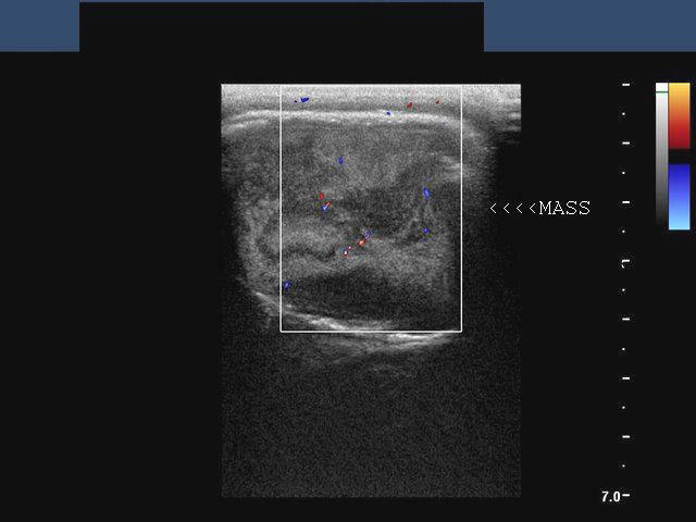
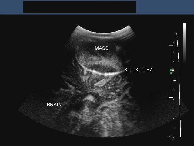
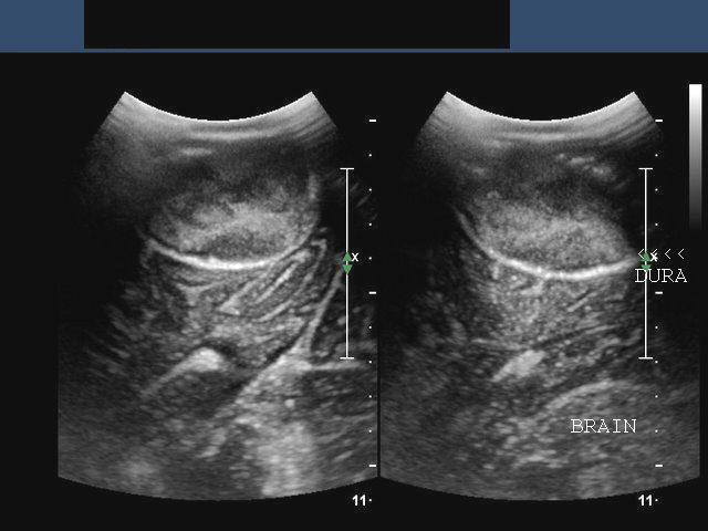
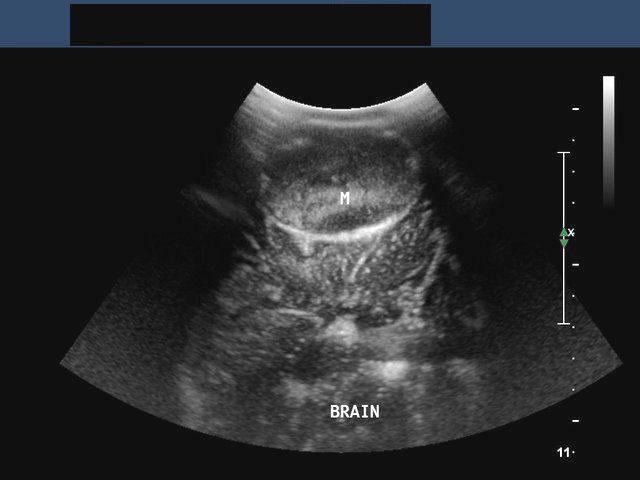
Dermoid mass or CNS Dermoid
This middle aged female patient presented with a mass in the parieto-occipital region. Sonography of the skull was done to image the mass which was close to the midline. Ultrasound images show a mass of mixed echogenicity with both solid and cystic components within the cranial vault (intracranial region), which has eroded the cranial vault to bulge outward. Particulate matter was seen floating the fluid component of the mass. The cerebral hemishperes appear to be compressed upon by the mass (M) via the dura mater. The meninges appear to be preserved though displaced inward by the mass. Color Doppler image (top row- left) suggests poor vascularity of the intracranial mass. These ultrasound and Color Doppler images suggest a dermoid tumour inside the cranium (CNS dermoid). Dermoids are known to occur in such locations though, are rare. The other diffferential diagnoses are meningioma and epidermoid cyst. But the midline location favors a diagnosis of dermoid cyst. Ultrasound images are courtesy of Dr. Ravi Kadasne, UAE.
Reference: http://emedicine.medscape.com/article/339797-imaging(free article and images).
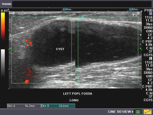
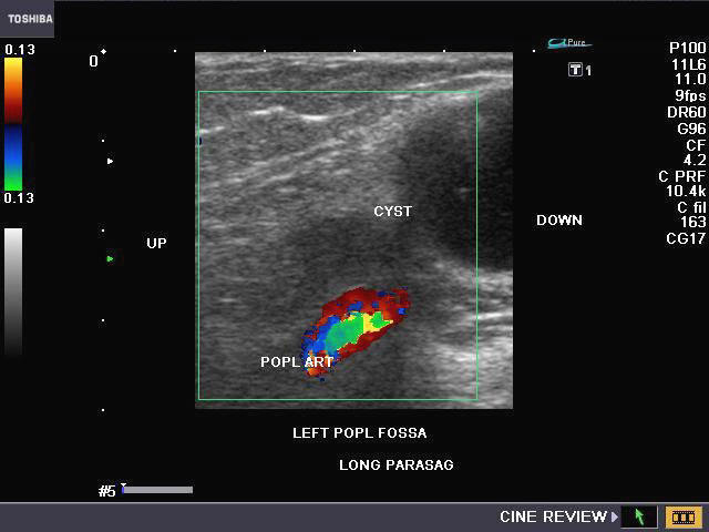
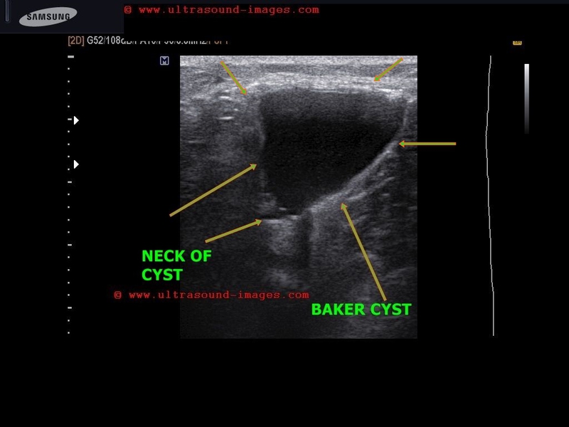
Baker cyst


Case-2-Baker-cyst
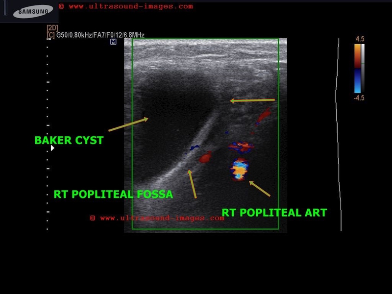
Large cystic lesion in left popliteal fossa (case-1)
This middle aged female patient presented with a large mass in the left popliteal region which was seen on extension of the knee joint. Ultrasound images of the popliteal fossa show a large cystic mass of 5.7 x 2.8 cms. with few echogenic particles within a primarily clear fluid. The cyst is in close relation to the left knee joint; however, these images did not reveal clear communication with the synovial space of the knee joint. The popliteal artery was visualized on Color Doppler imaging, and was clearly separate from the cyst, ruling out the possibility of aneurysm of this vessel. Final diagnosis: Baker cyst (also called Baker's cyst).
Case-2 (above) shows a Baker cyst of the right popliteal fossa. Note the beak of the cyst or neck which leads to the synovial cavity of the right knee joint (the speech bubble sign).
Reference: http://emedicine.medscape.com/article/387399-overview
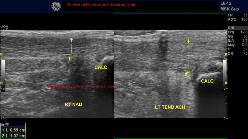
Rupture of the Achilles Tendon or Tendo achilles tear
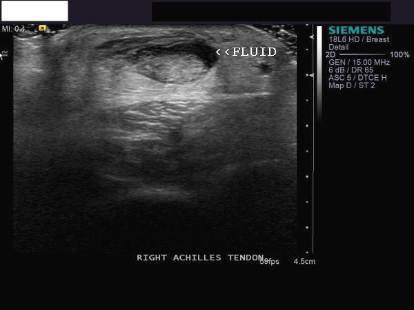
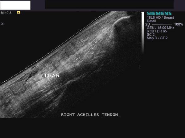
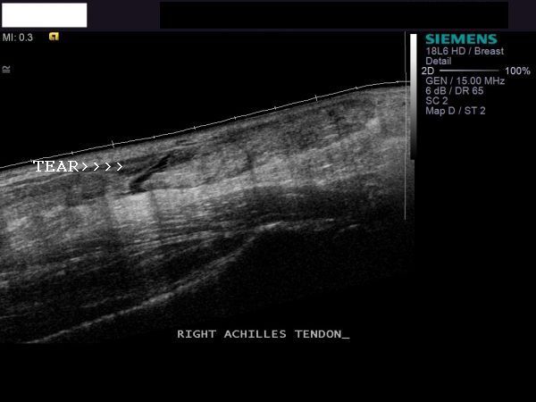
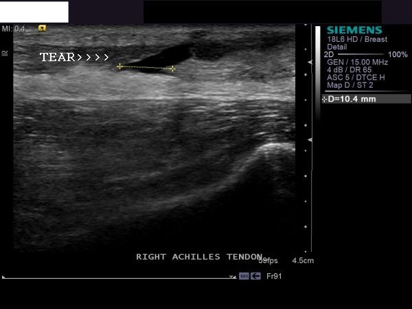
This patient had severe pain in the lower calf. Ultrasound examination of the tendo-achilles showed a full thickness tear of the Achilles tendon. This is seen as the linear anechoic area extending through the tendon (long section ultrasound images of Achilles tendon). Transverse section (image on top left) shows fluid and blood around the tendon, seen as anechoic area. All above images are courtesy of Shlomo Gobi, Israel.
Reference: http://emedicine.medscape.com/article/85024-overview
http://www.emedicinehealth.com/achilles_tendon_rupture/article_em.htm
Bicipital tendonitis or Biceps tendonitis

Axial section Long section Axial section Long section
This patient presented with pain and tenderness in the left shoulder. Ultrasound images show the tendon of the long head of biceps which is thickened and nodular due to inflammation with surrounding fluid (anechoic space around the tendon). These findings suggest biceps tendonitis. The axial section ultrasound images show considerable fluid within the bicipital groove. Images courtesy of Shlomo Gobi, Israel.
References: http://emedicine.medscape.com/article/96521-diagnosis
Glomus tumor (subungual location)


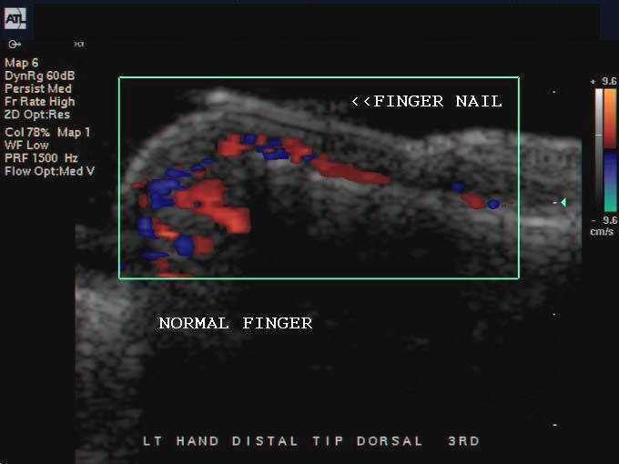
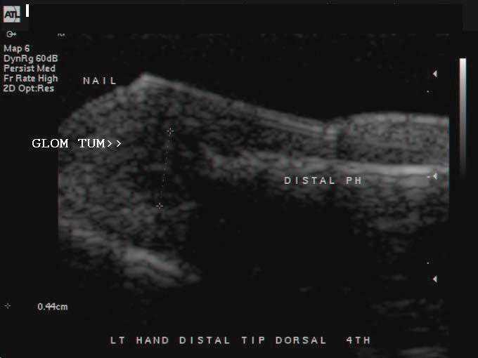
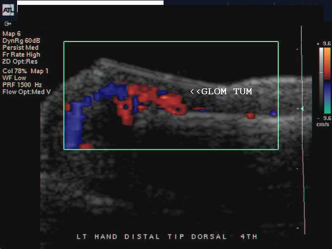
The fourth finger of this patient shows a subungual soft tissue mass (between the fingernail and the distal phalanx). This mass appears very vascular on color Doppler ultrasound imaging. Transverse and longitudinal ultrasound/ Color Doppler images show the vascular mass very clearly, increasing the width between the nail and the underlying phalangeal bone. These findings are typical of glomus tumor of the finger. The normal 3rd finger (distal part) is shown for comparison. Glomus tumor is a vascular mass arising from the glomus body, a nervous tissue nodule, that is important to sense temperature. The glomus tumor commons occurs in the tips of the first 4 fingers, usually in the subungual location as described above. All above images are courtesy of Shlomo Gobi, Israel.
Reference: Ultrasound imaging of glomus tumor (good article and images).
Jersey finger or rupture of the tendon of the flexor digitorum profundus muscle

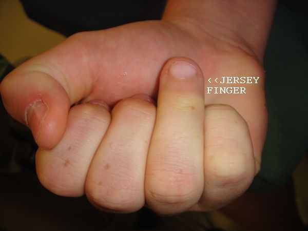
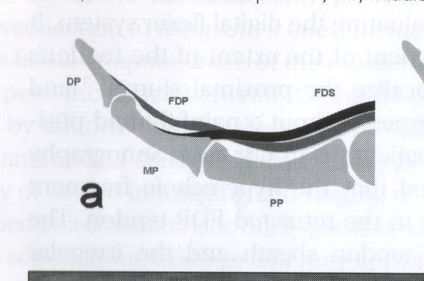
This young boy suffered a sports injury to the 4th finger whilst playing basketball. The result was inability to flex the distal phalanx of the 4th finger of the right hand. The snap of the boy's right hand shows the affected 4th finger. The ultrasound images of the 4th finger show the rupture or avulsion of the tendon of the flexor digitorum profundus muscle just at the site of its insertion into the palmar aspect of the distal phalanx. This condition is called a Jersey finger and is the result of acute injury to the distal phalanx resulting in sudden forced extension of the affected finger (distal phalanx) when the finger was in a flexed condition. The picture in lower right shows the normal anatomy of the finger.( FDP= flexor digitorum profundus tendon; DP= distal phalanx. PIP= proximal interphalangeal joint; DIP= distal interphalngeal joint).(Ultrasound images and case study of Jersey finger are courtesy of Shlomo Gobi, Israel).
Reference: http://en.wikipedia.org/wiki/Jersey_Finger
Full thickness tear of supraspinatus tendon
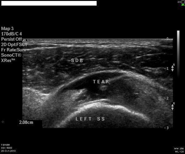
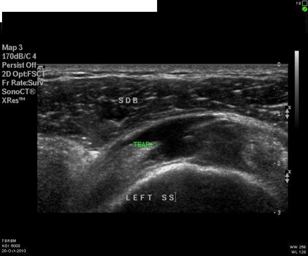
This patient had severe pain in the shoulder. Sonography of the shoulder was done. Ultrasound images show anechoic region within the supraspinatus tendon, just above the shoulder joint. The anechoic region extends through the full thickness of the supraspinatus tendon. This appearance is typical of full thickness supraspinatus tear. Ultrasound images of supraspinatus tear are courtesy of Mr. Shlomo Gobi, Israel. (SDB= subdeltoid bursa; SS= supraspinatus muscle).
Reference: http://emedicine.medscape.com/article/401595-overview
http://www.ajronline.org/cgi/reprint/184/1/180.pdf
Myositis ossificans
Long section- left hamstring muscles

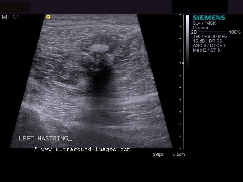
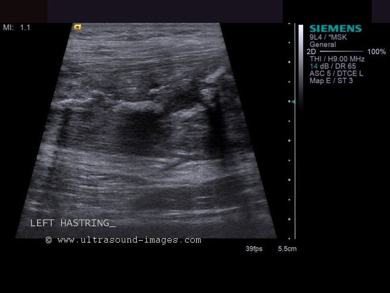
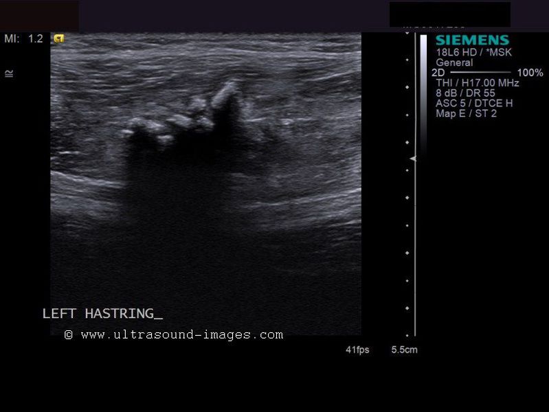
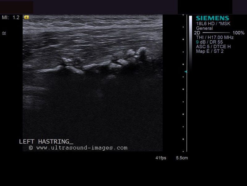

This patient had a blunt injury to the posterior part of left thigh. There was persistent pain and tenderness in this region for a few months. Ultrasound images of the left hamstrings show multiple calcific, echogenic lesions in the region, within the muscles. There is posterior acoustic shadowing behind the lesions which totally measure 4.8 cms. in length. These ultrasound findings are diagnostic of myositis ossificans of the left hamstrings. Myositis ossificans is the formation of bone within a long standing hematoma in the soft tissues, usually the result of trauma. Certain individuals are genetically susceptible to the formation of bone (ossification) of a hematoma. Color Doppler did not reveal any vascular changes in this lesion. Ultrasound images of myositis ossificans are courtesy of Mr. Shlomo Gobi, Israel.
References:
http://orthopedics.about.com/od/sportsinjuries/g/myositis.htm
http://www.nejm.org/doi/full/10.1056/NEJMicm1005605
Suprapatellar effusion (suprapatellar hematoma)

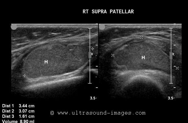
This child had a knee injury following which he had swelling and pain around the knee joint. He underwent sonography which showed a large collection of echogenic fluid (H) in the suprapatellar region; this collection measured 3 x 3 cms. with poor communication with the right knee joint synovial cavity. Knee injuries frequently result in the formation of hemorrhagic effusions which may extend into the suprapatellar bursa. In most cases, the suprapatellar bursa communicates freely with the synovial cavity of the knee joint. In a minority of patients, the suprapatellar bursa is separate from the knee joint resulting in loculation of fluid following trauma. These ultrasound images suggest suprapatellar hemaotma (H) or hemorrhagic effusion. The hematoma (H= hemorrhagic effusion) is seen deep to the quadriceps tendon and just above the patella, superficial to the distal end of femur. Ultrasound images are courtesy of Dr. Ravi Kadasne, MD, UAE.
References:
http://www.ajronline.org/cgi/reprint/175/5/1313.pdf (free article and images).
Imaging of bursae (clinical imaging science)
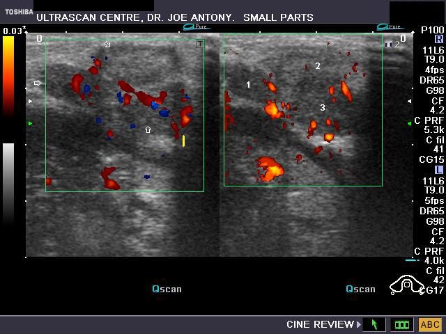
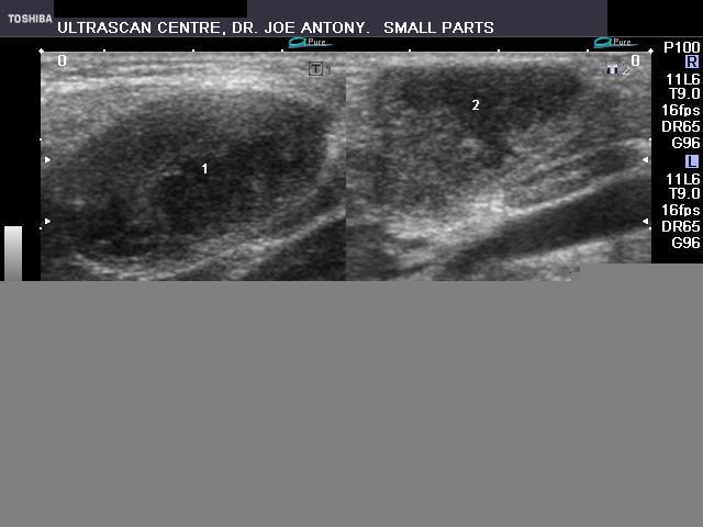
Tuberculous lymph node enlargement (tuberculous lymphadenitis)
Case-1: Enlarged submental and middle cervical lymph nodes
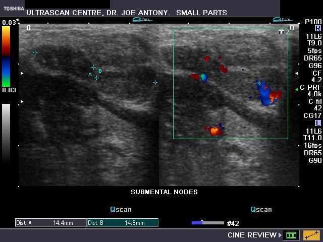

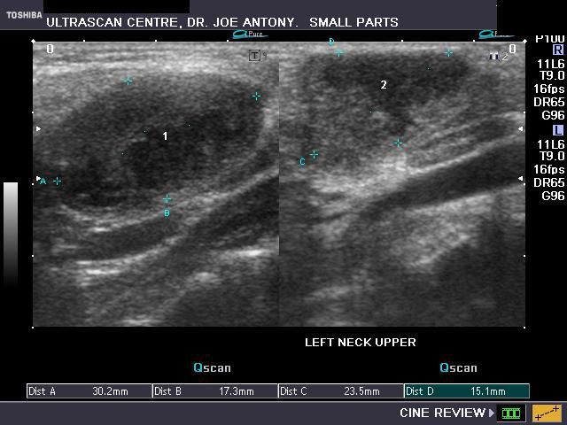

The above color Doppler and B mode ultrasound images show multiple (three in number) enlarged submental lymph nodes in a child. The nodes appear rounded and are in close relation (attached or matted) to each other. The lymph nodes are markedly hypoechoic and show peripheral vascularity on color Doppler ultrasound. In addition, these nodes also show echogenic margins. These ultrasound findings favor a diagnosis of tuberculous lymphadenitis. In addition this child also had enlarged middle group of deep cervical lymph nodes.
These sonographic images show 2 markedly enlarged lymph nodes with poor vascularity and cystic changes within them. These findings are seen mainly in tuberculous lymphadenitis and occasionally in metastases to lymph nodes. Very often, it may not be possible to accurately distinguish the main differentialdiagnoses of lymph node enlargement- ie: tuberculosis, lymphoma and metastases. It is here that clinical history and symptoms can help to diagnose the cause. In such cases biopsy of the lymph node may be indicated. The snap of the child's neck shows the swelling in the left lateral region anterior to the sternomastoid muscle. In this case, the diagnosis based on these ultrasound images was tuberculous lympadenitis.
References:
case-2-cervical lymphadenitis
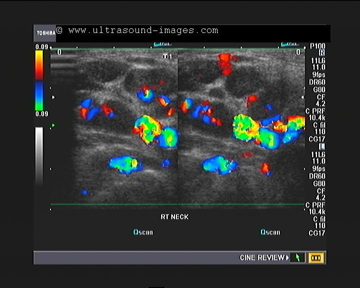
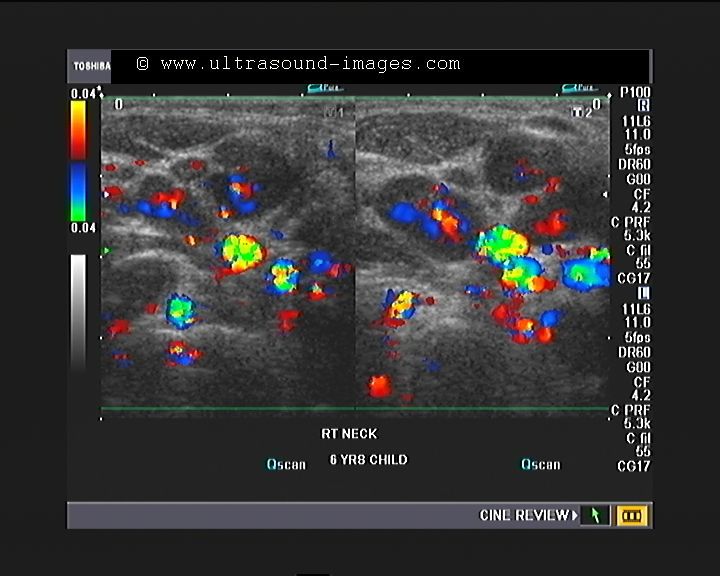
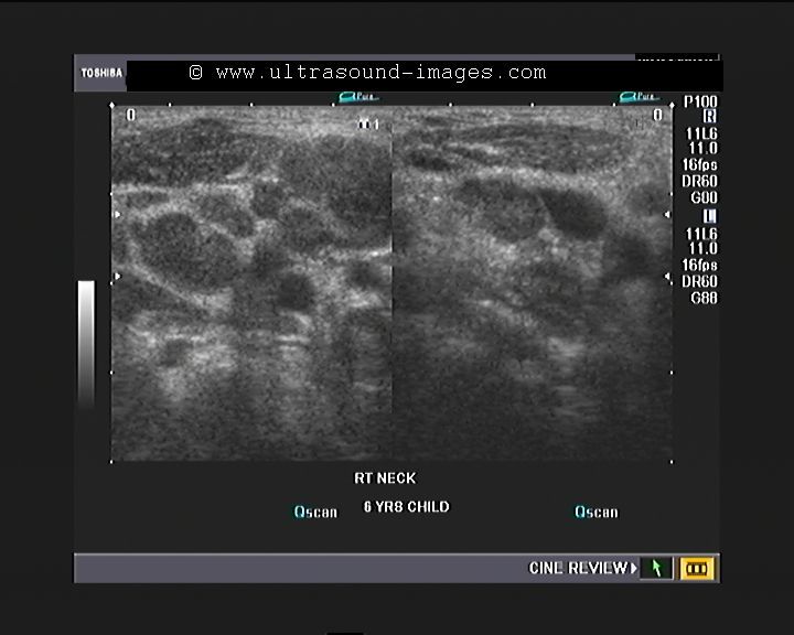
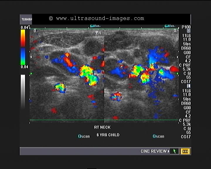
This six-year-old male child had pain and swelling in the right side of neck just below the angle of the mandible. Ultrasound and colour Doppler images show markedly vascular and enlarged hypoechoic cervical lymph nodes. I could count more than 7 to 8 such lymph nodes in this region. The marked vascularity of the lymph nodes suggests active bacterial infection and inflammation of the nodes. The possibility of bacterial or tuberculous lymphadenitis is the diagnostic opinion in this case. There is no gross evidence of liquefaction of the nodes or any other such evidence of abscess formation.
Divarication of recti
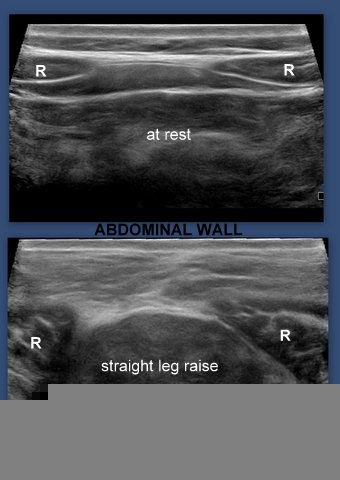
This patient complained of bulging of the midline in the abdomen. Ultrasound imaging of the abdominal wall showed typical bulging of the linea alba (the aponeurosis or fibrous tissue sheath that connects the two halves of the rectus abdominis muscles) with thinning of the linea alba on leg raising. These ultrasound images show these features diagnostic of divarication of the recti (also known as diastasis recti). Ultrasound images of divarication of recti are courtesy of Dr. Ravi Kadasne, MD, UAE).
References:
1) IJRI article on ultrasonography of divarication of recti
2) Wikipedia article on divarication of recti (diastasis of recti)
3D ultrasound image of foreign body
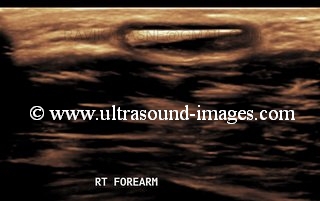
A foreign body is often seen well on 2D ultrasound imaging, if one uses the high resolution transducers available today. But what happens when we use a 3D ultrasound system? This 3D ultrasound image of a foreign body in the forearm (a date thorn) reveals the thorn beyond any doubt, and shows a small fluid collection around the object, suggesting focal edema.
This 3D ultrasound image of foreign body is courtesy of Dr. Ravi Kadasne, MD, UAE.
inguinoscrotal hernia-3D ultrasound imaging
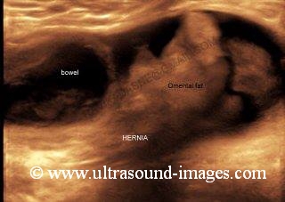
This is just another inguino-scrotal hernia, but the difference is that this is a 3D ultrasound image of this type of hernia. This image shows the contents of the inguino-scrotal hernia, including bowel and omental fatty tissue. The actual defect in the abdominal wall can be gauged in stunning detail, and the information obtained from a 3D sonographic image such as this can actually help change decisions on the surgical approach. This image is courtesy of Dr. Ravi Kadasne, MD, UAE.
xiphoid process- 3D ultrasound
Can bone and cartilage be imaged in 3D ultrasound studies? This 3D ultrasound image of the xiphoid process of the sternum (image courtesy of Dr. Ravi Kadasne, MD, UAE) testifies affirmatively to this fact. The xiphoid process is the terminal and lower end of the sternum and is cartilaginous in nature.
Read more on this topic at:
http://en.wikipedia.org/wiki/Xiphoid_process
torn adductor longus muscle

This young male football player developed acute pain in the right groin for which imaging was advised. Ultrasound imaging shows tear of the right adductor longus muscle with retraction of the muscle fibres proximal to the rupture of the adductor longus muscle. An anechoic collection suggestive of a haematoma (H) is seen just distal to the torn adductor longus muscle (see ultrasound images above). These ultrasound images are courtesy of Ravi Kadasne, MD. the adductor longus muscle is frequently involved in sports injuries involving the thigh muscles. The tear of this muscle is often caused by exertion of sudden stress during vigourous exercise or sports. The ruptured adductor longus muscle fibres are seen to retract proximally with fluid or blood seen to collect in the vacant space (see images above).
References:
imaging of adductor longus muscle tear
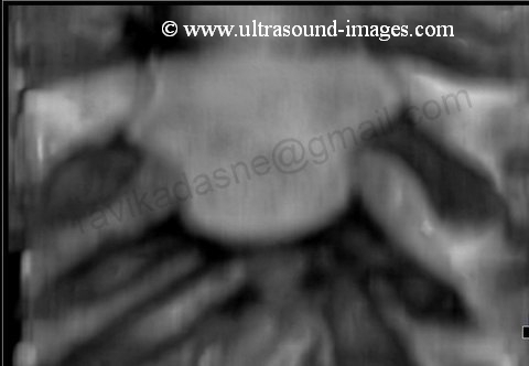
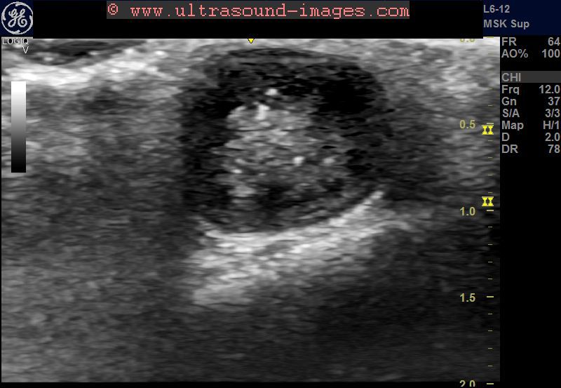
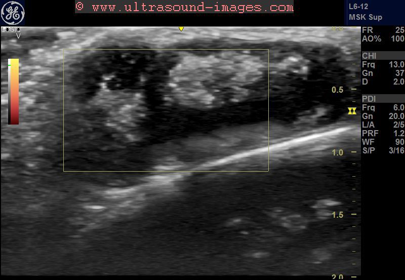
pilonidal cyst
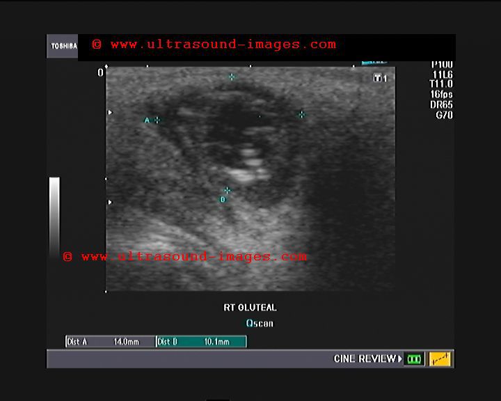
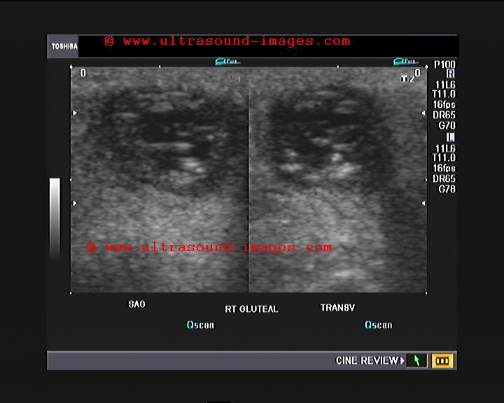
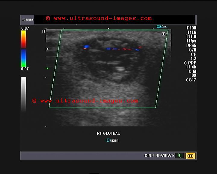
This 16 year old female child showed a nodule (2 weeks duration) on the gluteal region within the natal cleft. Ultrasound images show a cystic lesion containing particulate matter. The lesion is small (14 x 10 mm.) and also shows echogenic linear streaks (hairs) within it. This suggests pilonidal cyst. Pilonidal cysts are typically seen in this location in 15 to 35 year age group. Rupture of the cyst may cause a pilonidal sinus or fistula.
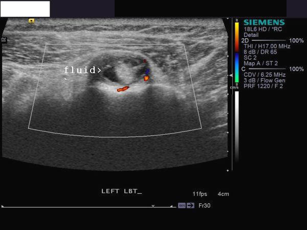
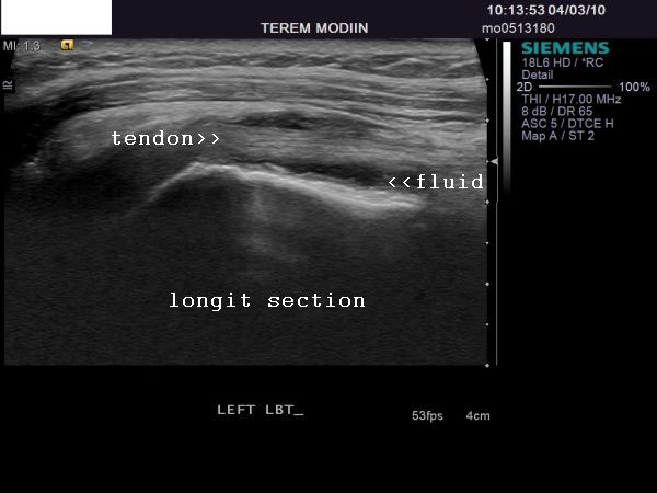
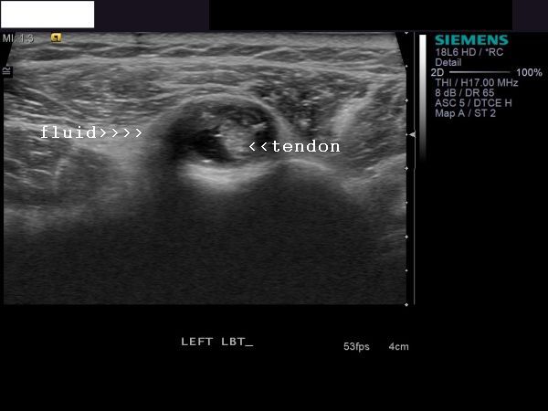
Achilles-tendinosis-or-tendinitis


This 25 yr old lady has a history of strenous physical activity followed by pain in the Lt. heel. Ultrasound images show the following features:
1) edematous hypoechoic and swollen Left Achilles tendon in its distal part close to insertion into the Left calcaneum.
2) loss of fibrillar detail of the strands of Achilles tendon on the left side s/o micro trauma.
3) On color and Power Doppler ultrasound there is mild hyperemia of the abnormal left Achilles tendon. All these findings point to a diagnosis of Left Achilles tendinosis or Achilles tendinitis. Unlike partial or complete rupture of the Achilles tendon, Achilles tendinosis or tendinitis can be corrected by conservative treatment including physiotherapy and medication
References: 1) Achilles tendinosis- ultrasound imaging- AJR
2) Sonoworld article on sonography of Achilles tendinosis or tendinitis
LSCS-scar-endometriosis
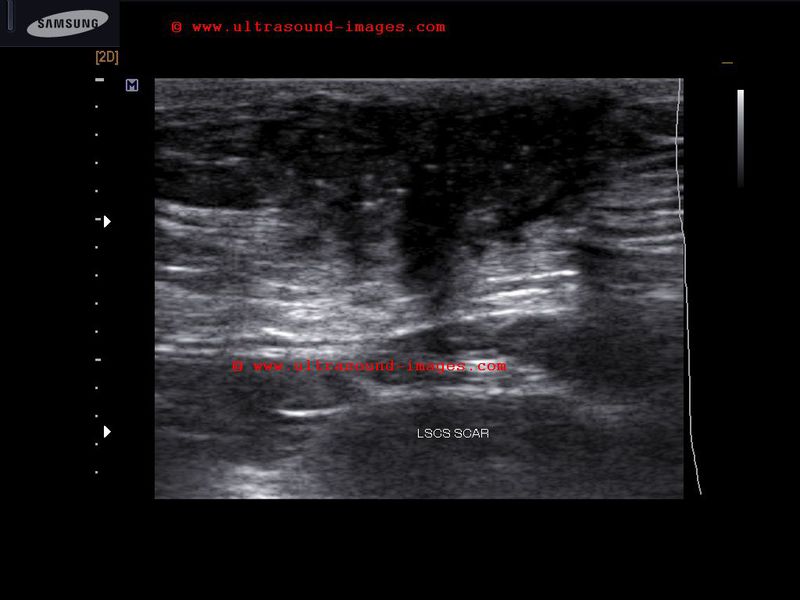
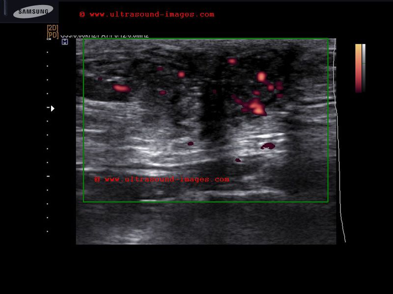

This 35 yr old lady underwent LSCS surgery 4 years ago. Presently she has cyclical pain (during her menses) localized to the lower abdomen and pelvis. A hyperpigmented patch was noted over a part of the LSCS scar on the abdominal wall and a hard nodular lesion was felt on palpation. Ultrasound image above shows a hypoechoic irregular mass infiltrating part of the anterior surface of the rectus abdominis and is located within the subcutaneous layer of the abdomen within the region of the LSCS scar. Power Doppler images above also show significant internal vascularity within the lesion. These ultrasound signs and correlation with the symptoms of cyclical pain during menses confirm this to be LSCS scar endometriosis.
Cesarean section in this case would have transplanted endometrial cells from within the endometrium during surgery, into the scar of the LSCS resulting in the development of endometriosis of the scar.
References: ultrasound imaging of LSCS scar endometriosis
epigastric-hernia

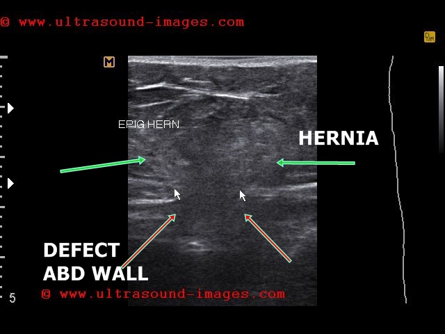
Ultrasound images and video above show a large epigastric hernia in the upper abdomen.
Hernia sac with abdominal contents including small bowel seen passing through the wall defect on coughing in a to and fro manner. Arrows point to the defect in abdominal wall in above video.
trichilemmal-cyst-pilar-cyst


Middle aged female with cystic swelling of scalp in frontal region.
Ultrasound images show:
= cystic lesion measures 2.5 x 1.6 cms. in frontal part of scalp
= central part of the cyst shows echogenic matter s/o cholesterol crystals and calcification
= on examining the lesion: local alopecia and absence of punctum
= this differentiates the pilar cyst or trichilemmal cyst from sebaceous cyst
= also trichilemmal cyst or pilar cyst is painless and does not communicate intracranially
= treatment: surgical excision
© 2026, Joe Antony, MD All images and content in this site are copyrighted. www.ultrasound-images.com When visiting this site, we may use cookies to improve the performance of this site. Use of this site means you accept the use of cookies. Read our privacy policy at this page: https://www.ultrasound-images.com/privacy-policy/