
gb mucocele
Contents of this page
Mucocele of the gall bladder


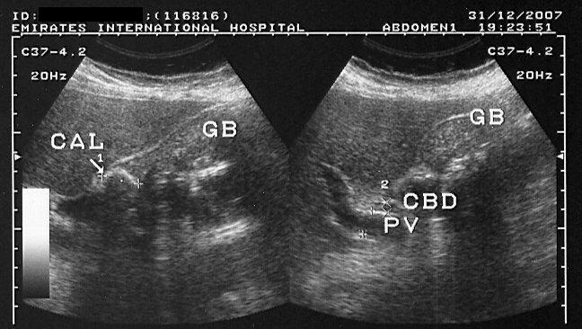
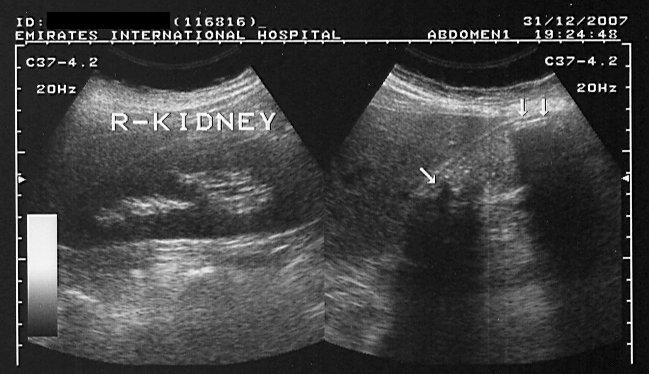
Sonography of the gall bladder in this patient revealed
1) huge, distended gall bladder, measuring 10 x 3 cms.
2) a large gall bladder calculus of 24 mm. lodged at the neck, producing obstruction in outflow.
3) multiple smaller calculi are seen in the gall bladder (sizes- 5 to 10 mm.)
4) large amount of turbid fluid, with fine particulate matter in the Gall bladder lumen.
5) normal wall thickness suggesting absence of cholecystitis.
These ultrasound images suggest mucocele of the gall bladder. Mucocele is caused by chronic obstruction of the gall bladder causing it to be overdistended.Bile is slowly absorbed from the lumen and replaced by mucous secretions from the wall. Images taken using a Toshiba Powervision color doppler machine, courtesy of Dr. Ravi Kadasne, UAE.
Reference: http://www.emedicine.com/med/topic2817.htm(free article).
Pyocele of gall bladder following acalculous cholecystitis
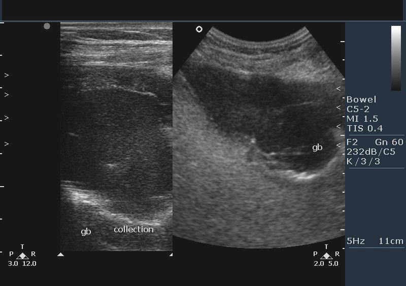

This patient had pain in the right hypochondrium and fever. Sonography of the gall bladder shows: a large over-distended gall bladder with echogenic fluid (purulent material) within its lumen. The walls of the GB (gall bladder) show layering (thickening or edema). No evidence of gall bladder calculi is present. Septate structures (linear echoes) within the gall bladder suggest evidence of sloughing of the mucosa. These ultrasound images suggest a diagnosis of Pyocele of the gall bladder following acalculous cholecystitis. This ultrasound case study is courtesy of Vikas Shukla, MD, India.
Reference: http://emedicine.medscape.com/article/195165-diagnosis
© 2026, Joe Antony, MD All images and content in this site are copyrighted. www.ultrasound-images.com When visiting this site, we may use cookies to improve the performance of this site. Use of this site means you accept the use of cookies. Read our privacy policy at this page: https://www.ultrasound-images.com/privacy-policy/