
Ultrasound images of diseases of the colon

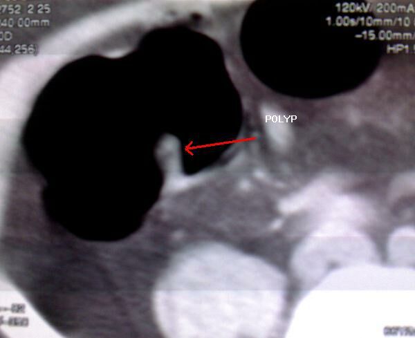
CLOSE-UP view of the CT image
Colonoscopic view of the polyp
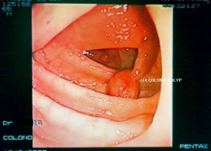
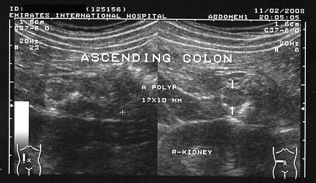
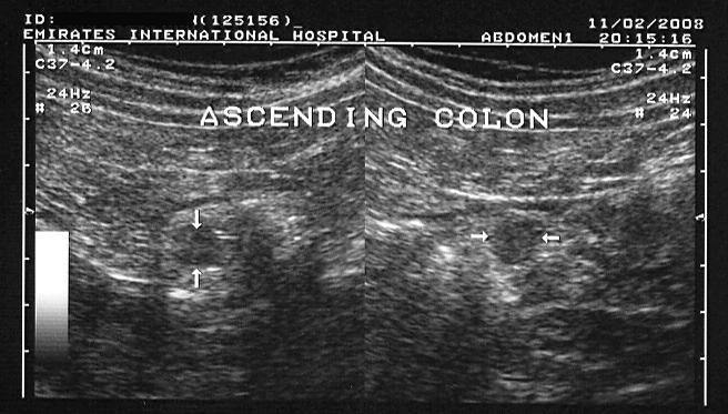
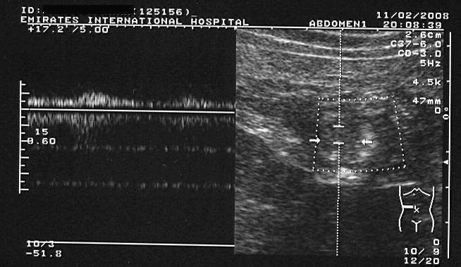
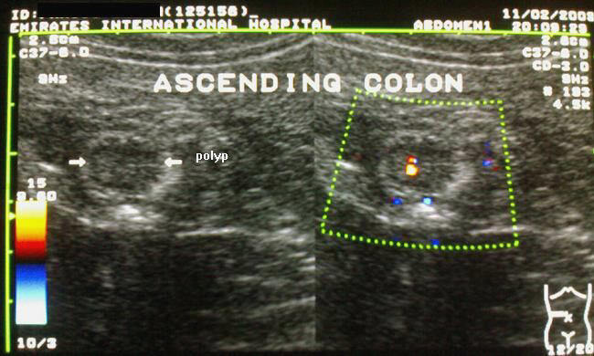
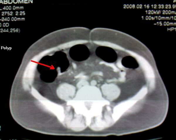
This 55 yr. old male patient underwent routine sonography of the abdomen. The scan reveals a rounded, solid, soft tissue lesion (isoechoic with the colon) of size 17 x 10 mm, in the lumen of the ascending colon. An atypical target sign is also seen. Color doppler shows a feeder vessel supplying the mass lesion. These ultrasound images suggest colonic polyp. Sonographic images taken using a Toshiba Powervision color doppler machine, courtesy of Dr. Ravi Kadasne, UAE. The polyp was also visualized on CT scan (see images above) and also on colonoscopy (above).
Reference:
1)http://en.wikipedia.org/wiki/Polyp_(medicine)(free article).
© 2026, Joe Antony, MD All images and content in this site are copyrighted. www.ultrasound-images.com When visiting this site, we may use cookies to improve the performance of this site. Use of this site means you accept the use of cookies. Read our privacy policy at this page: https://www.ultrasound-images.com/privacy-policy/