
Ultrasound images of fetal abdomen
Contents of this page
- Fetal gall bladder sludge
- Double bubble sign of duodenal obstruction
- Fetal ovarian cyst -fetal dermoid cyst vs. twisted ovarian cyst or torsion of ovarian cyst in fetus
- Echogenic fetal bowel
- Fetal dermoid cyst or mature cystic teratoma
- dilatated fetal bowel
- omphalocele
- hepatosplenomegaly-with-hypoechoic-liver
- echogenic-fetal-bowel-case-2
- fetus-in-fetu
- Omphalocele-3D-ultrasound-case-2
Fetal gall bladder sludge
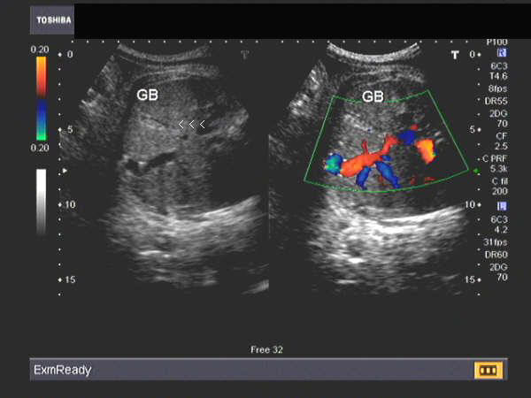
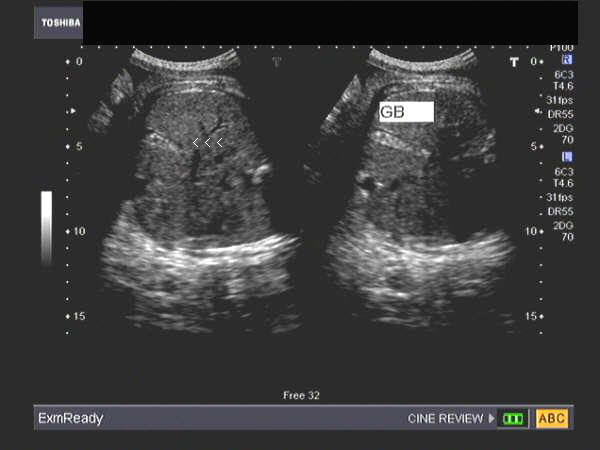
Echogenic particulate matter in the fetal Gallbladder
Sonography of this 3rd trimester fetus revealed echogenic debris within the gall bladder. These ultrasound images of the fetal gall bladder are diagnostic of biliary sludge. This sonographic finding does not appear to be of any clinical significance, as the sludge usually disappears after birth.Ultrasound images of fetal gall bladder sludge are courtesy of Dr. Jaydeep Gandhi, Mumbai, India.
Double bubble sign of duodenal obstruction
Download my E book for Amazon Kindle (download free Kindle reader for i Phone or Android)
Assorted ultrasound cases- by Joe Antony, MD
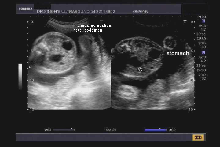
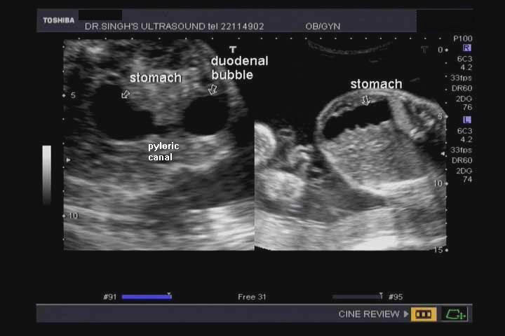
Sonography of the fetal abdomen (see pictures above), show 2 anechoic lesions (double bubble sign). The first bubble is formed by the fluid filled fetal stomach, the second being formed by the distended fetal duodenum. The obstruction is likely to be at the 2nd part of the duodenum. These ultrasound images (double bubble sign) suggest the likelihood of duodenal atresia. This sign may also be seen in duodenal stenosis or annular pancreas. These ultrasound images were taken using a Toshiba Nemio 30 ultrasound system, by Dr. Prasenjeet Singh, Delhi, India.
Reference: http://radiology.rsnajnls.org/cgi/reprint/220/2/463.pdf (free article and images).
3D-ultrasound-double-bubble-sign
Case-2:
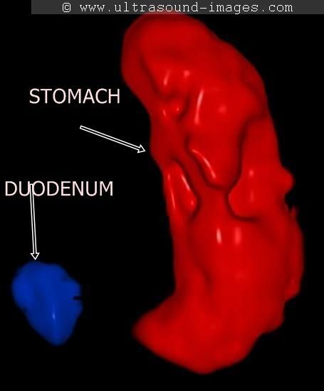
This 3D ultrasound image with color coding shows the fetal stomach and duodenum seen showing the double bubble sign. The duodenum is the smaller bubble to the left of the image (in blue) whilst the stomach bubble is seen color coded in red (to the right of the image). This is the classic double bubble sign of duodenal obstruction (this 3D image is courtesy of Mayank Chowdhury, MD).
Fetal ovarian cyst -fetal dermoid cyst vs. twisted ovarian cyst or torsion of ovarian cyst in fetus
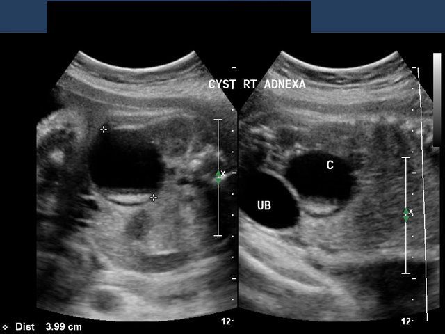
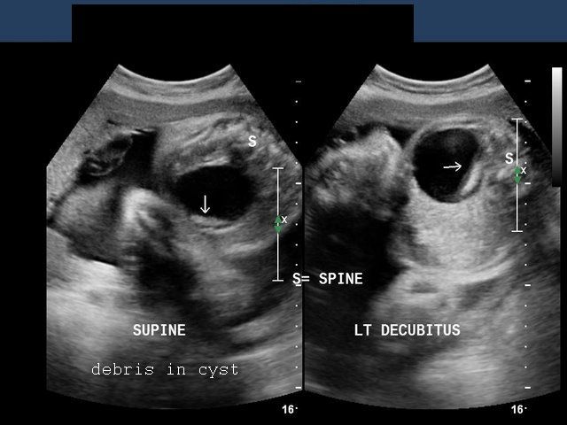

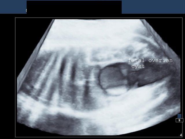
These ultrasound images of a 32 week old female fetus show a relatively large (3.9 cms.) cystic lesion in the right adnexal region, in close relation to the urinary bladder (UB). The cyst shows anechoic fluid content with echogenic debris that sediments towards the dependent part of the cyst (note the changes with change in posture). This type of appearance in a fetus is typical of ovarian cyst with solid debris which may be blood as in twisted ovarian cyst (torsion of ovarian cyst in fetus) or a fetal dermoid cyst of the ovary. The last ultrasound image (lower right) shows a 3-D image of the cystic lesion. All 4 ultrasound images are courtesy of Ravi Kadasne, MD, UAE. He used a Philips IU 22 ultrasound system for these images.
Reference: http://www.thefetus.net/page.php?id=524
Echogenic fetal bowel
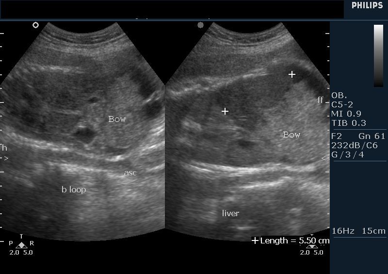
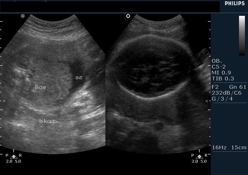
Hyperechoic fetal intestine
Sonography of the fetal abdomen done on this 28 week fetus showed markedly hyperechoic bowel and fetal ascites. The normal fetal intestine is isoechoic or hypoechoic with the fetal liver. (Bow = fetal bowel; as= fetal ascites). The bowel echogenicity is compared with that of the fetal iliac bone and graded from grade-1 (less echoic) to grade-3 (more echoic or hyperechoic than fetal iliac crest). Hyperechoic bowel in fetus is strongly associated with poor fetal prognosis- fetal growth retardation and anomalies and fetal death. This fetus did not survive. Ultrasound images courtesy of Dr. Vikas Shukla, MD, India.
Reference: http://radiology.rsna.org/content/188/2/527.abstract
Fetal dermoid cyst or mature cystic teratoma
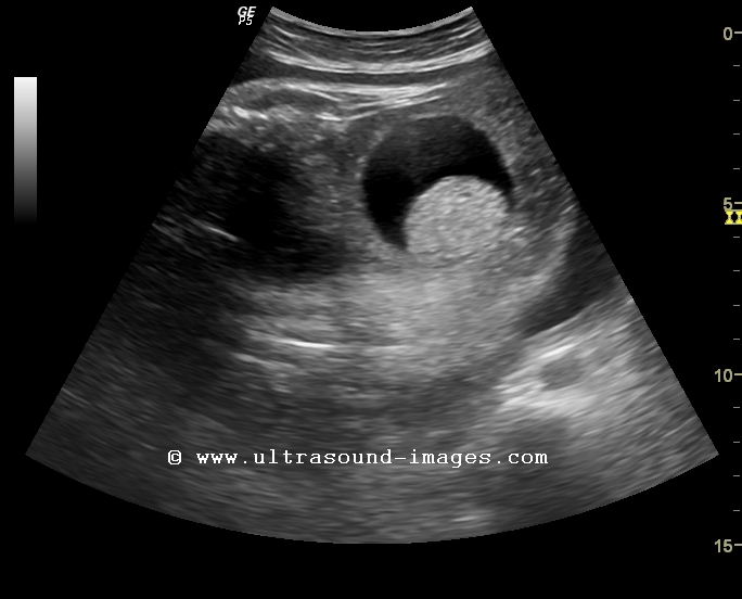
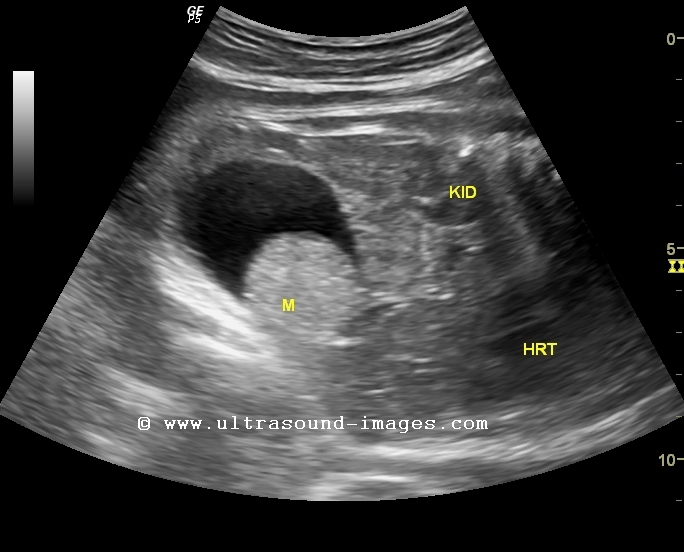
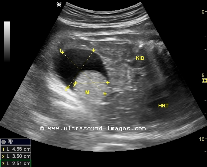


This 34 week fetus shows a large cystic lesion (4.5 cms.) arising from the pelvis with a solid, echogenic rounded mass within it (see ultrasound images above). This is the typical sonographic appearance of a sebum plug within a mature cystic teratoma or dermoid cyst. In ultrasound images above, ST= stomach, LK= left kidney, BL= fetal urinary bladder, M= mass.
For more on this topic visit:
Ultrasound imaging of fetal mature cystic teratoma or dermoid cyst
dilatated fetal bowel
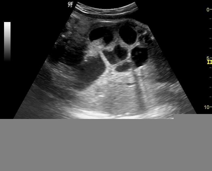
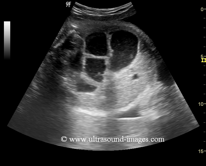
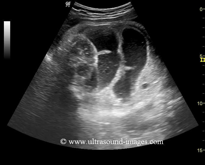
This 34 week old fetus shows dilated loops of small intestine with evidence of peristaltic waves. The diameter of the loops of small intestine is about 24 mm. Any diameter more than 7 mm is considered abnormal and suggests possible dilatation of the small intestine. The common causes of dilatation of the small intestine are meconium ileus, ileal atresia and jejunal atresia, volvulus etc. It can be difficult to distinguish between these various entities. However, the ultrasound images above, show absence of ascites which is usually present in cases of meconium ileus. The most common presentation seen with multiple loops of dilated small intestine as in the ultrasound images above is due to jejunal atresia.
References: http://www.jultrasoundmed.org/content/25/3/337.full.pdf
omphalocele
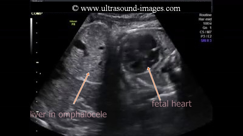
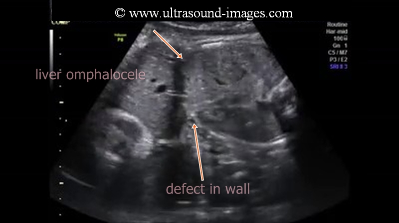
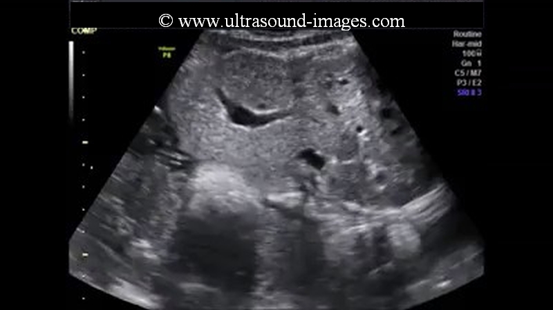
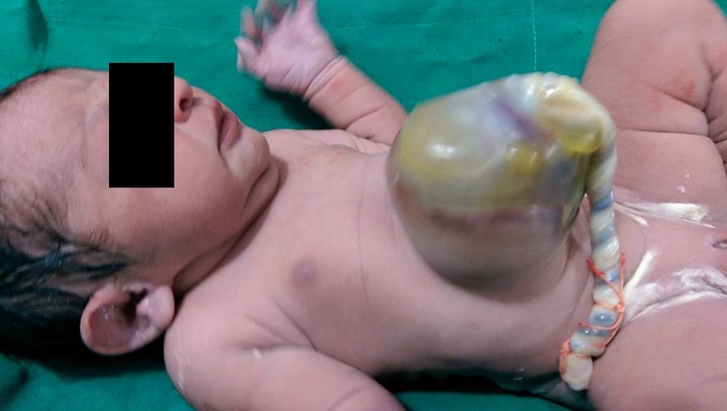
Omphalocele in fetus is characterised by the herniation of the liver and viscera including Fetal bowel into the amniotic cavity with the herniated structures enclosed in two layers of membrane namely the peritoneum and the amniotic membrane. The fetal abdomen shows a large defect in the midline of the abdomen with the umbilical cord inserted into the herniated sac. The presence of the umbilical vein in the herniated liver is usually diagnostic of omphalocele.
the chief differential diagnosis for omphalocele on ultrasound imaging include:
1. Gastroschisis: gastroschisis is characterised by a herniation of bowel and other visceral structures through the abdominal defect but lacking in a membranous covering of the herniated structures. Here, the fetal bowel is seen freely floating in the amniotic cavity and fluid.
2. Physiological fetal herniation of bowel ( this is seen at the umbilicus and is normal if the herniation does not persist beyond 12 weeks of gestation).
3. another important differential diagnosis is limb body wall complex in which there is a large defect in the abdominal wall usually on one side with associated severe anomalies of the other parts of the fetus.
4. Cloacal extrophy is also a less important differential diagnosis for omphalocele.
In the ultrasound images of omphalocele above, (courtesy of Firoz Bhuvar, MD), the fetal abdomen is seen showing the classic defect in the abdominal wall with herniation of liver into a membrane covering it and with the cord inserted into the convexity of the hernial sac. The ultrasound and color Doppler video below also shows a panoramic view of the omphalocele in this fetus.
The neonate after delivery can be seen with the large omphalocele sac with the umbilical cord inserting into the sac.
References:
overview of how ultrasound imaging of omphalocele- Medscape reference
hepatosplenomegaly-with-hypoechoic-liver
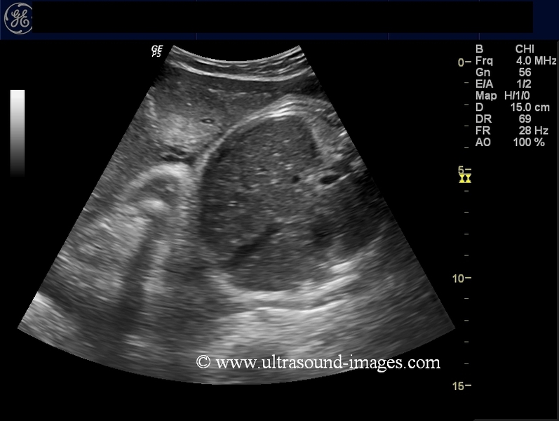
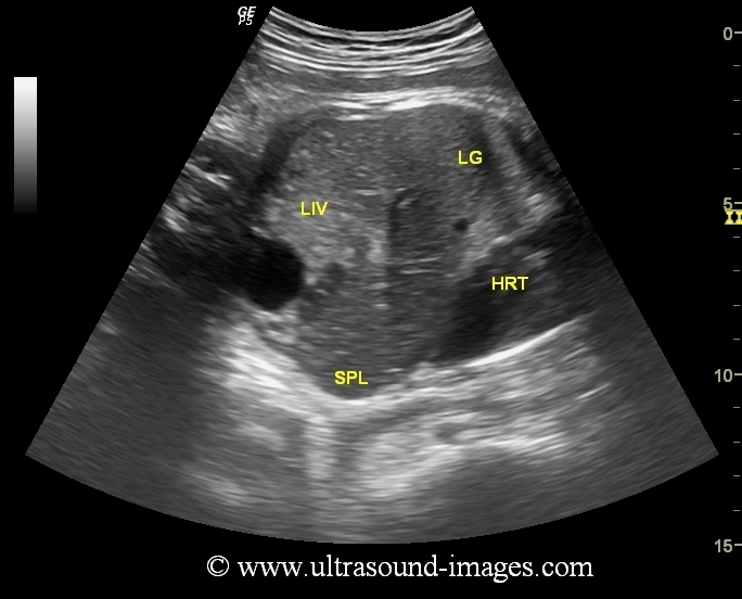
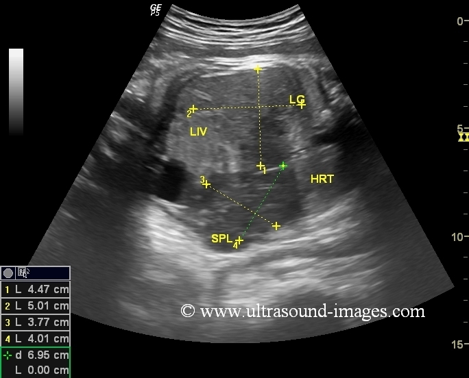

This third trimester fetus (34 weeks gestation age) shows hepatomegaly with moderate splenomegaly (see ultrasound images above). What is interesting is that the fetal liver is markedly hypoechoic. Further, color and spectral Doppler of the fetal middle cerebral artery and umbilical artery show no major abnormality. Moderate to severe hepatosplenomegaly with hypoechoic fetal liver as in this case suggests one of the following differential diagnosis as the cause of this condition: these include 1) TAM ( transient abnormal myelopoiesis), 2) fetal anaemia 3) TORCH infections. TAM or transient abnormal myelopoiesis is a Fetal disease characterised by a transient leukaemia like condition in the fetus and newborn. TAM is seen in babies with T21 chromosome and or down syndrome, and it is usually self-limiting with complete remission of the illness if the neonate survives till 4 to 8 weeks after birth. The alternate possibility of fetal anaemia appears less likely due to the normal middle cerebral artery Doppler findings.
The echogenicity of the Fetal liver can be compared to that of the adjacent right lung which shows its hypoechoic nature. Further, the ultrasound images above show prominent portal venous vasculature giving the liver a starry night appearance which is typically seen when the liver becomes hypoechoic.
However, TORCH infections cannot be ruled out as the cause for this gross hepatosplenomegaly in the fetus.
References:
1) differential diagnosis in Fetal hepatosplenomegaly- TAM
3) Causes of fetal hepatosplenomegaly
echogenic-fetal-bowel-case-2
This is yet another 2nd trimester fetus with the fetal bowel appearing to be hyperechoic as compared to the fetal iliac bone. As mentioned earlier on this page fetal bowel echogencity is compared to that of the adjacent fetal iliac crest to grade it from grade 1 to 3. Though this fetus did not show any other sign of aneuploidy, echogenic fetal bowel on ultrasound imaging is associated with poor prognosis in certain cases. In most fetuses echogenic fetal bowel is usually the result of ingestion of amniotic fluid containing blood usually the result of placental abruption or following amniocentesis. It may also be present in normal fetuses. Also known is the association with Trisomy 21 or Down's syndrome, thalassemia, cystic fibrosis and congenital viral infections. These ultrasound images of echogenic fetal bowel are courtesy of Firoz Bhuvar, MD.
References:
Ultrasound imaging and significance of echogenic fetal bowel- medscape
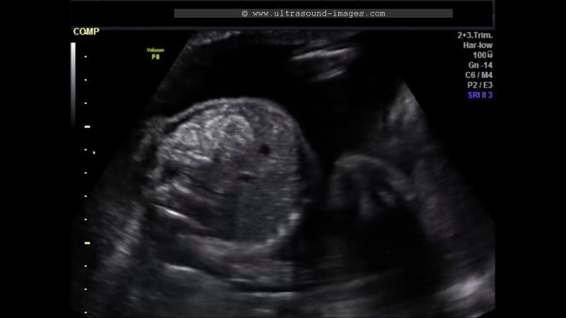
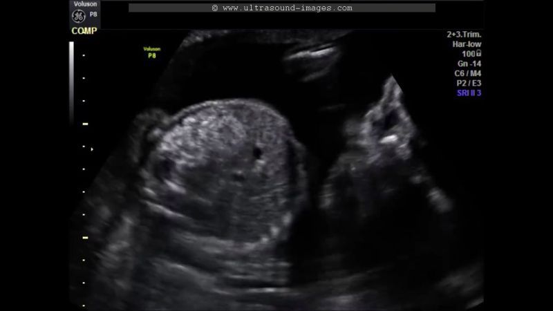
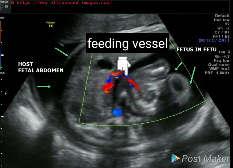
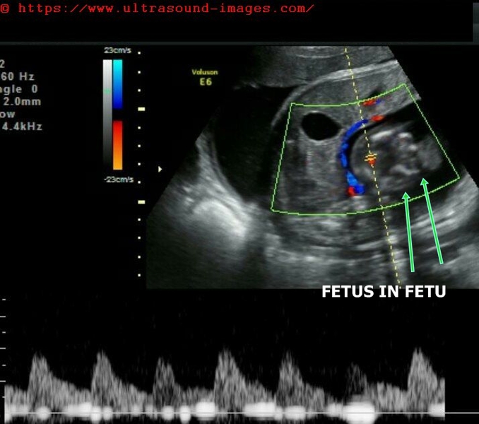
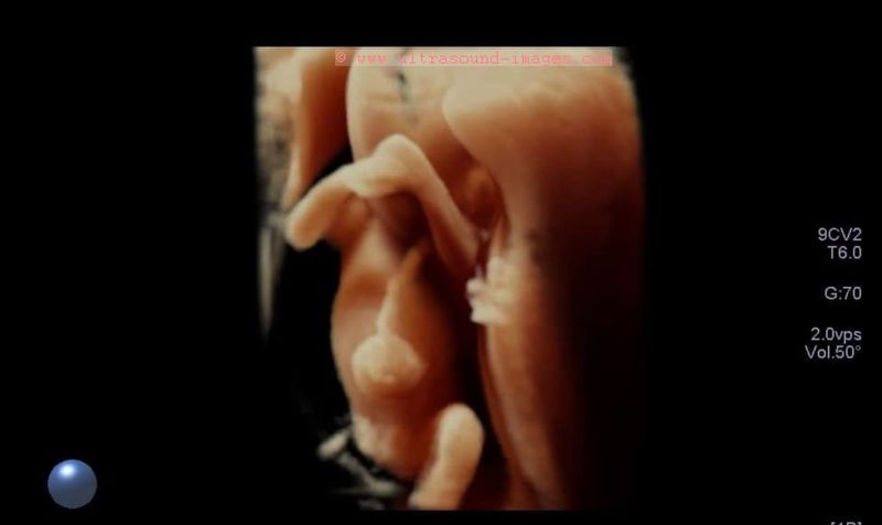
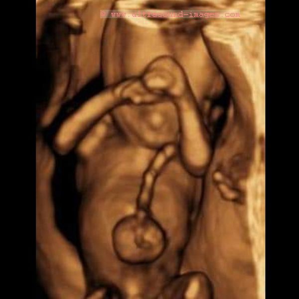
fetus-in-fetu
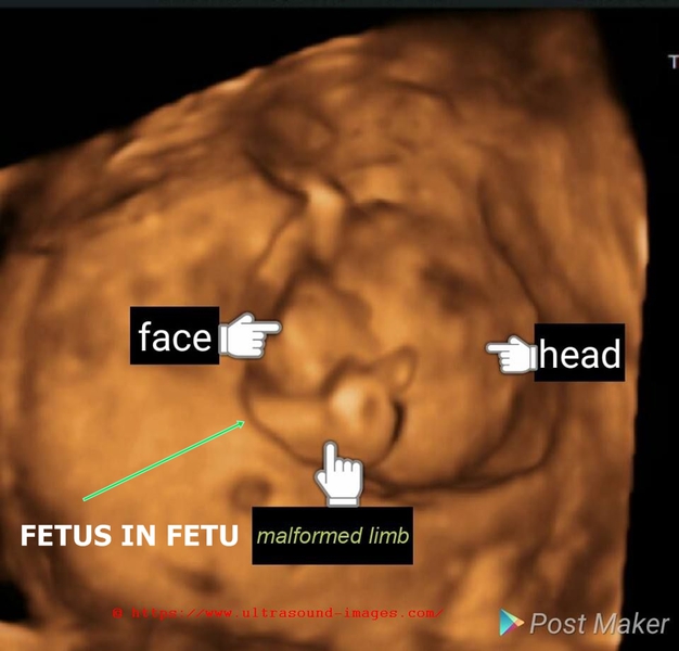
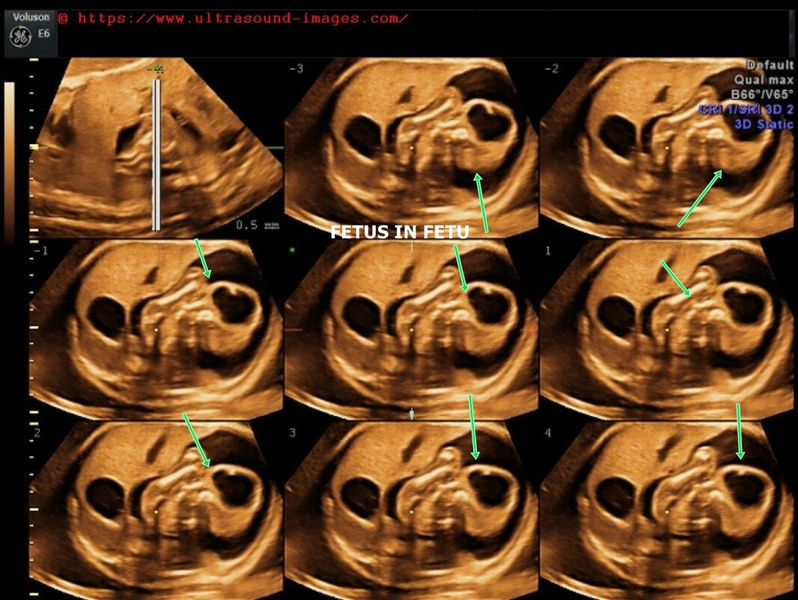

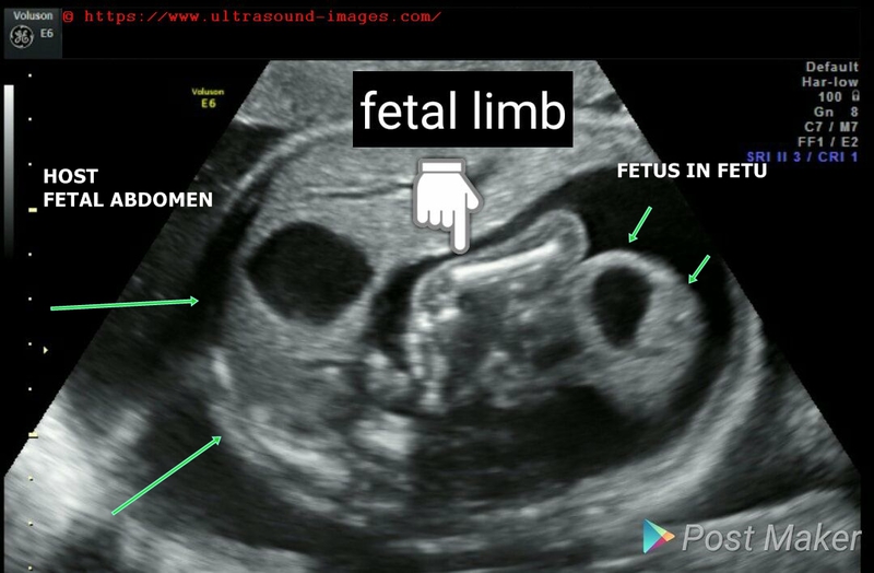

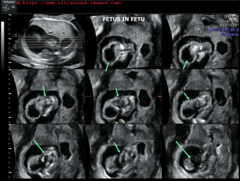
Fetus in fetu is a condition characterised by: (see 2D and 3D/ 4D ultrasound images above)
- partially developed parastitic fetal mass within host fetus, usually in the fetal abdomen (as in this case)
- anencephaly or hydranencepahly (as in this case) of the parasitic fetus or fetus in fetu
- first described in 1808 by George Young
- in this case fetus in fetu shows head parts and limb
- here feeder vessel seen supplying the parasitic fetus in fetu
- two theories about fetus in fetu- may be a monozygotic twin with host fetus wrapping itself around the fetus in fetu which usually then attaches itself inside the fetal abdomen of host fetal twin.
- 2nd theory- the fetus in fetu is a well developed teratodermoid tumor
- (2D and 3D/ 4D ultrasound images of fetus in fetu are courtesy of Dr. Rami Barakat, MD)
Omphalocele-3D-ultrasound-case-2
This is a 19 week fetus showing an omphalocele with probably bowel content.
3D ultrasound video and images of omphalocele (exomphalos) are courtesy of Dr. Vishal Kumat, MD.
A Toshiba/ Canon Aplio ultrasound system was used to capture these 3D images of exomphalos (omphalocele).
© 2026, Joe Antony, MD All images and content in this site are copyrighted. www.ultrasound-images.com When visiting this site, we may use cookies to improve the performance of this site. Use of this site means you accept the use of cookies. Read our privacy policy at this page: https://www.ultrasound-images.com/privacy-policy/