
Ultrasound images of Renal calculi
Contents of this page
- Renal calculus
- 3-D Ultrasound image of renal calculus
- Staghorn calculus
- Multiple renal calculi
- Case-2: Multiple, small renal calculi
- Nephrocalcinosis
- Anderson-Carr kidney
- 3D ultrasound image of calculus in renal pelvis
- 3-D ultrasound of multiple renal calculi
- nephrocalcinosis-Sjogrens-syndrome
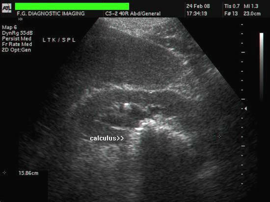
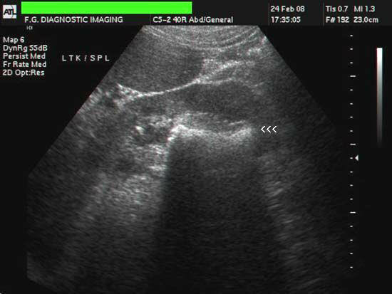
Renal calculus
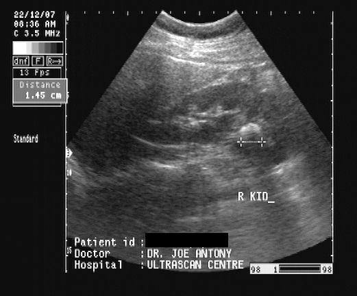
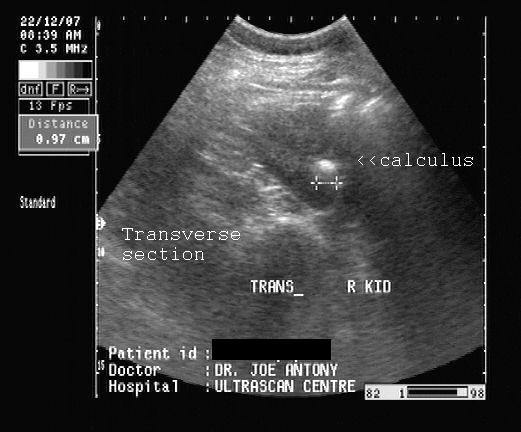
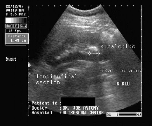
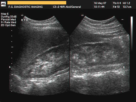
Kidney Stone
This patient had pain in the right lumbar region. Sonography of the kidneys reveals an markedly echogenic lesion in the lower calyx of right kidney. An acoustic shadow is seen posterior to the lesion. These ultrasound images are diagnostic of a renal calculus (measuring 15 x 10 mm. in size) in the lower calyx. Absence of pelvicalyceal dilatation suggests that there is no obstruction to urinary flow. It is always useful to scan the renal calculus and kidney in two planes to get an accurate measurement in two axis. Ultrasound images taken using Pie Scanner 100 Falco, by Dr. Joe Antony, MD, Cochin, India.
3-D Ultrasound image of renal calculus
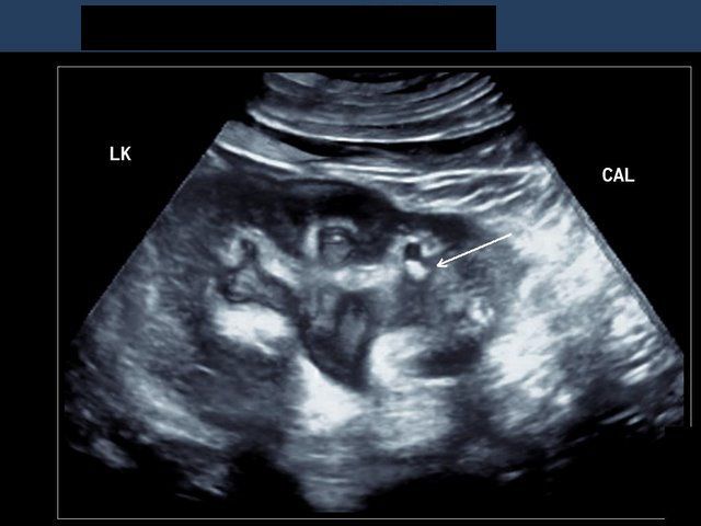
This 3-D (3 dimensional) ultrasound image of the left kidney shows a renal calculus in the lower calyx (arrow). 3D sonography has better ability to demonstrate calculi and rule out other causes of echogenic lesions in the kidney. This ultrasound image is courtesy of Dr. Ravi Kadasne, UAE. Picture taken using Philips iU 22 ultrasound system.
Staghorn calculus

Sonography of very large renal calculi
This huge calculus was discovered on ultrasound imaging of the left kidney. The calculus measuring almost 7 cms. is seen occupying the lower half of the left renal pelvis and the adjacent calyces. There is also mild dilatation of the renal pelvis due to urinary tract obstruction. Color doppler imaging shows the typical twinkling artifact near the calculus. These images suggest a staghorn calculus. Staghorn calculi are also called struvite calculi or triple phosphate calculi. Images courtesy of Shlomo Gobi, Israel. He used an ATL Philips HDI 3000 machine for these ultrasound images.
Reference: http://www.emedicine.com/MED/topic2834.htm(free article and images)
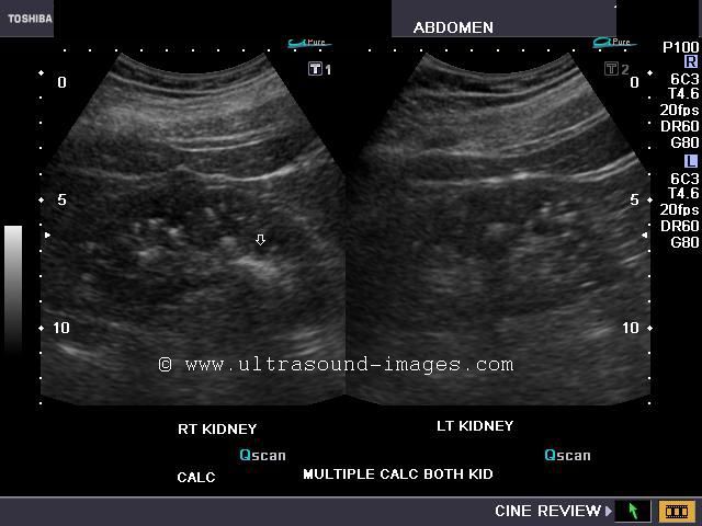
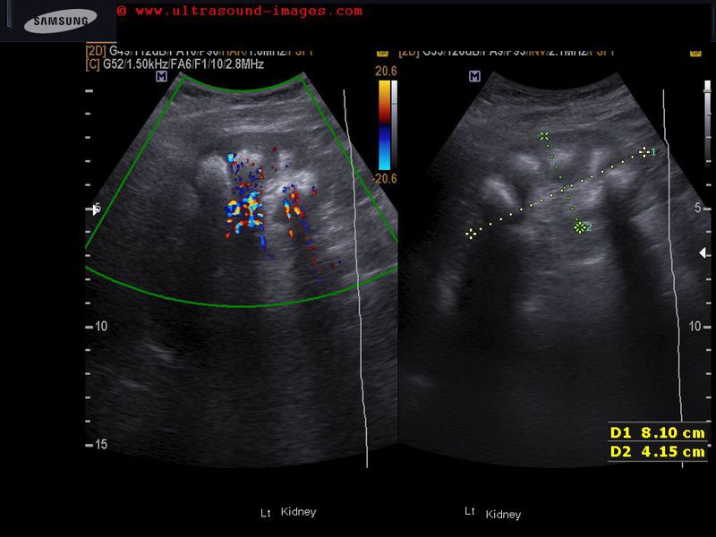
Multiple renal calculi


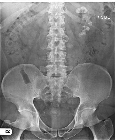
The above ultrasound images show multiple calculi in the left kidney. The calculi appear to be located in the renal calyces. X-ray of the abdomen shows the calculi in the left renal region, as radio-opacities. Sonography shows no pelvicalyceal dilation or hydronephrosis despite extensive urolithiasis or nephrolithiasis (stone formation) in left kidney. It is often not possible to be sure (on ultrasound) if these are separate calculi or part of a staghorn calculus. However, here the X-ray image confirms absence of staghorn calculus. Images courtesy of Gunjan Puri, MD, India.
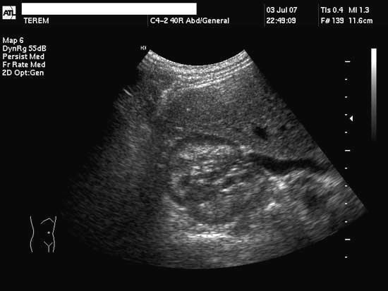
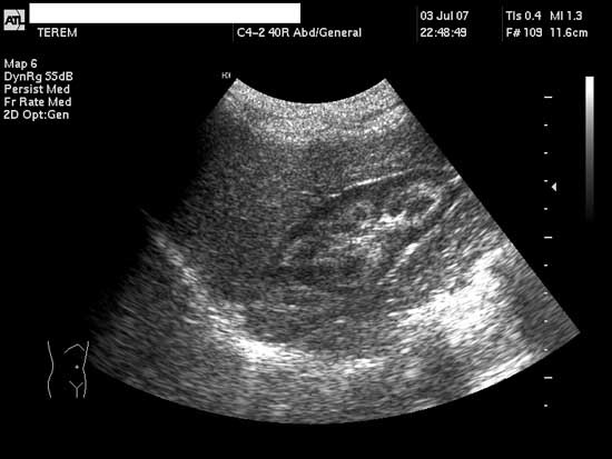
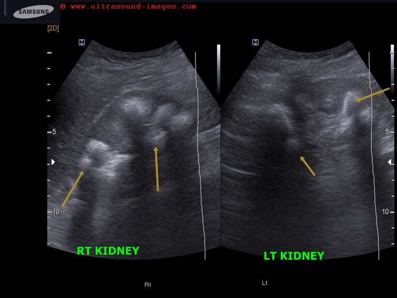
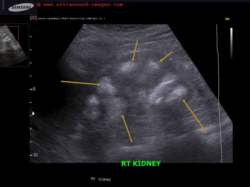
Case-2: Multiple, small renal calculi
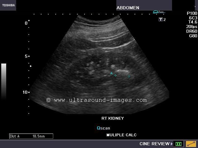
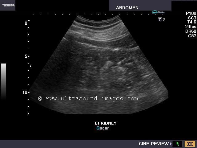
This elderly male patient has a history of stone disease. But the most striking feature of the ultrasonography of the kidneys in this patient (see ultrasound images above), is not the solitary large calculus (19 mm.) in the right kidney, but the multiple small stones distributed throughout the calyces of both kidneys. These numerous kidney stones measured from 2 to 4 mm. in size and number more than 10 per kidney. Such appearances can mimic the renal conditions like nephrocalcinosis and medullary sponge kidneys. But medullary sponge kidneys usually produces a bunch of grapes appearance for the calculi. This was not seen here.
References:
article on sonography of renal calculi(free article and images)
http://www.medicinenet.com/kidney_stone/article.htm(for the patient)
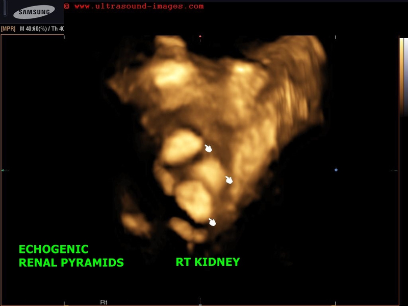
Nephrocalcinosis

Sonography of the kidneys in this patient reveal markedly echogenic (hyperechoic) renal pyramids with the central parts also affected. Ultrasound images also reveal renal calculus formation. These images suggest presence of medullary nephrocalcinosis.
Anderson-Carr kidney

The above 3 ultrasound images reveal nephrocalcinosis with involvement of the rim of the pyramids, a variant called the Anderson-Carr kidneys. The central part of the pyramids is relatively hypoechoic. All images above courtesy of Shlomo Gobi, Israel. Machine used here is the ATL-Philips HDI 3000.
Reference:http://www.emedicine.com/Radio/topic470.htm (free article and images).
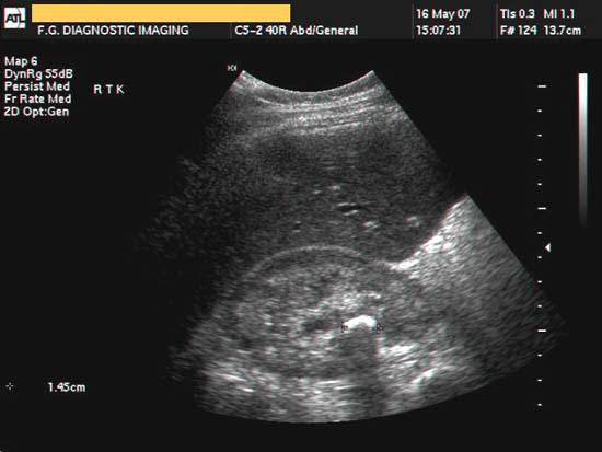
3D ultrasound image of calculus in renal pelvis
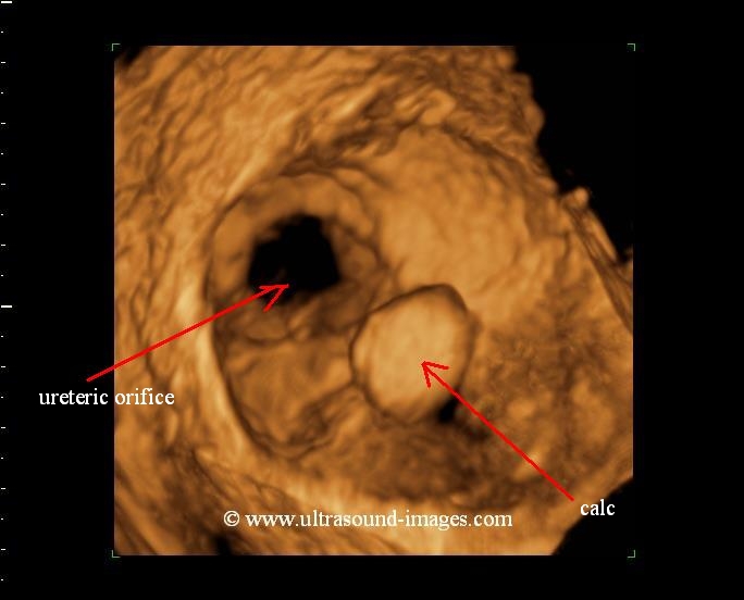
3D ultrasound imaging is a great tool to visualize renal calculi and their relation to surrounding structures. This 3D ultrasound image (courtesy of Dr. Sanjiv Bhalla, MD, India) reveals a spherical calculus within the renal pelvis. The pelvi-ureteric junction (UPJ or PUJ) is seen with the orifice of the ureter imaged here.
3-D ultrasound of multiple renal calculi
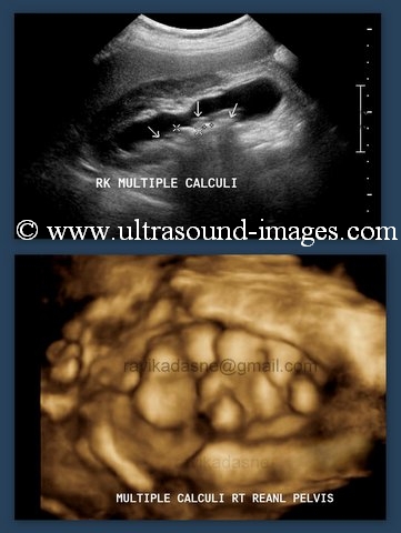
This is also another 3D ultrasound image of renal calculi- but in this case the right renal pelvis is distended and filled with multiple calculi. Image is courtesy of Dr. Ravi Kadasne, MD. Observe how the depth visualization helps show true dimensions of the renal calculi. The most imminent danger is that of one or more of these renal pelvic calculi slipping into the right ureter and causing partial or complete urinary obstruction.
nephrocalcinosis-Sjogrens-syndrome
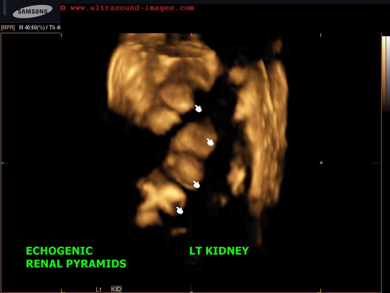
This middle aged lady is a known case of Sjogrens syndrome. Ultrasound images show multiple dense echogenic foci within the renal pyramids on both sides s/o nephrocalcinosis.
In this case nephrocalcinosis is the result of long standing renal tubular acidosis.
Nephrocalcinosis is the process of deposition of calcium salts within the renal pyramids.
Due to dense calcium deposition in this case there is also acoustic shadowing posterior to the kidneys.
Also shown are 3D/ 4D ultrasound images of nephrocalcinosis. The calcific pyramids are well displayed showing typical changes of nephrocalcinosis on 3D ultrasound.
References: Sjogrens syndrome and nephrocalcinosis
© 2026, Joe Antony, MD All images and content in this site are copyrighted. www.ultrasound-images.com When visiting this site, we may use cookies to improve the performance of this site. Use of this site means you accept the use of cookies. Read our privacy policy at this page: https://www.ultrasound-images.com/privacy-policy/