
Sonography of kidneys-2
Contents of this page:
Lupus-nephritis

Download my E book for Amazon Kindle (download free Kindle reader for i Phone or Android)
Assorted ultrasound cases- by Joe Antony, MD
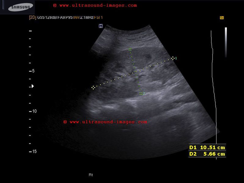
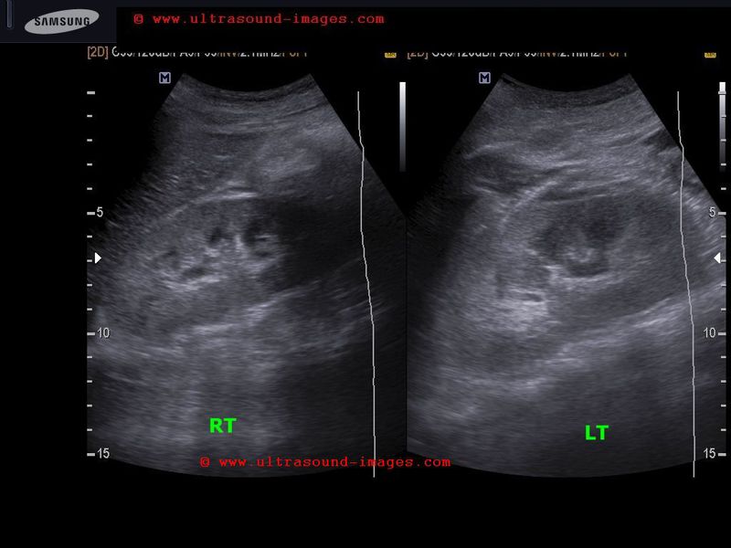
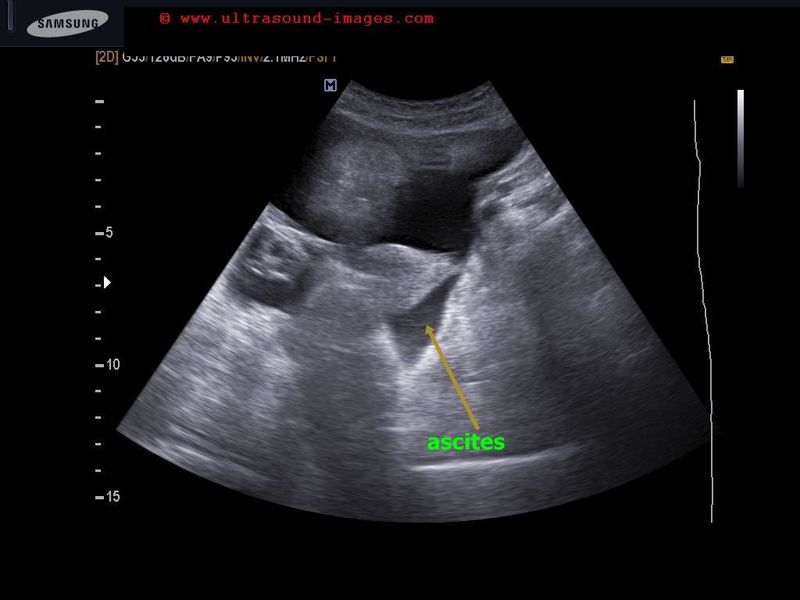

-This 15 yr old female patient showed mild pleural effusion and ascites on ultrasound imaging of the abdomen. Other ultrasound findings (see images above):
-kidneys appear enlarged and edematous with loss of cortico-medullary differentiation
-increased echogenicity of the renal cortex.
-also present: abuminuria and serum creatinine of 1.4 mg %
On renal biopsy it was diagnosed as membranous lupus nephritis or SLE (Systemic lupus erythematosus) involving the kidneys. Lupus nephritis is graded from 1 to 6 depending on clinical and pathological findings.
In chronic lupus nephritis, the kidneys may show typical songraphic features of medical renal disease with small echogenic kidneys.
References: sonography of lupus nephritis
perinephric-fluid-abscess
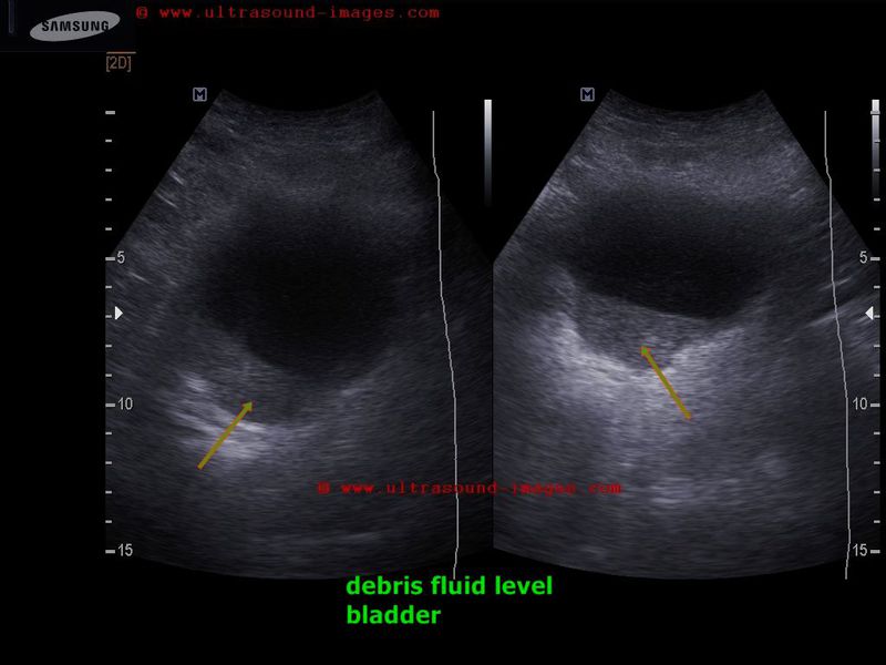
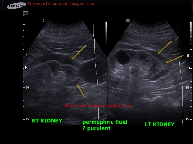
Elderly male patient with severe UTI (urinary tract infection).
Here ultrasound scan shows sediment containing particulate matter in urinary bladder.
Also a perinephric hypoechoic collection around the lower half of both kidneys.
This suggests bilateral perinephric abscess.
D/d -urinoma or extravasated urine
-hemorrhagic fluid (perinephric hematoma)
-lymphatic fluid
Looking to the history of severe UTI and findings, the diagnosis of perinephric abscess is likely.
References: Perinephric fluid or abscess-ultrasound imaging
dromedary-hump-left-kidney


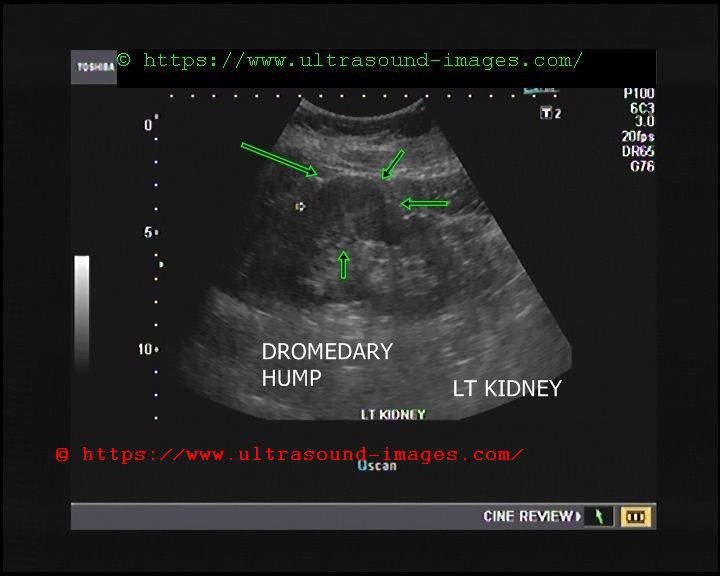
Dromedary hump of left kidney:
= a hump or lump bulging from the superior and lateral spect of the kidney
= always affects the left kidney and is due to the spleen compressing upon the left kidney
= name dromedary hump is derived from the hump seen in dromedary camels
= D/d: must be distinguished from renal masses. Here (dromedary humps) margins are well defined and show normal renal vessels on color Doppler ultrasound imaging
= clinically not significant; follow up will show no change in characteristics on ultrasound/ sonography
renal-cortical-cyst-Bosniak-type-2F
© 2026, Joe Antony, MD All images and content in this site are copyrighted. www.ultrasound-images.com When visiting this site, we may use cookies to improve the performance of this site. Use of this site means you accept the use of cookies. Read our privacy policy at this page: https://www.ultrasound-images.com/privacy-policy/