
Ovaries
Contents of this page
- Functional cysts of the ovary
- Hemorrhagic ovarian cysts
- Ovarian dermoid cyst or Cystic teratomas
- Case-2: Mature cystic teratoma or dermoid cyst of left ovary
- Endometrioma or Endometriotic cyst of ovary or Chocolate cyst of the ovary
- Rupture of hemorrhagic ovarian cyst
- Ovarian hyperstimulation syndrome (OHSS)
- Polycystic ovaries (polycystic ovary disease- PCOD/ polycystic ovarian syndrome)
- torsion of ovary or ovarian torsion
- peritoneal inclusion cyst
- cumulus-oophorus
Functional cysts of the ovary
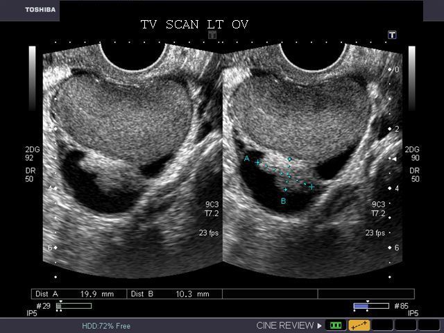
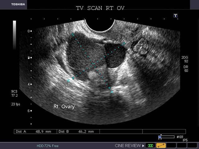
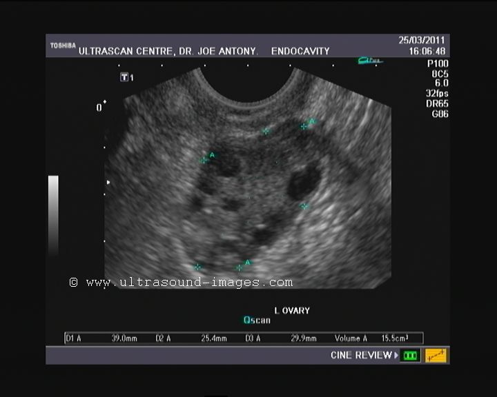
A) Follicular cysts
This young female patient underwent sonography for non-specific pain in the lower abdomen. Ultrasound images of the pelvis show bilateral ovarian cysts which show absence of internal nodules, septae or debris. These findings are typical of follicular cysts of the ovaries. Follicular cysts are functional cysts and are enlarged ovarian follicles that have not ruptured (ovulated). They are usually unilateral. These ultrasound images were taken using a Toshiba Nemio-XG ultrasound system.
Reference:
1) http://emedicine.medscape.com/article/255865-overview
2) http://drjoea.googlepages.com/ovary
B) Corpus Luteal cysts
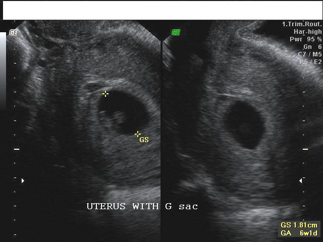
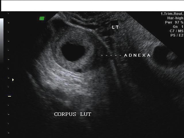
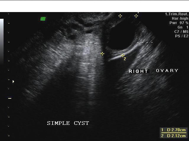
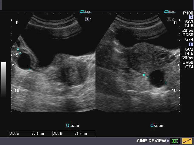
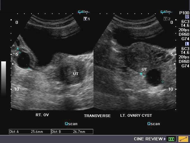
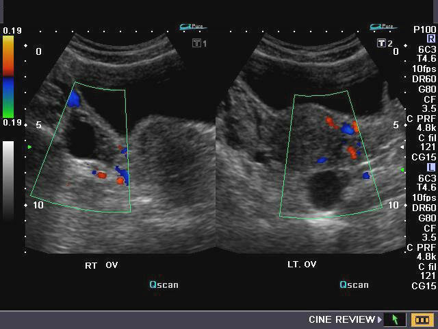
Uterus with gestation sac and embryo Lt. adnexal cystic mass- Luteal cyst (Lt. ovary) Rt. ovarian simple cyst
This patient had an early gestation with embryo and intrauterine gestation sac. Sonography of the adnexal regions also showed a cystic lesion of the right ovary, which was thin walled and showed no septae or nodules within it, suggestive of a simple cyst (functional) of the right ovary. However, the left adnexal region showed a thick walled cystic lesion with echogenic walls. This appearance can easily be due to an ectopic pregnancy. Both ectopic gestations and corpus luteal cysts show similar features including the presence of "ring of fire" or ring of vessels around the lesion (on Color Doppler imaging). The left ovary was not seen separate from the left adnexal cyst; also there was no evidence of significant fluid in the cul de sac; besides, the presence of intrauterine pregnancy lead to the diagnosis of a left ovarian Corpus Luteal cyst. Ultrasound images are courtesy of Dr. V. Ganesan, MD, India.
Reference:
http://emedicine.medscape.com/article/255865-diagnosis
http://www.drspock.com/article/0,1510,5335,00.html
http://www.emedicinehealth.com/ovarian_cysts/article_em.htm
Hemorrhagic ovarian cysts
Hemorrhagic cysts of the ovary are the result of bleeding from vascular tissues within the walls of the freshly formed corpus luteum or luteal cyst. The hemorrhagic cyst of ovary can present in various ways, mimicking a number of more ominous diseases or cysts of the ovary. These include ovarian cancers to serous or mucinous cystadenomas. Sometimes, rupture of the hemorrhagic ovarian cyst can result in a medical emergency (see below).
a) Hemorrhagic cyst of ovary resulting from Ovulation induction
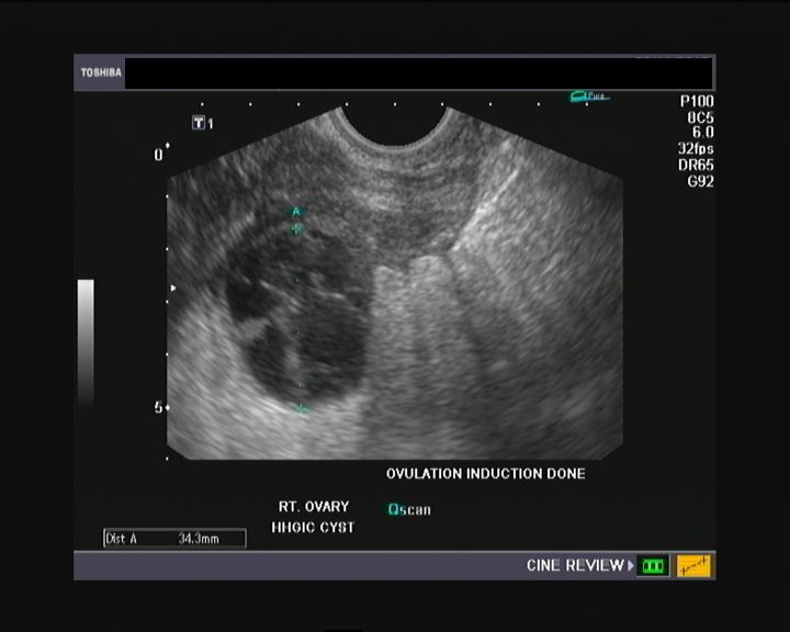
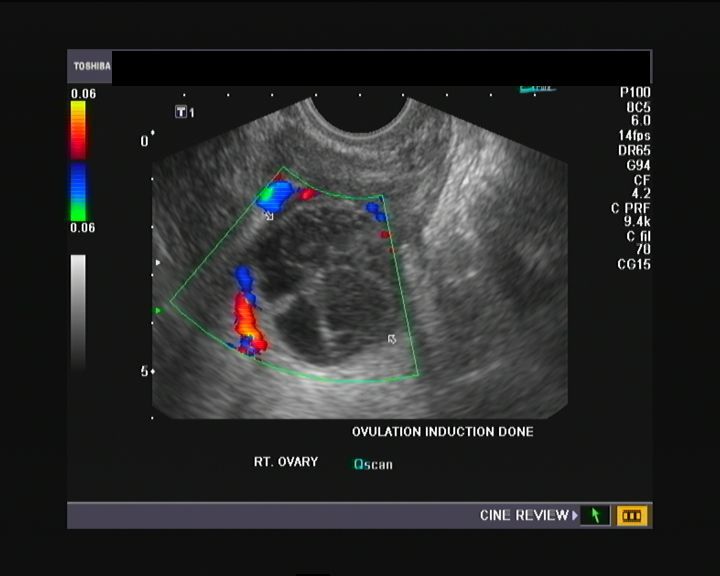
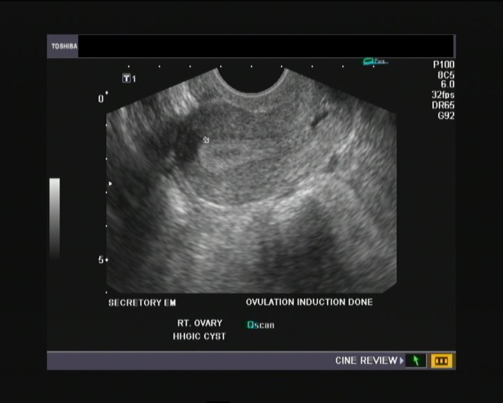
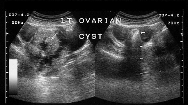
This young nulliparous female patient undwerwent ultrasonography following ovulation induction. The right ovary shows a typical hemorrhagic cyst formed from the corpus luteum. The first image (top row- left) is a transabdominal ultrasound image showing fine fibrinous strands within the cystic mass in the right ovary. Transvaginal ultrasound and color Doppler images confirm these findings. The uterus shows typical secretory changes in the endometrium suggesting post ovulatory phase.
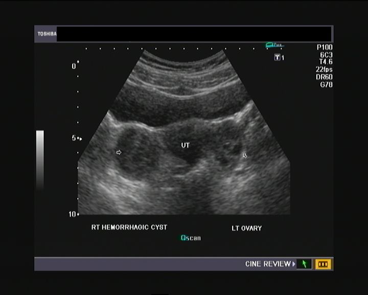
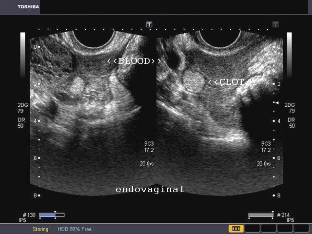
b) Hemorrhagic cyst of ovary with co-existing chocolate cyst/ endometrioma
This patient has a co-existing chocolate cyst with a hemorrhagic cyst in the same (right) ovary. The cyst on the left half of the ultrasound image is a hemorrhagic cyst. Note the fine fibrinous strands within the cyst suggesting clot formation. The cyst on the right half of the image is homogenous with fine echoes throughout the ovarian cyst. This is a typical appearance of an endometrioma (chocolate cyst). Ultrasound image is courtesy of Dr. Gunjan Puri, MD, India.
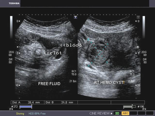
c) Hemorrhagic cyst of ovary with ruptured ectopic pregnancy

This female patient has a left ovarian hemorrhagic cyst (see ultrasound image above-left). In addition, there is a large collection of free fluid with particulate matter in the pelvis. The right fallopian tube is thickened with a ring shaped mass. This suggests that there is significant hemorrhage into the pelvis due to a ruptured ectopic pregnancy (right tubal ectopic gestation). The left ovarian hemorrhagic cyst appears intact, ruling out ruptured hemorrhagic cyst. Both above ultrasound images are courtesy of Gunjan Puri, MD, India.
References:
http://www.jultrasoundmed.org/content/21/8/879.full.pdf(free article on hemorrhagic cysts of ovary)
Ovarian dermoid cyst or Cystic teratomas
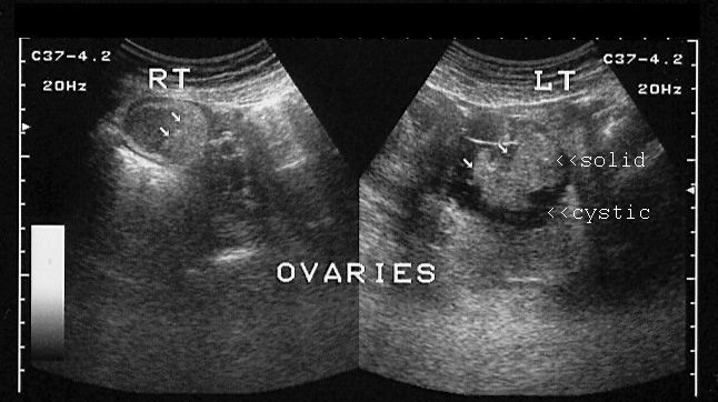
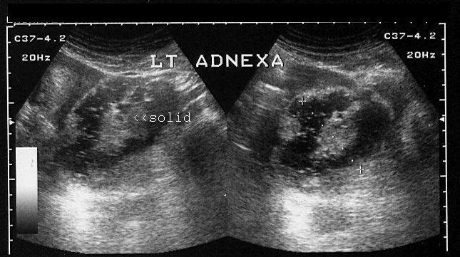
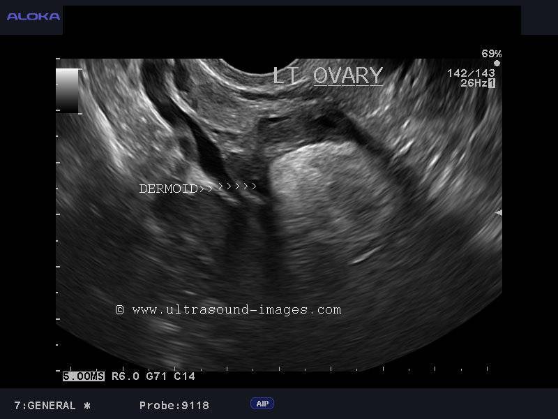
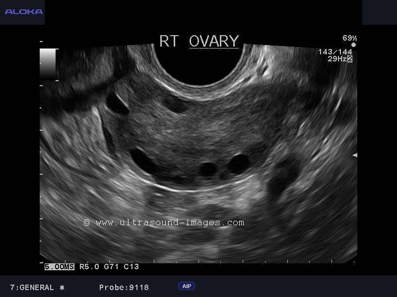
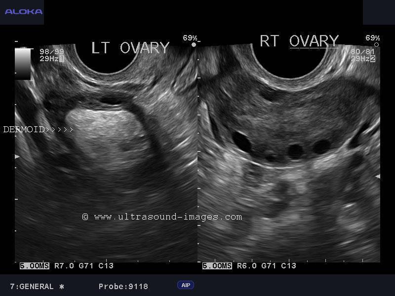
These ultrasound images reveal bilateral ovarian complex masses that contain both solid and cystic components. The right ovary shows a cystic mass with a solid, highly echogenic "dermoid plug". This is a solid nodule containing fat and various tissues including hair. Posterior acoustic shadowing is seen. The left ovary shows a dermoid plug and, in addition, a "dermoid mesh" is also seen, an irregular echogenic solid mass within the cyst. Echogenic debris is seen floating within the fluid. Images courtesy of Dr. Ravi Kadasne, UAE.
Case-2: Mature cystic teratoma or dermoid cyst of left ovary

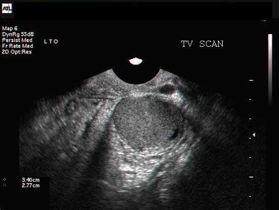
Left ovarian dermoid cyst: Echogenic mass in left ovary: Normal right ovary: Left ovarian dermoid:
These ultrasound images show a typical dermoid cyst of the left ovary. Transvaginal images of the left ovary show an echogenic mass of the left ovary with attenuation of the ultrasound beam posteriorly. The echogenic mass contains sebum and other fatty material giving rise to hyperechoic appearance of the lesion. The differential diagnosis in this case include- hemorrhagic cyst of the ovary with organized hematoma. Here the hematoma would show acoustic enhancement posterior to the lesion, rather than the attenuation (shadowing) seen in dermoid cyst. Bowel gas can also produce the appearance of an echogenic lesion in the ovary. However, the dirty shadowing seen in bowel gas and also peristalsis would help distinguish bowel gas. Images are courtesy of Dr. Jaydeep Gandhi, MD, India.
References: Ultrasound and radiology of ovarian masses- radiographics
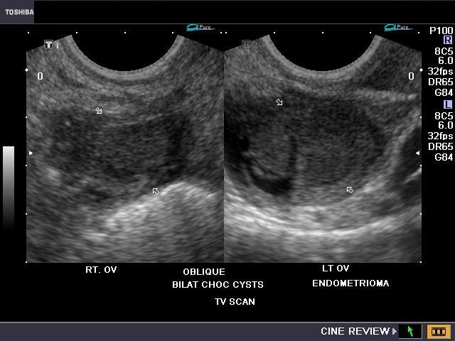
Endometrioma or Endometriotic cyst of ovary or Chocolate cyst of the ovary
case 1:
(Ultrasound images of chocolate cysts of ovary, courtesy of Gunjan Puri, MD, India)
case 3

(Ultrasound image of endometrioma by Joe Antony, MD, India).
Chocolate cysts or endometrioma of the ovary are caused by bleeding from ectopic endometrial tissue within the ovary. Some researchers believe that normal tissue within the ovaries might undergo changes which results in bleeding within the ovary. All the above ultrasound images (4 different cases) show small to large cystic lesions within the ovaries. The cysts show diffuse, low level echoes within them with poor flow on Doppler imaging. The main differential diagnosis in these cases is hemorrhagic cyst where fine strands are seen coursing within the cyst. Also note that endometriomas show homogenous appearance of the echogenic (altered blood) content within them. Chocolate cysts can be of varying sizes and cause considerable pain, especially during menses. Usually chocolate cysts show absence of septae or calcification and can be mistaken for solid masses. MR imaging can be helpful in clinching the diagnosis of this disease.
Reference:
http://emedicine.medscape.com/article/403435-media
Rupture of hemorrhagic ovarian cyst
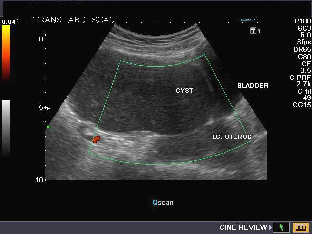
Transabdominal scan images
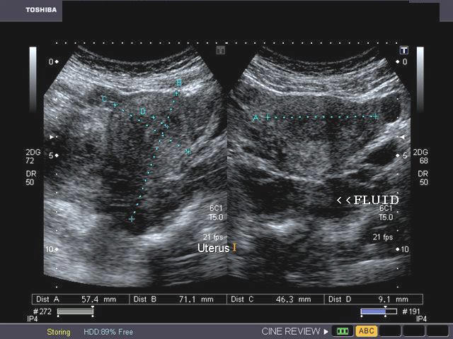
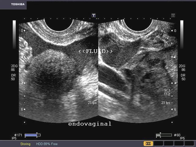
This female patient presented with pain in the pelvis and tenderness. Transabdominal sonography of the pelvis showed a large amount of free fluid with particulate matter in the cul de sac and around the right adnexal region. The right ovary showed remnants of what appeared to be a ruptured hemorrhagic corpus luteal cyst. Endovaginal imaging showed large amount of fluid with debris posterior to the uterus. The left ovary appeared normal. Lab tests and patient history excluded the possiblity of ectopic pregnancy. It was hence concluded that this was a ruptured right hemorrhagic ovarian cyst with free blood in the pelvis. The rounded echogenic mass seen near the fluid appears to be an organized blood clot. Ultrasound images are courtesy of Gunjan Puri, MD, India.
Reference:
1) http://www.jultrasoundmed.org/cgi/reprint/21/8/879 (free article and images)
2) http://www.ajronline.org/cgi/reprint/173/5/1301.pdf (free article and images)
Ovarian hyperstimulation syndrome (OHSS)
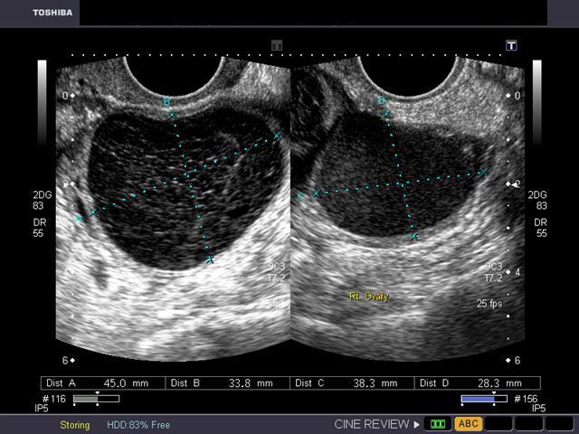
Case-1
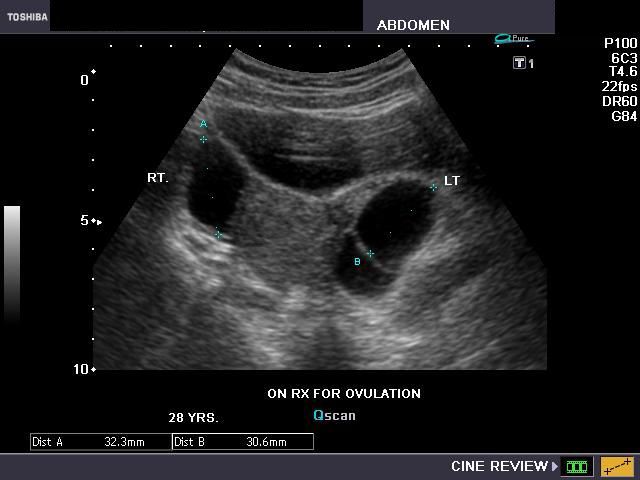

This young adult female patient was examined to evaluate the uterus and ovaries. She was under treatment for infertility and was using gonadotropins. Ultrasound images of the ovaries show grossly enlarged ovaries with large cysts (measuring 2.6 to 3 cms.) in both ovaries. These ultrasound findings are diagnostic of OHSS or ovarian hyperstimulation syndrome.
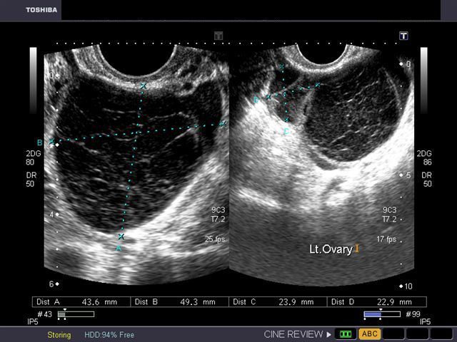
Case-2
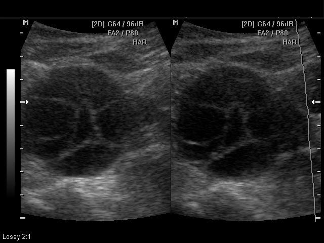

The above 2 ultrasound images again show hyperstimulated ovaries. Both ovaries are grossly enlarged and cystic. (Images are courtesy of Dr. Arun Mahajan, MD, India)
Case-3
The above images are courtesy of Dr. Dilraj Gandhi, MD, Delhi, India. Grades of OHSS: There are 3 grades of ovarian hyperstimulation based on sonography and clinical features: 1) mild OHSS- ovaries are less than 5 cms. in diameter and patient has mild abdominal dsicomfort. 2) moderate OHSS- ovaries measure 5 to 10 cms. in diameter 3) severe OHSS- ovaries measure greater than 10 cms. in diameter.
References:
http://radiopaedia.org/articles/ovarian-hyperstimulation-syndrome-1(free images/ radiology of OHSS)
http://emedicine.medscape.com/article/1343572-overview (free article and grades of OHSS)
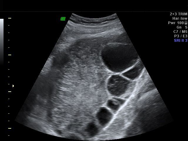
Case-4:Transvaginal ultrasound images of ovarian hyperstimulation syndrome
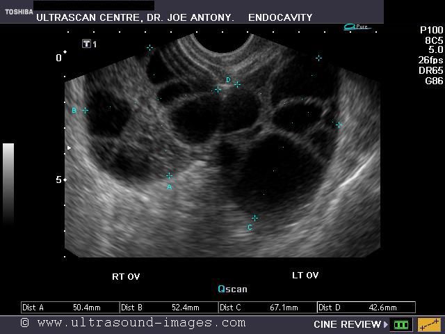
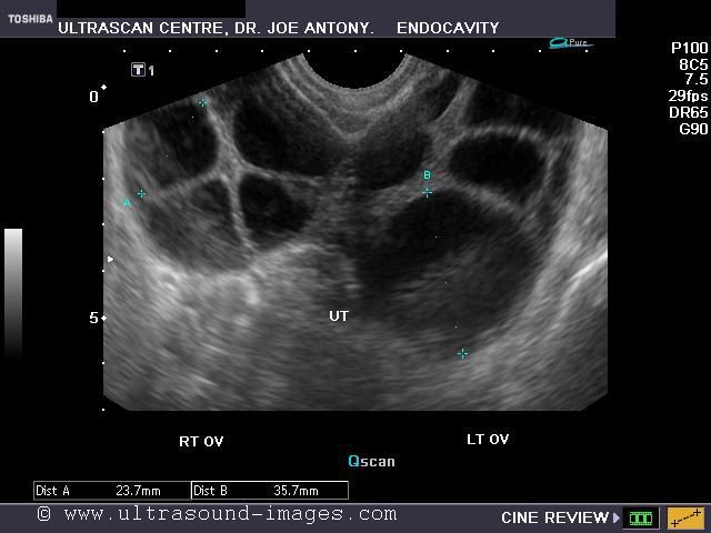
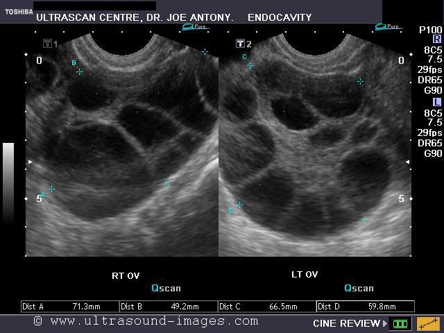
This young adult female patient showed multiple large theca lutein cysts of both ovaries, arranged in spoke-wheel pattern (ultrasound images above) which were the result of use of gonadotropins in the management of infertility. The cysts vary in size from 2 to 4 cms. with the ovaries massively enlarged (each ovary measures up to 7 cms. in size). This can be classified as grade-2 hyperstimulation of the ovaries (ovarian diameter from 5 to 10 cms.). There is not evidence of ascites. The color Doppler image of the ovaries shows vessels passing along the margins of the cysts. One of the complications of such enlarged ovaries in OHSS is torsion and in certain cases rupture of the ovaries, both of which are medical emergencies. Ovarian hyperstimulation syndrome is known to occur more frequently in patients of pre-existing Polycystic ovaries (PCO).
References: Ultrasonography in obstetrics and gynecology- Peter Callen.
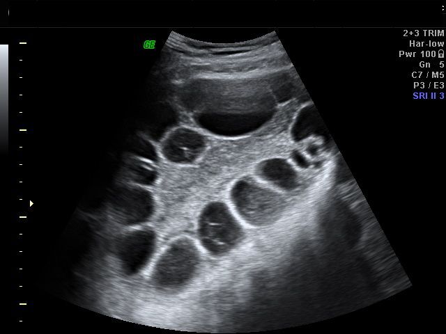
Polycystic ovaries (polycystic ovary disease- PCOD/ polycystic ovarian syndrome)
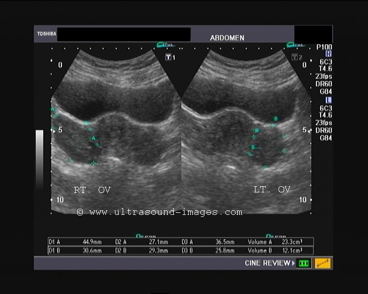
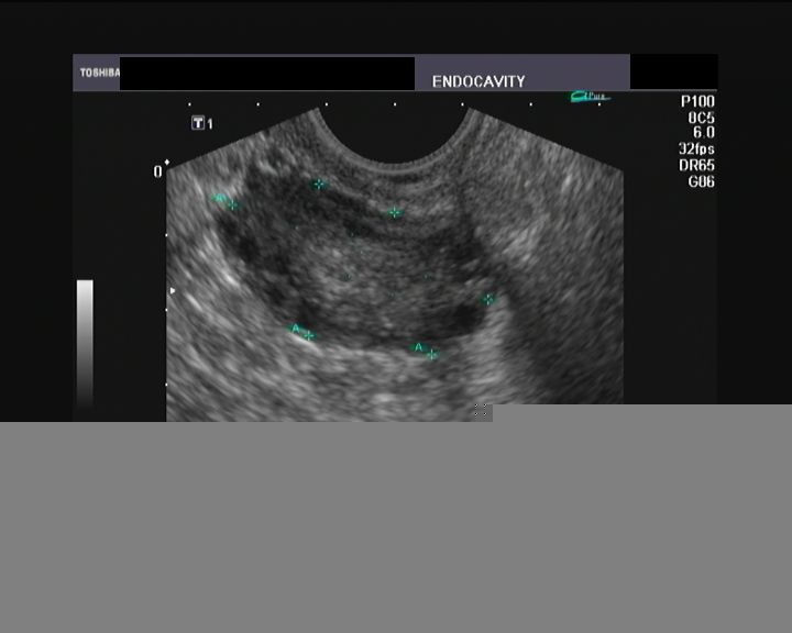
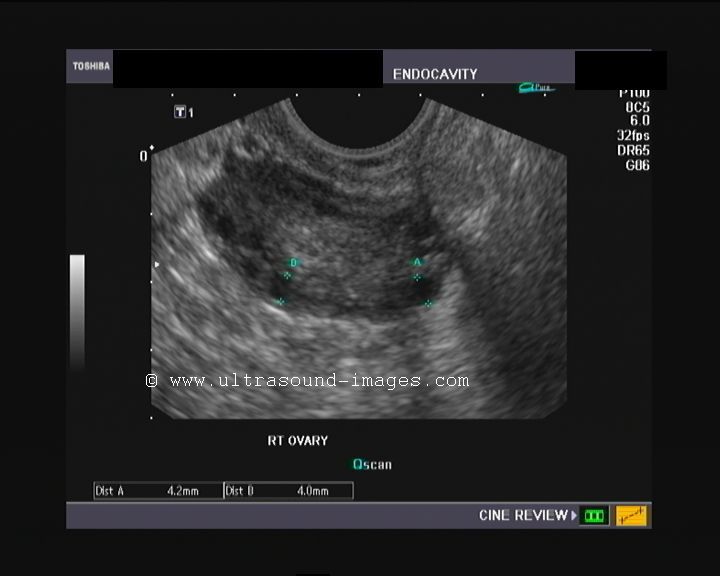
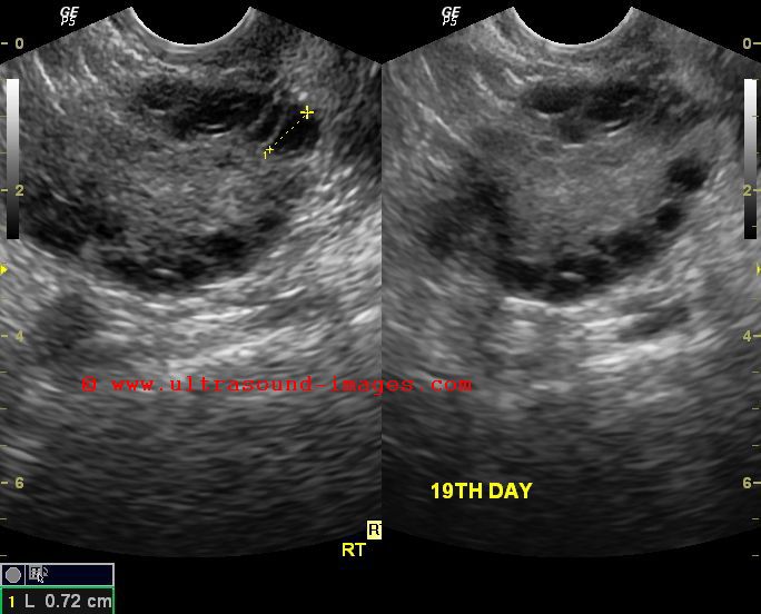

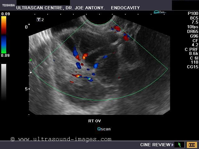
This middle aged lady has a history of irregular menses. Transabdominal ultrasound shows (see top row-left), enlarged ovaries on both sides. However, no definite follicles were visualized. The uterus was normal in size. The transvaginal ultrasound images show surprising details of the affected ovaries. The sonographic findings include: a) Enlarged ovaries- the volume of these ovaries ranged from 12 to 15 cc. This is due to stromal proliferation. b) The ovarian stroma (parenchyma) appears echogenic. c) Multiple follicles of small size are seen along the rim of the ovaries. d) The ovarian follicles are less than 10 mm. in size (each of the follicles averaged at 4 to 5 mm. in size). e) There are more than 10 follicles per ovary (here we could count at least 12 to 15 follicles per ovary). The arrangement of the follicles along the rim of the ovary is called a necklace sign and is diagnostic of PCOD or polycystic ovary disease. Usually, clinical findings associated with PCOD include- hirsuitism, obesity, irregular menses and infertility. These conditions associated with polycystic ovaries are together labelled PCOS (polycystic ovarian syndrome).
Lower row middle and right images shows yet 2 other patients with PCOD. Multiple small follicles seen in both cases.
References: Ultrasound videos of polycystic ovary disease
torsion of ovary or ovarian torsion
case-1:

Case-2:

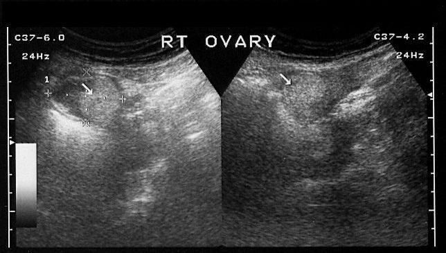
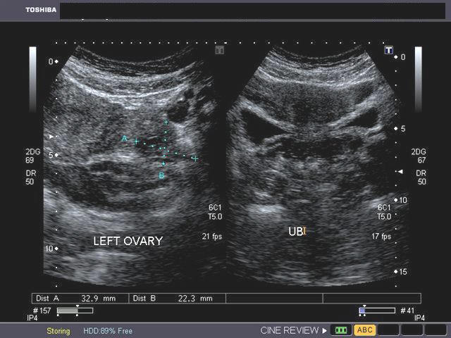
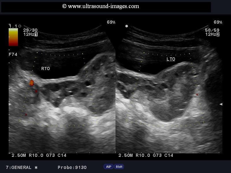
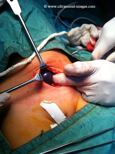
Case-1 (top):This young girl (6 years age) had severe left pelvic pain. Ultrasound images show an enlarged left ovary with lack of significant blood flow on power Doppler imaging. The right ovary appeared normal. These ultrasound images suggest torsion of the left ovary. This was surgically confirmed, with the left ovary seen to be gangrenous. The normal right ovary is seen as a pink healthy organ (see snapshot) compared to the gangrenous left ovary. These images of torsion of ovary are courtesy of Dr. Jaydeep Gandhi, MD, India.
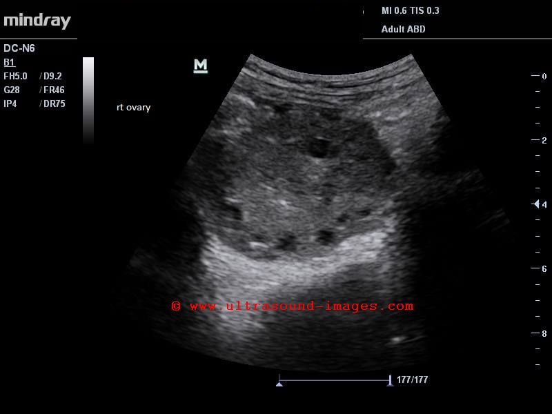
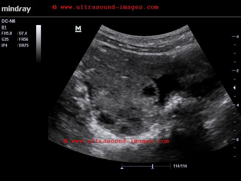
Case-2: (lower set of images) also shows torsion of Rt. ovary with swollen edematous ovary with marked increase in ovarian volume. Absence of flow signals on color Doppler further confirms the diagnosis of Rt. ovarian torsion.
(2nd set of images- case -2): courtesy of Dr. Moin Kamran, MD.
References:
RSNA article on ovarian torsion
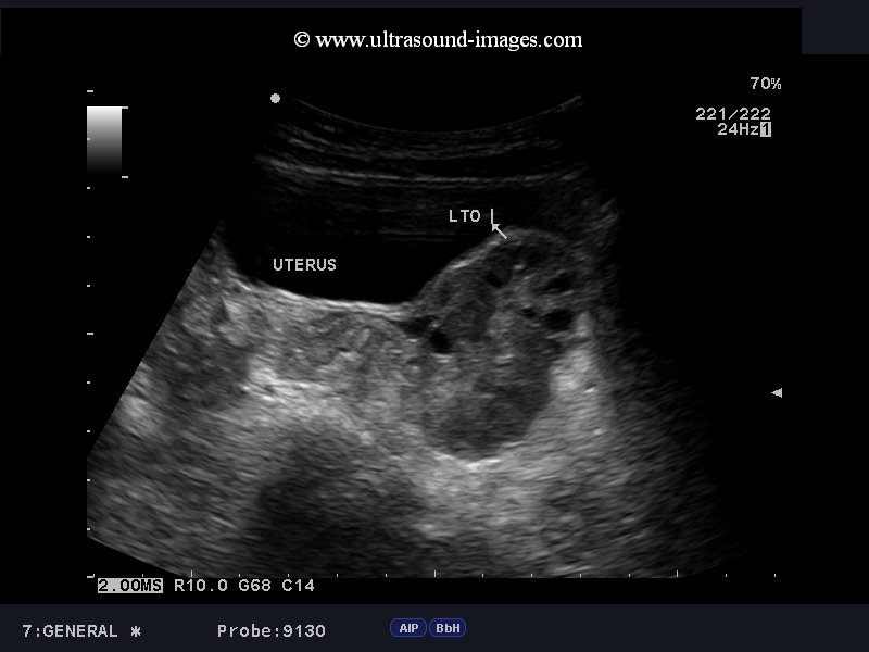
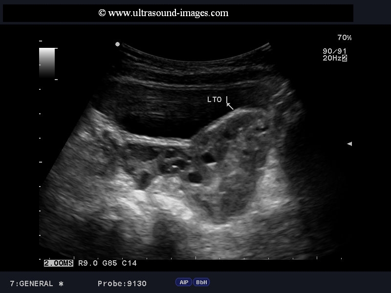
peritoneal inclusion cyst
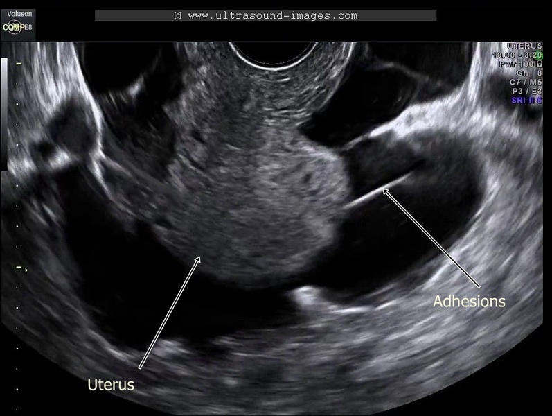
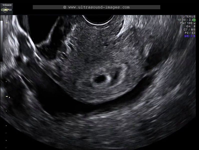
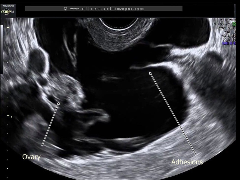
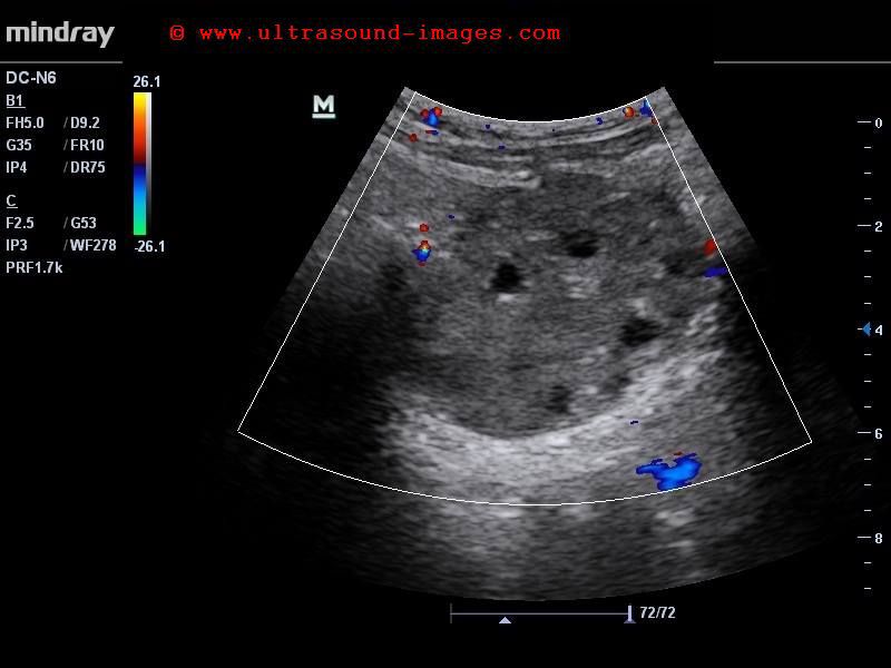
This young adult female patient has a past history of ectopic pregnancy for which she underwent surgery. she presented with symptoms of early pregnancy with pelvic pain. Ultrasound shows presence of intrauterine gestation sac with multiple adhesions within the pelvis with pockets of ascitic fluid. The right ovary appears to be surrounded by multiple adhesions and fluid resulting in an appearance like a spider in a web. These ultrasound image findings are diagnostic of peritoneal inclusion cyst. This is a condition resulting from trauma to the peritoneum usually the result of surgery with resultant accumulation of peritoneal fluid and multiple adhesions. The ovaries appear normal in structure and are well preserved. These ultrasound images of peritoneal inclusions cyst are courtesy of Ajay Garg, MD.
The principal differential diagnosis in such cases of peritoneal inclusion cysts include ovarian cystic masses such as serous or mucinous cystadenoma of the ovaries. Also, peritoneal inclusion cysts must be differentiated from more ominous conditions such as ovarian malignancies. Careful study of the ovaries, usually via the transvaginal ultrasound scan will help differentiate peritoneal inclusion cysts from ovarian lesions mentioned above.
References:
Sonographlc imaging of peritoneal inclusion cysts-AJR article
cumulus-oophorus
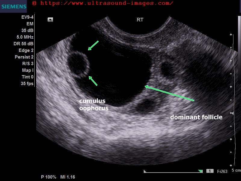

=cumulus oophorus is a collection of fluid and granulosa cells around the mature ovum within the dominant follicle of the ovary (in the ultrasound images above-- arrows point to the cumulus oophorus in right ovary).
= on sonography the cumulus oophorus is seen as a daughter cyst or cyst within cyst within the dominant follicle of the ovary.
= during ovulation the cumulus oophorus releases the mature oocyte or ovum into the fallopian tube.
= ultrasound images above show the cumulus oophorus in the right ovary.
= cumulus oophorus of the same case is seen in the ultrasound video above.
= cumulus oophorus must be distinguished from serous cystadenoma of the ovary and other cystic ovarian lesions. Transvaginal ultrasound scan will show typical dominant follicle containing the cumulus oophorus
© 2026, Joe Antony, MD All images and content in this site are copyrighted. www.ultrasound-images.com When visiting this site, we may use cookies to improve the performance of this site. Use of this site means you accept the use of cookies. Read our privacy policy at this page: https://www.ultrasound-images.com/privacy-policy/