
Ultrasound images of multifetal pregnancy
Contents of this page
- Sextuplets
- Multifetal reduction
- The twin peak sign of Diamnioitic Dichorionic twins
- Conjoined twins (Siamese twins)- a case of thoraco-omphalopagus
- Acardiac twin or severe TTTS (severe twin to twin transfusion syndrome)
- twin-yolk-sacs
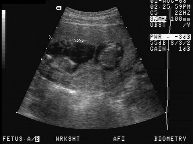
Sextuplets
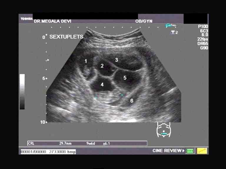
This ultrasound image shows 6 separate gestation sacs with embryos within them. Image courtesy of Dr. Megala Devi, Chennai, India.
Multifetal reduction
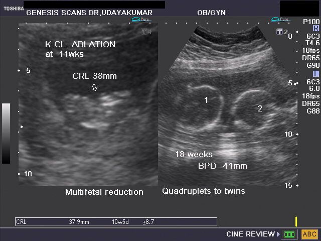
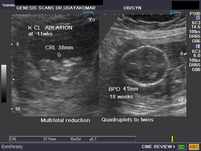
The above ultrasound images reveal reduction of early quadruplet gestation by ablation (using KCl) of 2 fetuses, converting it successfully to a twin gestation (image on right half). Images courtesy of Dr. Udaya Kumar, Chennai, India.
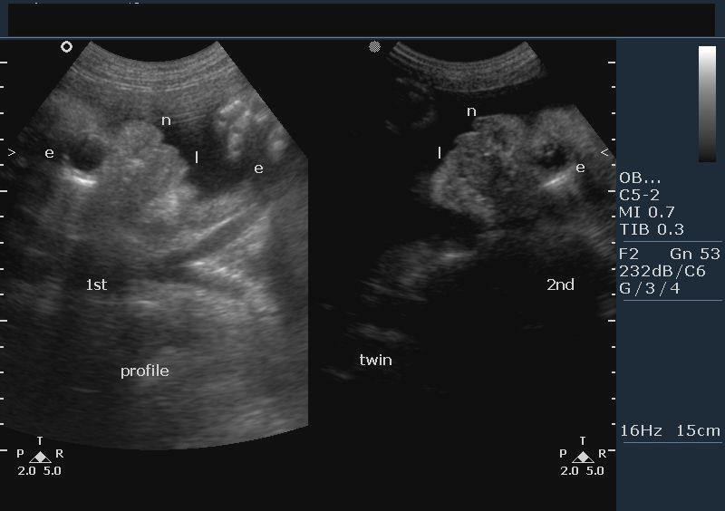
The twin peak sign of Diamnioitic Dichorionic twins
These ultrasound images demonstrate twin pregnancy at 13 weeks gestation. The intertwin membrane reveals that this is a diamniotic twin pregnancy. Also note the presence of the triangular wedge of placental tissue projecting into the intertwin membrane. This is called the Twin peak sign or the lambda sign which clearly suggests a Dichorionic twin pregnancy. Ultrasound images of Dichorionic gestation courtesy of Dr. Mohammed Sheriff, Tanzania.
Reference:http://radiology.rsnajnls.org/cgi/content/full/220/1/68(free article and images).
Conjoined twins (Siamese twins)- a case of thoraco-omphalopagus
Shared common heart
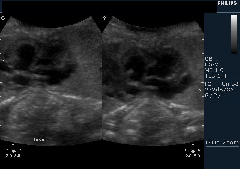
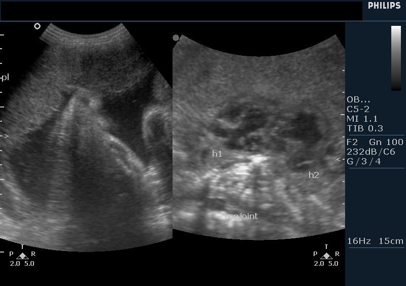

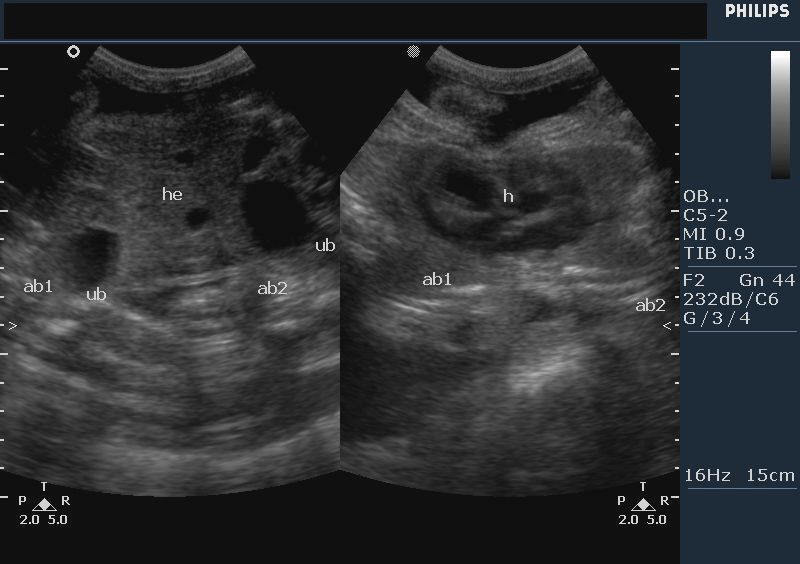
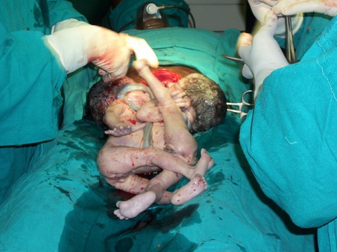
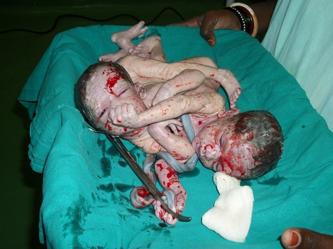
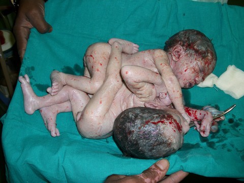
Conjoined twin fetuses were detected on ultrasound imaging during routine obstetric sonography. The ultrasound images above show a shared heart with markedly altered anatomy. The fetal abdomen were also fused to each other (see post natal snaps) with shared livers. The urinary bladders (UB) were seen to be separate. These ultrasound findings suggest thoraco-omphalopagus or fusion of the thorax and abdomen. This case study is courtesy of Dr. Vikas Shukla, MD, India.
Reference:
http://www.sonoworld.com/Fetus/page.php?id=315
http://sites.google.com/site/drjoea/obstetric-2
Acardiac twin or severe TTTS (severe twin to twin transfusion syndrome)
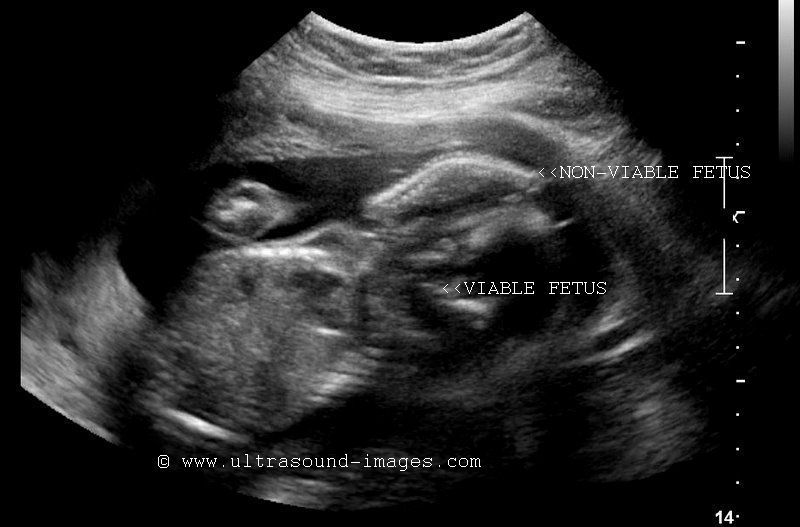

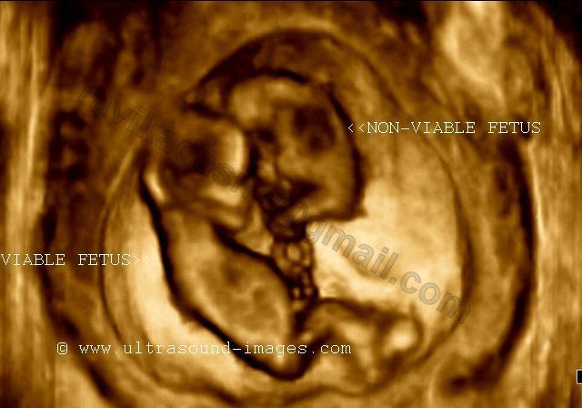
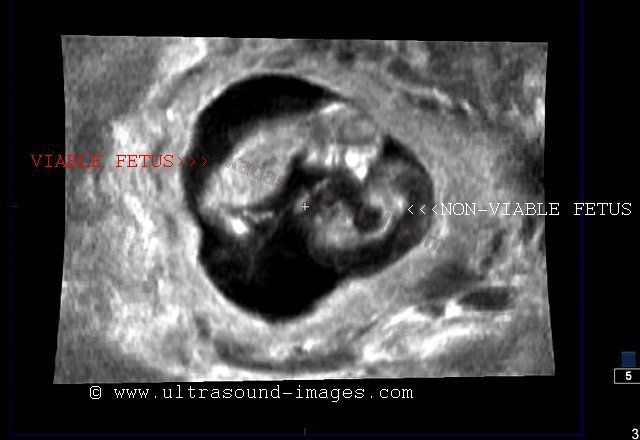
This 2nd trimester twin pregnancy (diamniotic- monochorionic) shows a viable fetus (normal fetus) with no evidence of associated congenital anomalies, and the twin fetus with severe congenital anomalies (non viable fetus). The non viable fetus shows very little normal tissue, but for part of a rudimentary spine, surrounded edematous soft tissue (fetal hydrops). There is clearly evident formation of inter-twin membrane between the two fetuses. The non viable twin is also called the acardiac twin because it has no identifiable cardiac structures; besides the fetal cranium or thorax are also not identified. These ultrasound images suggestive of a non viable fetus with severe anomalies, the most probable diagnosis being acardiac twin, a severe form of twin to twin transfusion syndrome (TTTS), or possible TRAP or twin reversed arterial perfusion syndrome. In this condition, blood flows via an ARTERIO-ARTERIAL-ANASTOMOSIS (AAA) on the placenta; this deoxygentated blood is carried to the non viable, and extremely abnormal twin (the recipient), from the donor twin, from its umbilical artery. This phenomenon and transformation of the non viable or recipient twin to the acardiac twin is called the TRAP sequence or Twin Reversed Arterial Perfusion syndrome. Images of acardiac/ non viable twin are courtesy of Dr. Ravi Kadasne, MD, UAE.
References:http://www.sonoworld.com/fetus/page.aspx?id=308 (article on acardiac twins)
twin-yolk-sacs
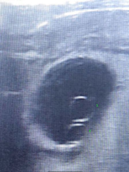
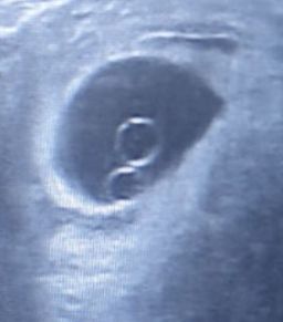
Twin yolk sacs in a 5 week pregnancy suggest early twin gestation, typically diamniotic. They are seen as two echogenic rings within the gestational sac on ultrasound. This finding helps predict multiple pregnancy before embryo visualization. Follow up scanning confirms viability, chorionicity, and detects possible complications early in clinical practice. Images courtesy of Dr. Golam, MD.
#TwinPregnancy #Ultrasound #EarlyPregnancy #YolkSac #Radiology #Obstetrics #FetalImaging
© 2026, Joe Antony, MD All images and content in this site are copyrighted. www.ultrasound-images.com When visiting this site, we may use cookies to improve the performance of this site. Use of this site means you accept the use of cookies. Read our privacy policy at this page: https://www.ultrasound-images.com/privacy-policy/