
Ultrasound images of placental pathology
Contents of this page
- Subchorionic cyst of the placenta
- Velamentous insertion of umbilical cord into placenta
- Vesicular mole (also called Molar pregnancy or Hydatidiform mole) in 1st trimester
- Placental calcification
- Placental mass (mass in placenta - Chorioangioma of placenta or Placental Chorioangioma)
- Case-2: Chorioangioma of placenta
- Succenturiate placenta
- Circumvallate placenta
- Placental venous lake
- Placenta previa
- Retained products of conception/ retained placenta
- Bilobed placenta: (bilobate placenta)
- partial molar gestation
- placental surface cysts
- placentomegaly-increased-thickness
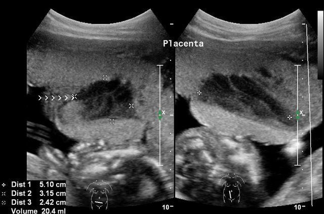
Subchorionic cyst of the placenta
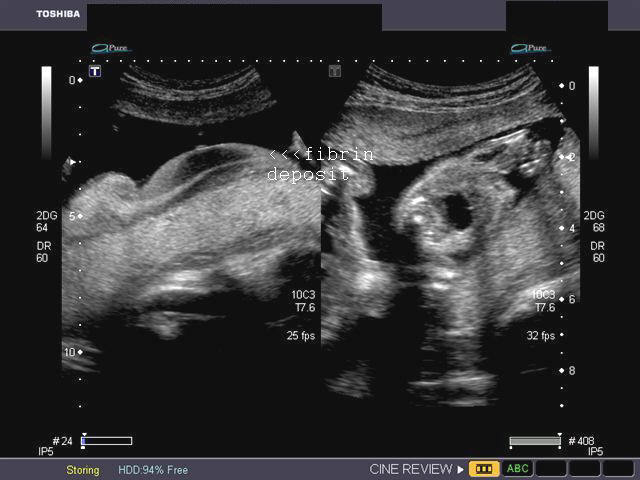
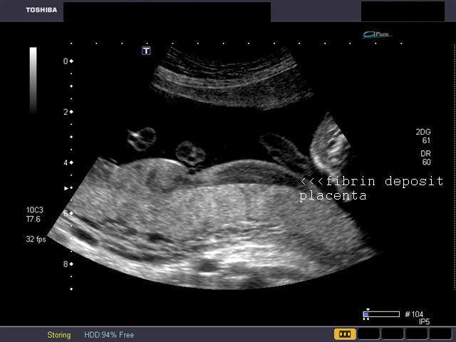
The two sonographic images above show a cystic lesion of the placenta, just below the placental surface. Few mobile echoes were seen within the lesion. No other abnormalities were seen in the placenta and the fetus. This finding is generally considered to be clinically of little significance. However some reports suggest an association with fetal IUGR (or intrauterine growth retardation). Diagnosis: subchorionic cyst of the placenta. Also known as membranous cyst, chorionic cyst. It is believed to be due to deposition of fibrin in the subchorionic region of the placenta. Images courtesy of Dr. Gunjan Puri, Surat, India.
Reference: 1) http://www.jultrasoundmed.org/cgi/content/full/21/6/641 (free article)-- rated excellent by us.
Velamentous insertion of umbilical cord into placenta
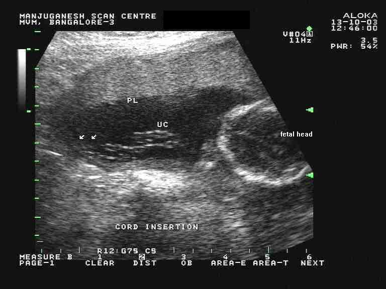
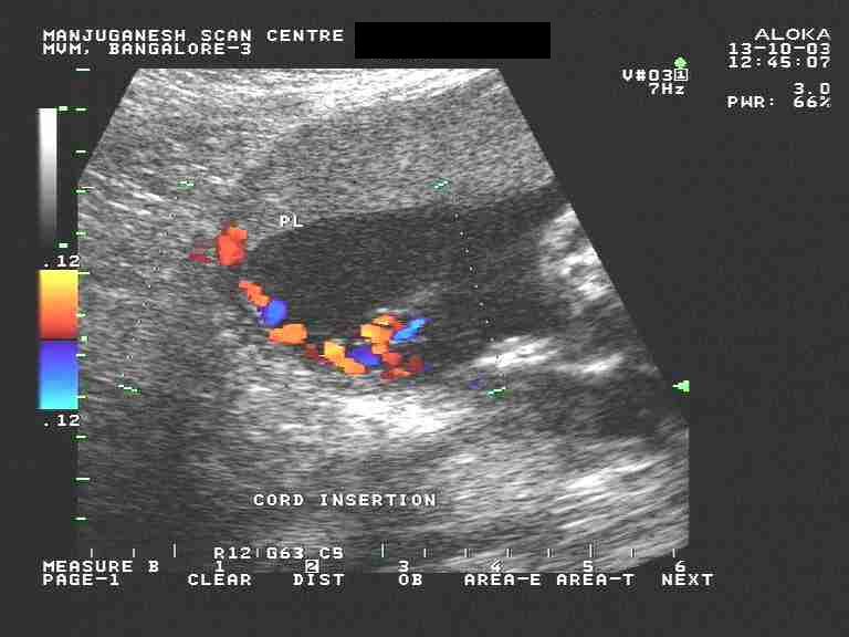
These ultrasound and color doppler images show the umbilical cord inserting into the placental membranes before reaching the placental tissue proper. This is the typical appearance on sonography, of velamentous insertion of the umbilical cord. Ultrasound images courtesy of Dr. Latha Natrajan, Bangalore, India.
Reference: http://www.jultrasoundmed.org/cgi/content/full/25/8/963 (free article and images).. excellent
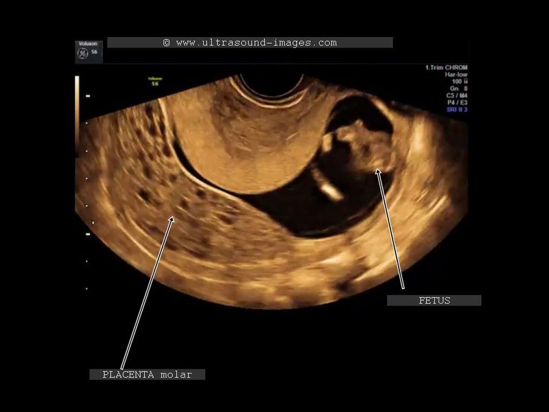
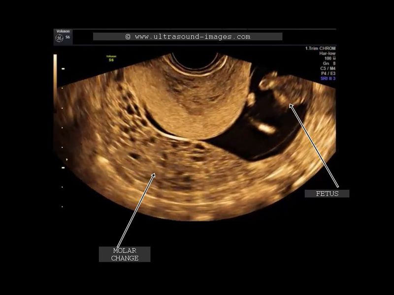
Vesicular mole (also called Molar pregnancy or Hydatidiform mole) in 1st trimester
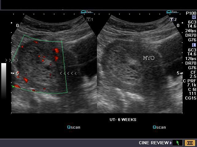

Sonography of the uterus was done in this 1st trimester pregnancy. Ultrasound and Power Doppler images reveal: a) Hyperechoic mass in the uterine cavity with multiple cystic spaces within it. b) Uterus is enlarged (bulky) c) The myometrium is hypoechoic compared to the contents of the uterine cavity. These appearances can be likened to a "snowstorm" or "moth eaten" look. d) Power Doppler images show increased flow within the enlarged uterus. These ultrasound images suggest a sonographic diagnosis of Vesicular Mole. Ultrasound images were taken using a Nemio- XG machine by Joe Antony, MD, India.
Reference:http://www.ajronline.org/cgi/reprint/160/1/137.pdf
Placental calcification
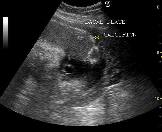
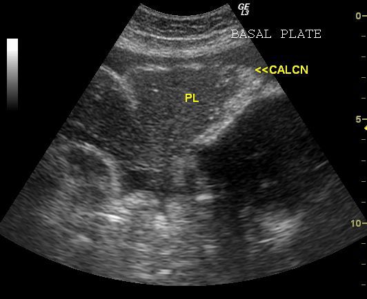
Calcific areas in placenta
This 3rd trimester pregnancy shows extensive calcification of the basal plate (uterine or maternal surface) of the placenta. There is also calcification along the placental septa and chorionic plate. The calcific deposits in the placenta are seen as hyperechoic foci with acoustic shadowing. Such calcium deposits in the placenta suggest macroscopic calcification as opposed to micro-calcification seen in the 2nd and early 3rd trimester. Clinically and pathologically, calcific changes of placenta have no significance.
More ultrasound images and short ultrasound video clips of placental calcification can be seen at:
http://cochinblogs.blogspot.com/2009/08/placental-calcification.html
Dense placental calcification - case2
The ultrasound images above show dense macro calcifcations within the placenta in this 34 week old gestation. Again, though this degree of calcification is unusual (both the density and number of calcific foci), it has little clinical significance. However, follow up sonography may be advisable to rule out possible fetal morbidity. Color Doppler image of the placenta shows normal flow within the tissue.
References: Placental imaging and sonography- Radiographics
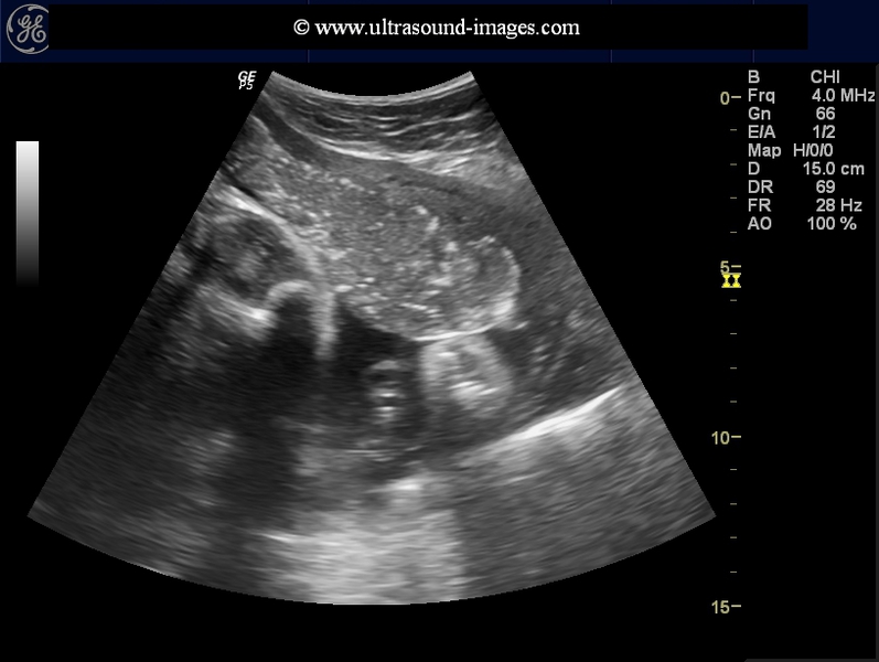
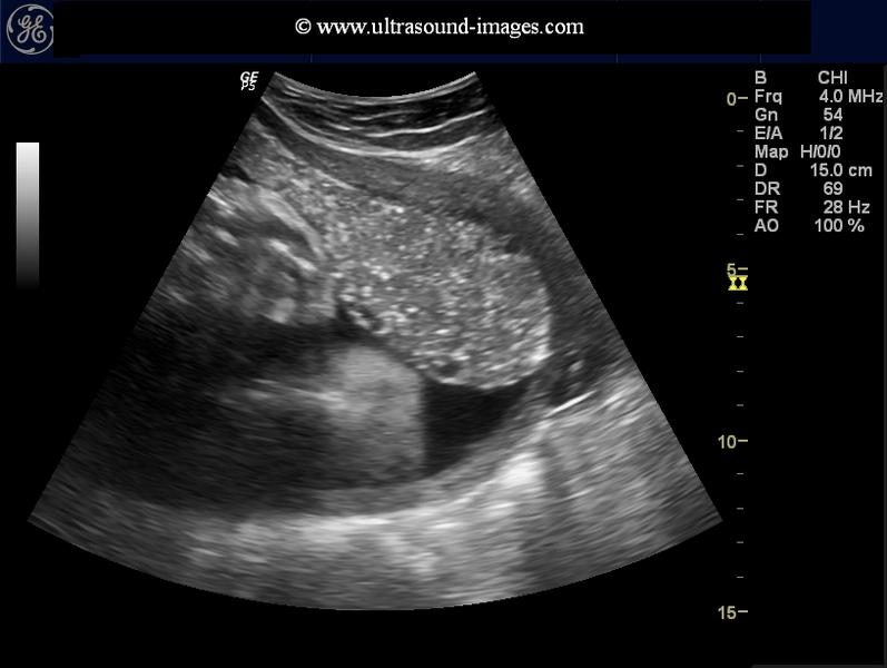
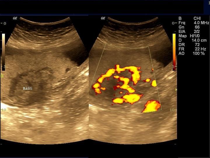
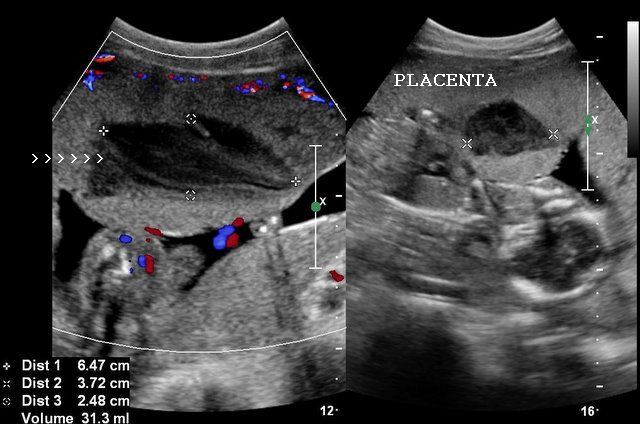
Placental mass (mass in placenta - Chorioangioma of placenta or Placental Chorioangioma)

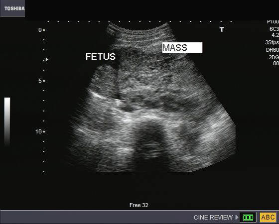

Placental mass (mass in placenta - Chorioangioma of placenta or Placental Chorioangioma)
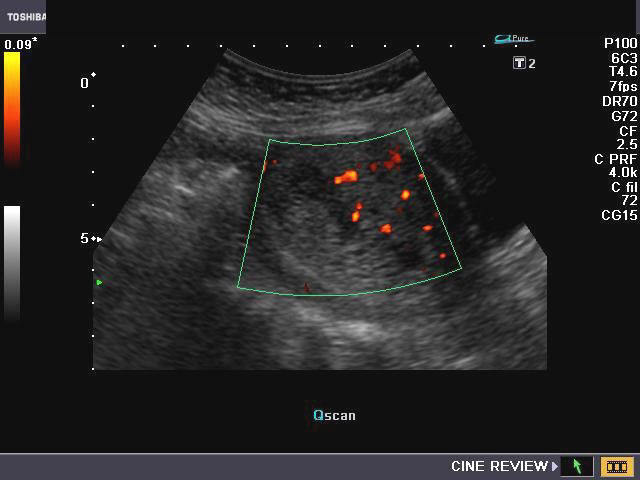
Sonography of the placenta in this 16 week pregnancy shows a large, solid mass, that is non calcific and shows mild vascularity on Power Doppler imaging. The mass is inhomogenous and shows many cystic spaces within it. This tumor of the placenta lies close to the cord insertion site. Flow seen on Power Doppler image suggests that this placental tumor is vascular and excludes placental hematoma. Ultrasound images of this type of placental mass are highly suggestive of placental chorioangioma. The other diagnostic possibility can be a hamartoma of the placenta. Chorioangioma of placenta is the commonest tumor of the placenta and is benign in nature. This mass measures 12 x 8 cms., an unusually large size for a chorioangioma, and can signify poor prognosis for this pregnancy. Images courtesy of Jaydeep Gandhi, MD, India. These ultrasound images were taken with a Toshiba Nemio-30 Ultrasound system.
Reference: http://radiographics.rsna.org/content/27/4/1187.full
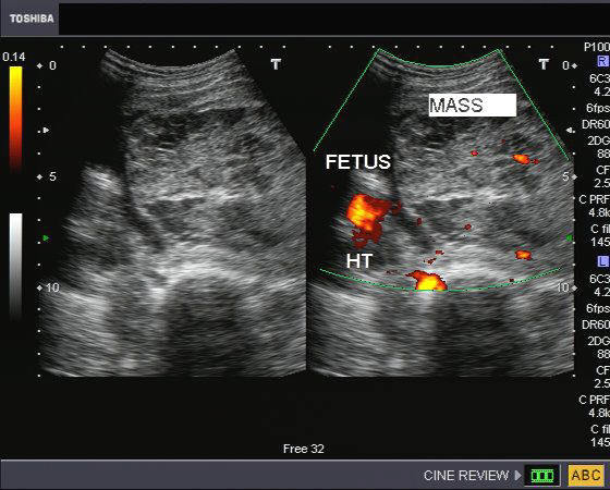
Case-2: Chorioangioma of placenta
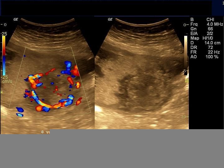

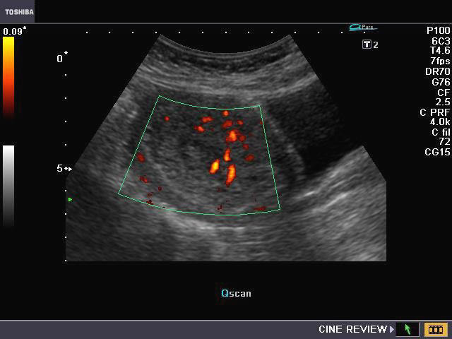
This was a 3rd trimester pregnancy with ultrasound imaging of the placenta showing what appears to be a hypoechoic, well defined but inhomogenous mass with marked vascularity on Power and color Doppler imaging. The vessels in the mass appear to be in continuity with the insertion of the cord which is very close to the edge of the placental mass. Though the mass appears to be deep within the placenta, the image on topmost row (left) shows the mass is bulging onto the amniotic surface- these ultrasound appearances are typical of a highly vascular mass like the chorioangioma of the placenta. Images are courtesy of Dr. Sanjiv K Bhalla, MD, India.
References: http://radiographics.rsna.org/content/29/5/1371.full.pdf

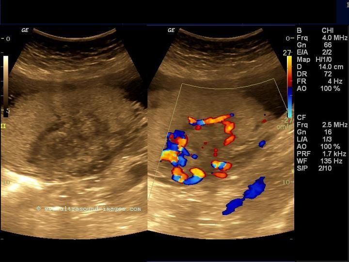
Chorioangioma of placenta
This is a classic , moderately vascular placental mass, again bulging into the amniotic cavity, again, having all the typical ultrasound features of chorioangioma of the placenta.
These ultrasound images of chorioangioma of the placenta are courtesy of Dr. Durr-e-Sabih, MBBS, FRCP.
Succenturiate placenta
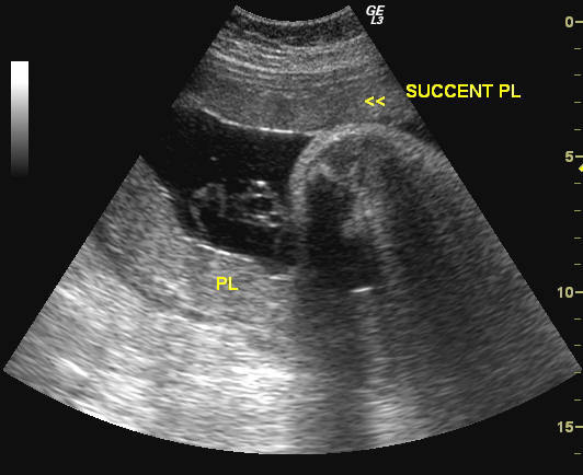
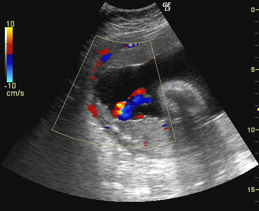
Synonyms: bilobed or bilobate placenta
This was a 3rd trimester pregnancy showing part of the placenta along the anterior wall of the uterus (SUCCENT PL), and the main part of the placenta along the posterior wall (PL). The sucenturiate lobe of placenta is connected to the main placenta by a string of blood vessels (see Color Doppler image on right). Both images taken on a GE, Logiq-3 ultrasound system.
Reference:http://www.sonoworld.com/Fetus/page.aspx?id=181
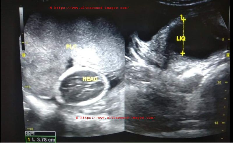
Circumvallate placenta
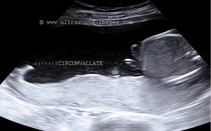
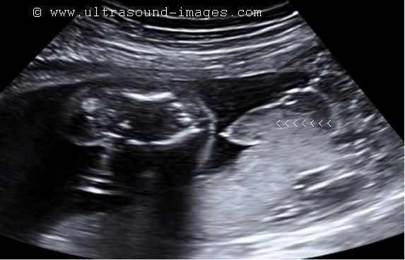

The above ultrasound images show a posterior placenta with infolding of the rim or margins of the placenta. This condition is a normal variant and is produced due to the fact that in this case, the chorionic plate (fetal surface) of the placenta is smaller than the basal plate (surface in contact with the uterine wall or decidua) of the placenta with resultant shouldering or infolding/ rolling of the placental margins. This condition is called circumvallate placenta and usually causes no harm to the fetus. However, it can sometimes be associated with increased chances of placental abruption and hemorrhage. Sometimes the free margin of the circumvallate placenta can mimic a fetal membrane such as amniotic bands which have more ominous implications.
References: Radiographics article on circumvallate placenta
Placental venous lake
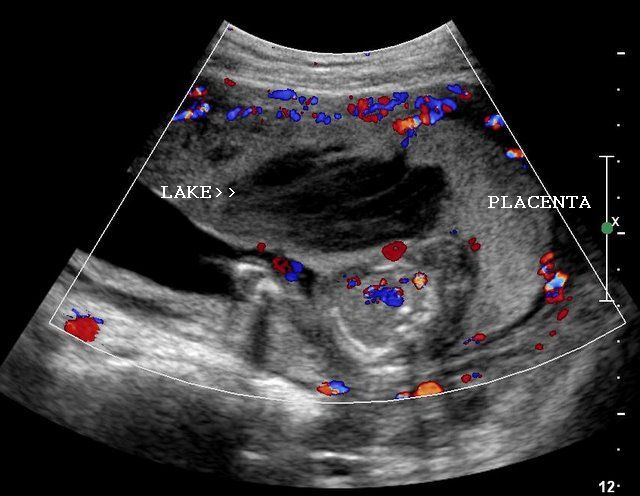
This placenta, in a 28 week pregnancy shows a large hypoechoic (almost anechoic), measuring 5 x 3.5 cms. in size. Some particulate matter was seen flowing through this area, which was closer to the fetal surface of the placenta. These ultrasound images suggest a typical appearance of a large venous lake in the placenta. Color Doppler image showed no major flow pattern within this placental lake. The fine, echogenic strands within the lesion appear to be nothing more than artefacts produced by slow flowing blood within the lesion. Ultrasound images are courtesy of Dr. Ravi Kadasne, MD, UAE.
Reference:
http://www.obgyn.net/ultrasound/ultrasound.asppage=/us/present/0206/moroder_placental_lakes
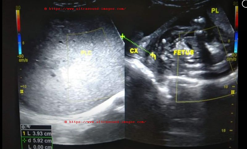
Placenta previa
This is a condition, wherein the placenta covers the internal os. Depending on the degree of coverage of the internal os, there are 3 grades of placenta previa.
Marginal placenta (marginal placenta previa): here is the tip of the placenta reaches the margin of the internal os.
Partial or incomplete placenta previa: here the internal os is partially covered by the lower part of the placenta.
Complete placenta previa: here the placenta completely covers the internal os.
Another condition is low lying placenta, wherein the placenta reaches into the lower uterine segment, but clearly away from the internal os.

Incomplete/partial placenta previa
The above ultrasound and color Doppler images show the lower margin of the placenta partially covering the internal os, suggesting partial placenta previa. One point to be noted is that placenta previa is diagnosed in the 2nd and 3rd trimester of pregnancy, and that normal uterine contractions can cause the placenta to be "pushed" lower down its normal position, creating an appearance of placenta previa (a false positive diagnosis of placenta previa). Hence it is advisable to repeat the ultrasound scan after 30 minutes to exclude a false diagnosis of this condition.

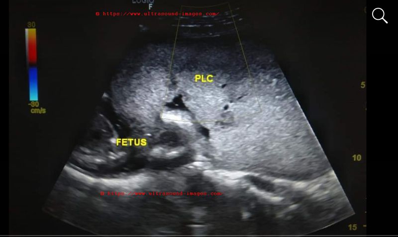
Complete placenta previa
This ultrasound image shows the placenta completely covering the internal os (INT OS), thus diagnostic of complete placenta previa. Follow up ultrasonography is advisable in all cases of placenta previa, to look for ascent of the placenta to a higher position due to the growth of the uterus. Such cases of placenta previa (both partial and complete) are in danger of hemorrhage (antepartum) and are advised rest to prevent this. Ultrasound image of complete placenta previa is courtesy of Dr. Vikas Shukla, MD, India.
Reference:http://emedicine.medscape.com/article/796182-overview
Retained products of conception/retained placenta


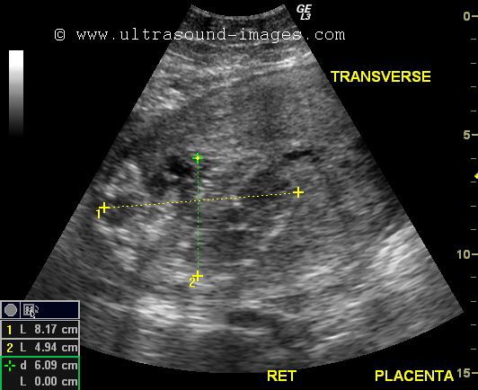
The above ultrasound images show a post partum uterus on transabdominal sonography. There is a hyperechoic mass within the endometrial cavity measuring 8 x 5 cms. The color Doppler ultrasound image shows poor vascularity of the mass and the endometrium. Transverse section ultrasound image of the post partum uterus shows that the mass is located more towards right half of the uterine cavity; also note that the endometrial mass is eccentric within the cavity- the anterior myometrium is thicker whilst the posterior wall of the uterus is thinner. The placenta was not expelled at the time of delivery. Thus this eccentric, markedly thick, inhomogenous mass is the retained placenta with a certain degree of placenta accreta being present. Absence of vascularity or poor flow does not rule out retained products of conception/ retained placenta. The single most important sign of retained products of conception is the large endometrial mass. Other signs of retained placenta or products include complex fluid or thickened endometrium (more than 10 mm.).
References:
http://www.jultrasoundmed.org/content/24/9/1181.full.pdf+html(RPOC-free article and images).
http://radiographics.rsna.org/content/29/5/1371.full.pdf+html(imaging of placenta - free article)
Bilobed placenta: (bilobate placenta)
This is a 3rd trimester pregnancy with ultrasound images showing two parts of the placenta along the anterior and posterior walls of the uterus, connected by a thin bridge of placental tissue. This kind of sonographic appearance is typical of bilobed placenta. Ultrasound images captured using a GE, Logiq 3, ultrasound system.
Reference:http://radiographics.rsna.org/content/29/5/1371.full
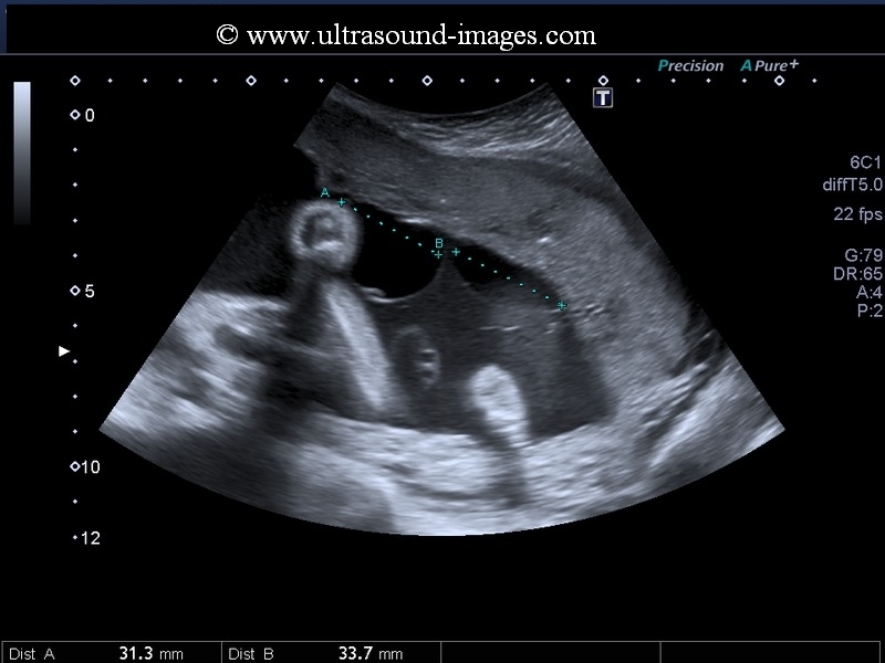
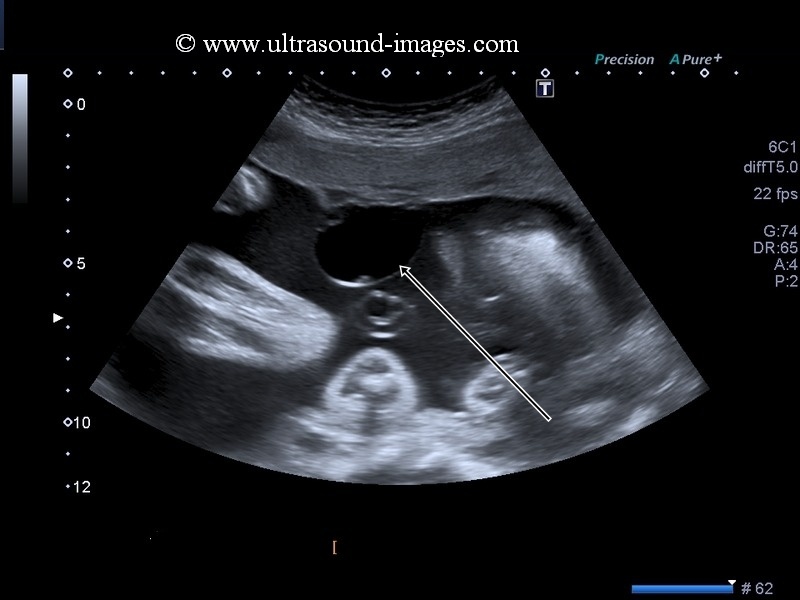
Partial molar gestation
A partial molar pregnancy or partial vesicular mole or partial hydatidiform mole is a pregnancy in which a molar change of the placenta is present along with presence of fetal parts or a complete fetus, which may or may not be viable. This 12 week fetus (see ultrasound images above) shows a viable fetus with multiple cystic lesions within the placenta. In addition, the placenta appears enlarged with increased thickness. Sometimes, it may not be possible to distinguish a partial mole from a complete mole if fetal parts are not visualized. These ultrasound images of partial molar pregnancy are courtesy of Dr. Anand Patel M.D.
References:
1. Sonographic imaging of partial hydatidiform mole- sonoworld article.
2. partial mole- ultrasound imaging- radiopaedia
Placental surface cysts
This 25 weeks old pregnancy shows, on ultrasound imaging of the placenta, a cystic lesion on the surface of the placenta bulging into the amniotic cavity. In fact, there are two cystic lesions of the placental surface each measuring approximately 3 cm in size- (see ultrasound images above). This sonographic appearance is said to be due to haemorrhage below the amniotic membrane on the surface of the placenta and has been termed as subamniotic haematoma or alternately as subchorionic cysts or chorionic cysts. In the early stages of haemorrhage beneath the amniotic membrane, the haematoma may appear echogenic; however with the passage of time it is anechoic or purely cystic in nature as in this case. These ultrasound images of subamniotic haematoma or placental surface cysts are courtesy of Durr-e-eSabih, MBBS, FRCP.
Medically, placental surface cysts do not have much clinical significance and are often an incidental finding. However certain studies suggest that placental surface cysts may be associated with intrauterine growth retardation or IUGR foetuses.
References:
Sonography of placental surface cysts- Journal of ultrasound
ultrasound imaging of subamniotic haematoma- sonoworld article
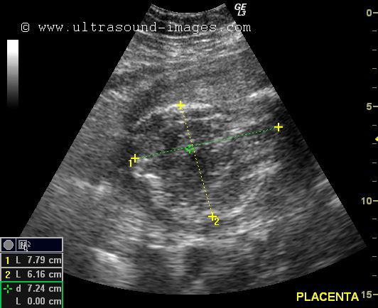
placentomegaly-increased-thickness
=Increased placental thickness beyond normal range is called placentomegaly
= this 2nd trimester pregnancy (see ultrasound images above) shows marked increase in placental thickness (placentomegaly)
= normal case- the placenta increases in thickness at a rate of appx. 1 mm. per week of gestation
= upper limit of placental thickness at term pregnancy is 40 mm.
= marked placentomegaly is associated with increased fetal mortality and morbidity
= seen in placental insufficiency and diabetic mothers
(images of placentomegaly courtesy of Dr. Yusef Ali, MD).
Reference: sonography of placentomegaly
© 2026, Joe Antony, MD All images and content in this site are copyrighted. www.ultrasound-images.com When visiting this site, we may use cookies to improve the performance of this site. Use of this site means you accept the use of cookies. Read our privacy policy at this page: https://www.ultrasound-images.com/privacy-policy/