
Ultrasound images of anomalies of fetal spine
Contents of this page
- Normal fetal spine
- Spina bifida in fetus
- Meningocele of Thoracic spine in fetus
- Meningocele of the lower lumbar spine in fetus
- Fetal caudal regression syndrome
- sacrococcygeal teratoma
- Fetal spinal cord and filum terminale
- 3-D sonography of iniencephly in fetus
- Hydrocephalus-with-lumbosacral-meningomyelocele
- spinal-arachnoid-cyst
Normal fetal spine
Ultrasound images of normal anatomy and appearances of fetal spine
Download my E book for Amazon Kindle (download free Kindle reader for i Phone or Android)
Assorted ultrasound cases- by Joe Antony, MD
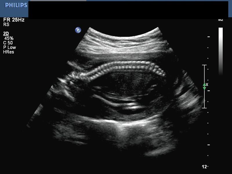
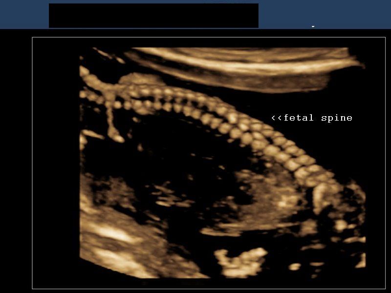
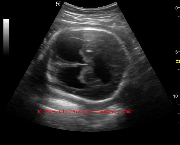
This 2-D, ultrasound image shows the sagittal section of the normal fetal spine in longitudinal section. (Image courtesy of Dr. Ravi Kadasne, UAE).
3-D Ultrasound images of normal fetal spine
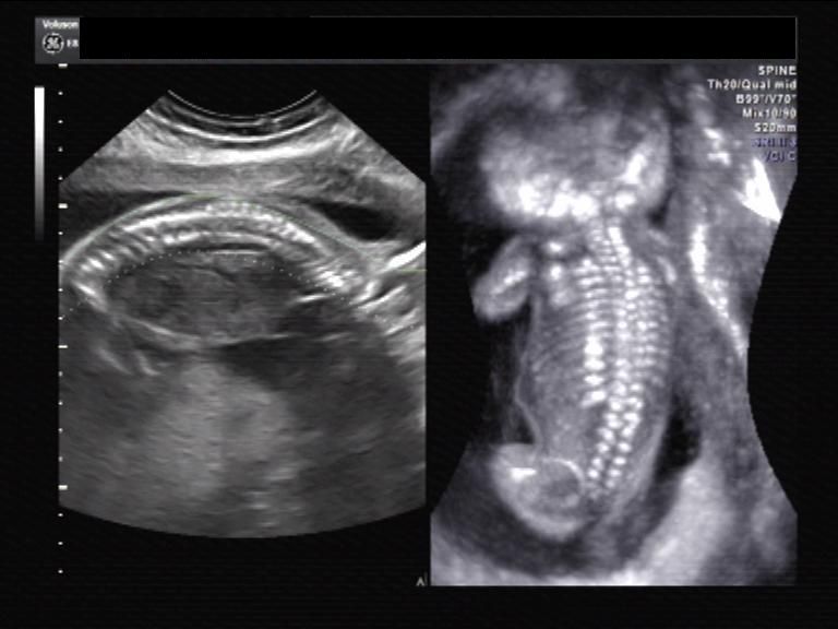
Sonography of fetal spine using 3-D/ 4-D ultrasound reveals greater detail of the fetal spine in 3-Dimensions. Ultrasound visualizes the ossified part of the fetal spine. The 3 main ossification centers in the fetal vertebrae are: a) the centrum b) the right neural process and c) the left neural process. The centrum forms the central part of the vertebral body. The postero-lateral parts of the vertebrae are formed by the right and left neural processes. (These 3-D ultrasound images are courtesy of Dr. PK Srivastava, India, and Dr. Ravi Kadasne, UAE).
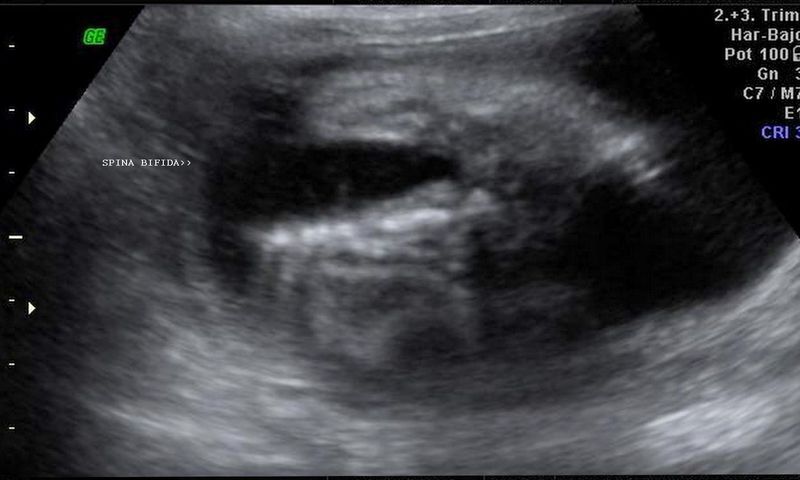
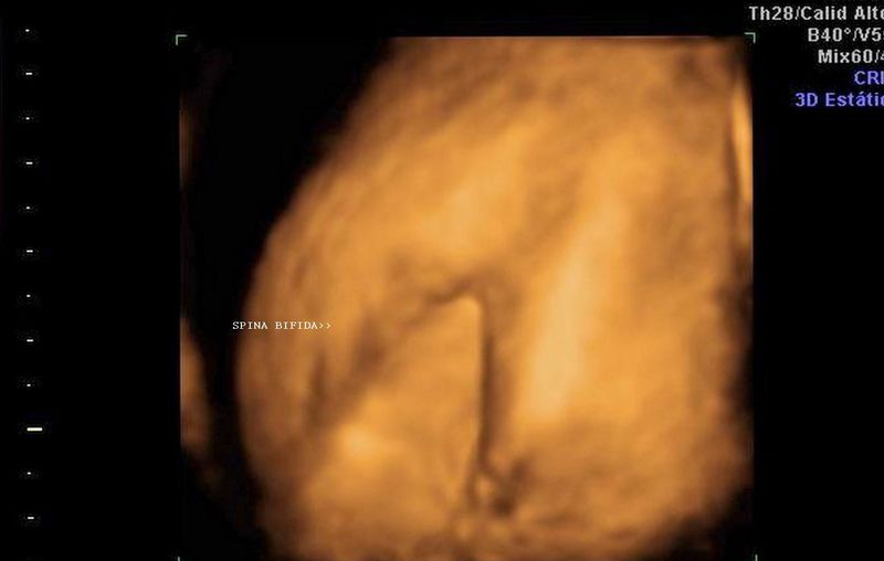
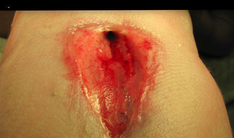
In this fetus , sonography of the lumbosacral spine shows a major defect in the posterior part of the fetal lumbar and sacral vertebrae due to failure of closure of the dorsal part of the vertebrae (the laminae and spinous processes). The ultrasound image in top row- Left, shows 2-D (B-mode) display of the large defect in long section. The image on top-row- Right, shows the same appearance in 3-D ultrasound. The post natal photograph of the area (lower back) shows the open spinal canal. This type of spina bifida is called spina bifida cystica. (Ultrasound images of spina bifida are courtesy of Dr. Martin Horenstein, Argentina).
References:
1) http://www.thefetus.net/page.php?id=135 (free article and images). 2) http://www.emedicinehealth.com/spina_bifida/article_em.htm#Spina%20Bifida%20Overview
Meningocele of Thoracic spine in fetus

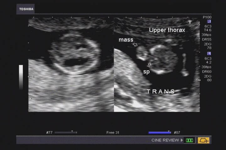
Neural tube defect and sac at the level of fetal thoracic spine in this 3rd trimester fetus showed a sac herniating from a defect (spina bifida) in the thoracic vertebrae. Sonography of the fetal brain showed ventriculomegaly. These ultrasound images suggest a diagnosis of meningocele of thoracic spine/ vertebrae in fetus. Meningocele of thoracic vertebrae is a rare entity. Ultrasound images are courtesy of Dr. Prasenjeet Singh, India.
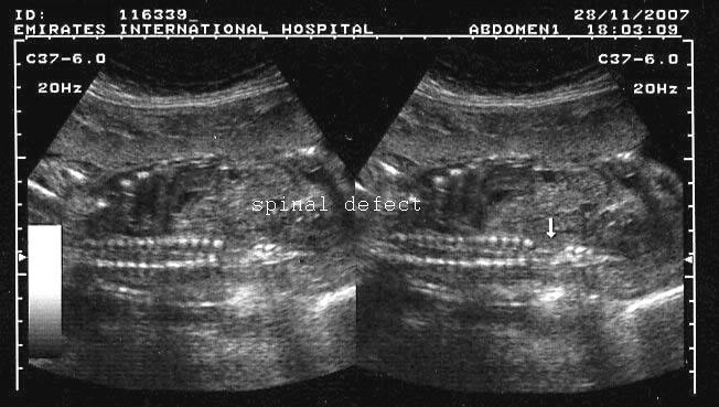
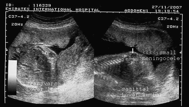
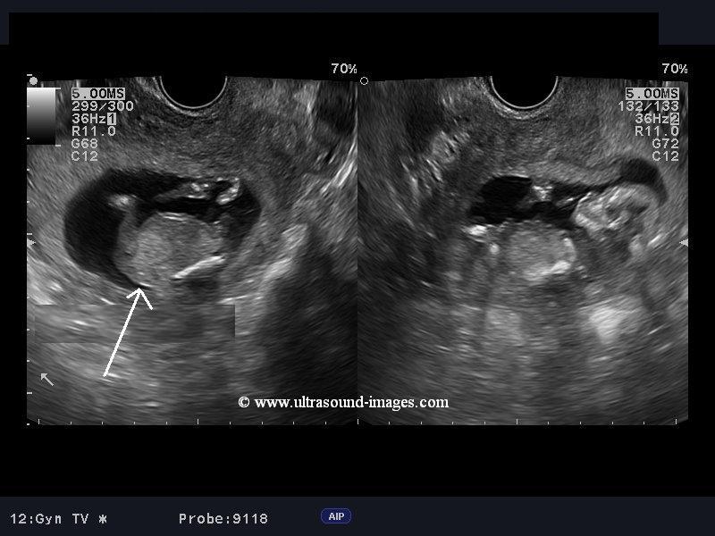
Meningocele of the lower lumbar spine in fetus

A: TRANSVERSE AND B: CORONAL
SAGITTAL SECTIONS SECTION SPINE

C: TRANSVERSE AND D:SHOWING THE
SAGITTAL SECTIONS CLUB FOOT

E:FETAL CLUB FOOT F: FETAL FOOT
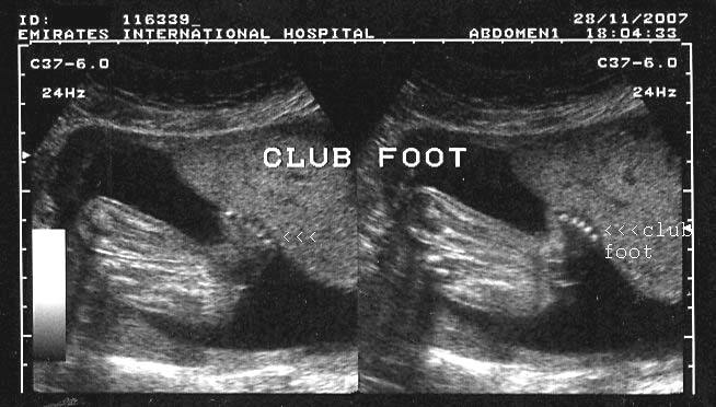
These are ultrasound images of a 2nd trimester fetus, which show a neural tube defect (spina bifida) of the lower lumbar vertebrae. There is also splaying of the vertebral laminae and widening of the interpedicular distance in this region. A small cystic soft tissue mass is seen overlying (dorsal) this part of the spine (meningocele). Also note the associated fetal club foot anomaly. Normally, the fetal foot is seen perpendicular to the plane of the fetal leg. In this case, the angle with the ipsilateral leg is altered and the foot is almost parallel to the leg. Ultrasound images are courtesy of Dr. Ravi Kadasne, UAE.
References: for more images and information on fetal club foot and neural tube defects, please see:
Fetal caudal regression syndrome


This 13 weeks old fetus shows a rare spinal anomaly. Part of the fetal spine below the thoracic vertebrae- that is to say the lumbar and sacral vertebrae are absent. Such an anomaly is called caudal regression syndrome with an incidence of as little as 1: 100000 pregnancies. In actual practice, this rare anomaly has been reported in around 300 cases so far. Caudal regression syndrome is usually diagnosed sonographically after late second trimester or the third trimester of pregnancy. However, with the use of transvaginal ultrasound, as in this case, caudal regression syndrome is now being diagnosed very early in pregnancy including in the late first-trimester fetus. Along with absence or hypoplasia of the lumbosacral spine, caudal regression syndrome may be associated with hypoplasia or maldevelopment of the lower limbs also. There is also frequent association of caudal regression syndrome with congenital anomalies of the gastrointestinal tract, uro-genital tract and the cardiovascular system.
These ultrasound images of caudal regression syndrome are courtesy of Dr Jaydeep Gandhi, MD.
References:
1) http://radiopaedia.org/articles/caudal-regression-syndrome
2) http://sonoworld.com/TheFetus/page.aspx?id=94
3) http://www.jultrasoundmed.org/content/21/10/1175.full.pdf
Sacrococcygeal teratoma
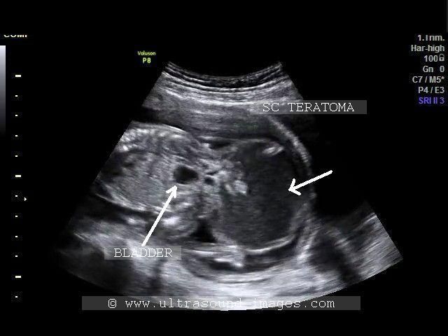
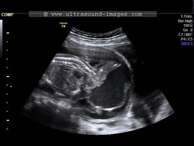
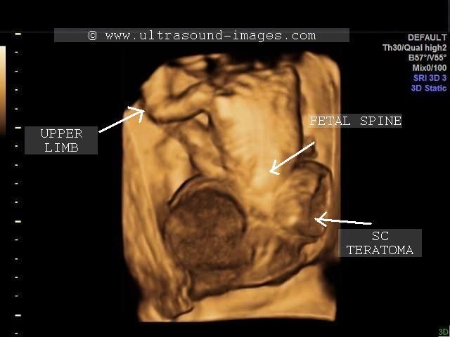

This fetus displays a huge complex but primarily cystic mass in the caudal end of the body towards the sacrum and coccyx. The mass extends below the gluteal region and expands laterally as well as anteroposteriorly and projects below the fetal body. the mass shows thick shaggy walls and contains echogenic fluid with particulate matter and also solid tissue. These and other findings mentioned earlier in the typical location is diagnostic of a sacrococcygeal teratoma in this fetus. The 3-D and B mode ultrasound images show characteristic features of sacrococcygeal teratoma in this late second trimester fetus. These ultrasound images of sacrococcygeal teratoma are courtesy of Dr. Firoz Bhuvar, MD.
The chief differential diagnosis of sacrococcygeal teratoma in fetus is lumbo-sacral meningocele or myelomeningocele. however, meningocele is typically located higher up the spine in the lower lumbar or upper sacral region. Besides the contents of the meningocele are usually clear fluid-originating from the CSF or cerebro-spinal fluid. Also, a meningocele contains thin walls unlike a sacrococcygeal teratoma.
References: http://sonoworld.com/fetus/page.aspx?id=530
Fetal spinal cord and filum terminale


The fetal spine must be carefully imaged as mentioned earlier in this page. However, oftne neglected is the fetal spinal cord must also be studied during sonography of the fetus. In these ultrasound images, the terminal part of the fetal spinal cord and filum terminale have been diplayed beautifully. Images of filum terminale are courtesy of Dr. Mayank Chowdhury, MD. He used the GE Voluson E8 for this study. Among the various anomalies with the fetal filum terminale that need to be excluded are tumors such as lipomas and cysts, as well as a well known entity called tethered cord syndrome. The filum terminale is fine fibrous strand that extends from the distal conus medullaris to the coccyx.
See: http://ultrasound-images.blogspot.in/2010/09/ultrasound-and-doppler-videos-of-normal.html
3-D sonography of iniencephly in fetus
This 15 week old twin pregnancy (the abnormal twin) shows certain characteristic ultrasound findings , including marked hyper extension of the cervical spine with severe cervical lordosis and an upward turned fetal face. in addition, the cervical and part of thoracic spine are absent with the occipital region of the fetal head in almost close contact with the dorsal aspect of the fetal spine. there also appears to be an occipital bone defect. All these ultrasound features are seen in the abnormal twin in the B-mode and 3D ultrasound images above. The above 3D and B-mode ultrasound images (courtesy of Durr-e-Sabih, MBBS, FRCP) suggest a diagnosis of fetal iniencephaly in the abnormal twin (on the left of the images). Two types of this condition (iniencephaly) have been described- apertus and clausus depending on the presence of an encephalocele or its absence. In our case described above, there is no evidence of a cephalocele, thus suggesting the clausus variety of iniencephaly.
References:
3-D ultrasound imaging of iniencephaly- Journal of ultrasound article
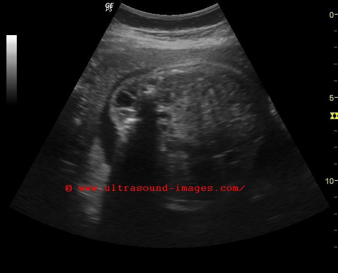
Hydrocephalus-with-lumbosacral-meningomyelocele
This 3rd trimester fetus shows an obstructive hydrocephalus with dilated lateral ventricles. In addition there is a lumbosacral meningomyelocele.
spinal-arachnoid-cyst
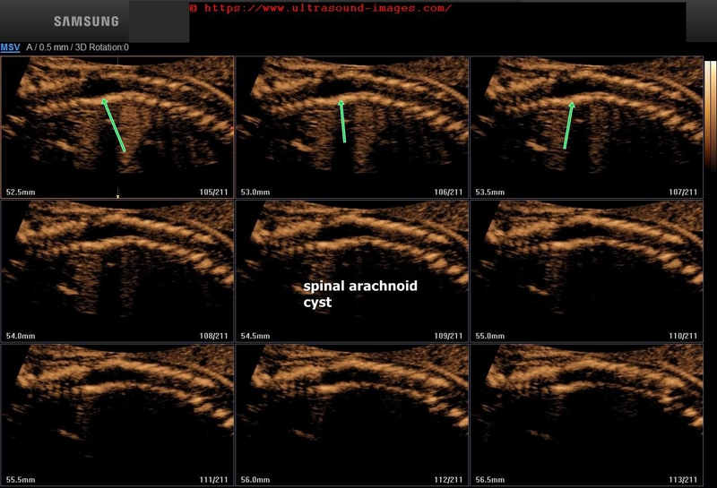
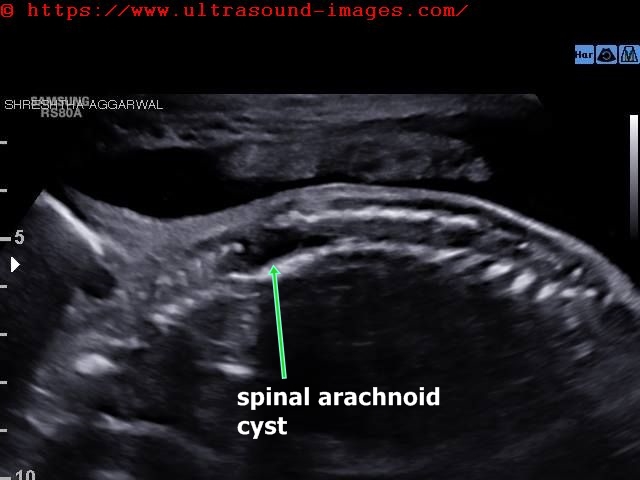
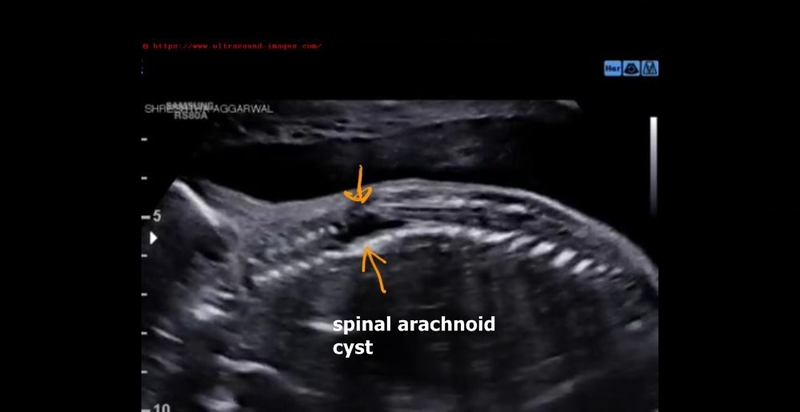
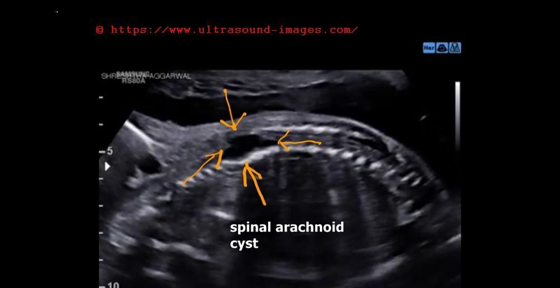
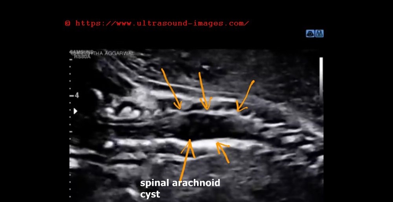
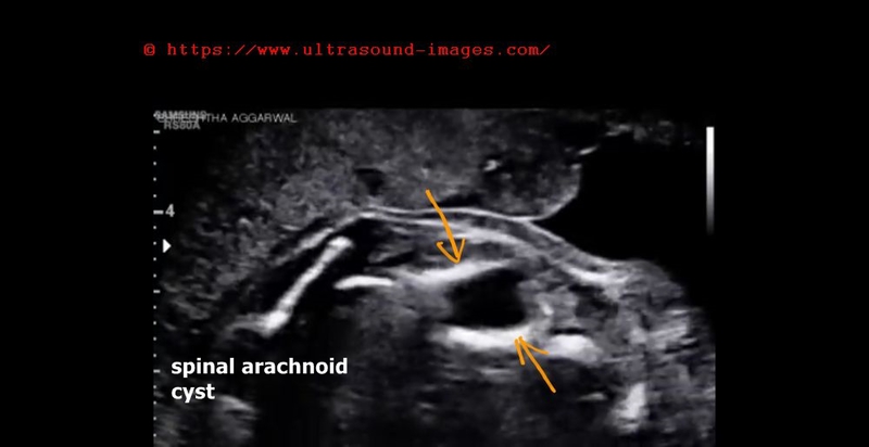
- Arachnoid cysts are benign cystic lesions formed due to splitting of the arachnoid membrane
- Case above shows an intra-spinal cystic lesion in fetal thoracic vertebrae at 24 weeks pregnancy
- Spina bifida occulta present (closed spinal dysraphism)
- Differential diagnosis includes thoracic meningocele
- Usually asymptomatic in neonates
- May cause spinal cord compression symptoms post natal period
- Cystic lesion contains cerebrospinal fluid
- Extremely rare in spine, (with the thoracic spine usually involved as in this case) as most arachnoid cysts are intracranial
- Images courtesy of Dr.Shreshtha Aggarwal MD, DGO, India, fetal medicine specialist.
- References:
- Researchgate article spinal arachnoid cysts
- E-medicine article on arachnoid cysts
© 2026, Joe Antony, MD All images and content in this site are copyrighted. www.ultrasound-images.com When visiting this site, we may use cookies to improve the performance of this site. Use of this site means you accept the use of cookies. Read our privacy policy at this page: https://www.ultrasound-images.com/privacy-policy/