
Ultrasound images of pathology of the eye and orbit
Contents of this page
- Trauma to eye- Intraocular foreign body
- Orbital trauma- Corneal ulceration
- ectopia lentis or luxation of the lens
- cataract-with-asteroid-hyalosis
Trauma to eye- Intraocular foreign body
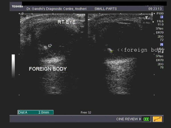
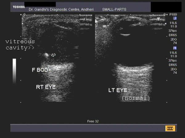
Sonography of the orbits was performed on this patient who sustained injury. Ultrasound images reveal a hyperechoic object in the vitreous cavity within the vitreous humor of the right eyeball. These sonographic findings are diagnostic of intraocular foreign body. Ultrasound image of the normal eye is shown for comparison. Images courtesy of Dr. Jaydeep Gandhi, Radiologist, Mumbai, India.
Reference:
1) http://www.emedicine.com/oph/topic648.htm
2) http://www.emedicinehealth.com/anatomy_of_the_eye/article_em.htm
Orbital trauma- Corneal ulceration
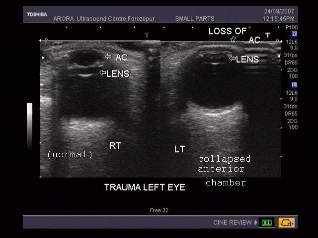
Ulcer of cornea
This patient had a penetrating injury to the left eye. These ultrasound images reveal the resulting severe corneal ulceration and collapse of the anterior chamber of the eyeball due to escape of aqueous humor. The vitreous humor is well preserved. The normal Rt. eye is shown for comparison. Images courtesy of Dr. Vikas Arora, Ferozepur, India.
ectopia lentis or luxation of the lens
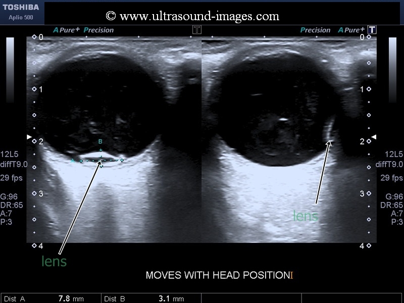
These ultrasound images (above) show the lens has been dislocated or luxated to the dependent part of the patients eyeball, close to the retina. This condition is called ectopia lentis or luxation of the lens. If partial displacement of the lens occurs, it is called subluxation of the lens. The commonest cause of luxation of the lens is physical trauma to the eyeball. With change in posture the luxated lens (arrows) can be seen to gravitate to the dependent part of the eyeball. These ultrasound images of luxation of the lens are courtesy of Dr. Durr-e-Sabih, MBBS, FRCP.
References:
cataract-with-asteroid-hyalosis
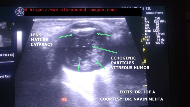
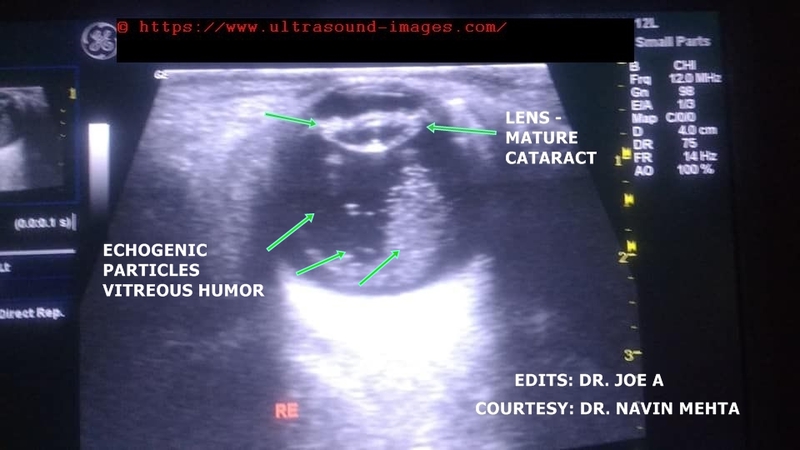

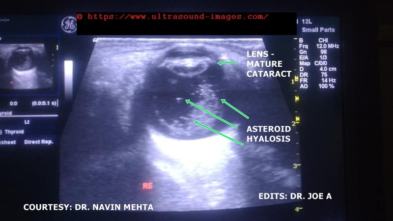
Ultrasound images of Rt. eye in elderly female patient show following features:
= echogenic and thickened lens of the eye--> diagnostic of mature cataract (see the ultrasound images with arrows pinting to the lens).
= multiple echogenic (bright) particles within the vitreous humor or vitreous chamber
= these particles are easily mobile on movement of the eyeball
= the particles donot produce shadowing. The mobile and sparkling nature (brightly echogenic) of the particles help differentiate asteroid hyalosis from vitreous hemorrhage.
= Significance- asteroid hyalosis is not clinically significant as the particles donot produce major interference in visual quality. However, abundant particles in the vitreous humor may cause irritation to the patient during vision. The particles are also a type of floater within the eye.
= composition: asteroid hyalosis particles are composed of calcium phosphates and lipids- the so called calcium soaps.
Above ultrasound images of cataract with asteriod hyalosis are courtesy of Dr. Navin Mehta, MD. India
© 2026, Joe Antony, MD All images and content in this site are copyrighted. www.ultrasound-images.com When visiting this site, we may use cookies to improve the performance of this site. Use of this site means you accept the use of cookies. Read our privacy policy at this page: https://www.ultrasound-images.com/privacy-policy/