
Ultrasound images of abnormalities of amniotic fluid
Contents of this page
- Polyhydramnios
- Hyperechoic amniotic fluid/ Liquor
- Free floating particles in Liqor amnii
- Severe oligohydramnios in late 2nd trimester
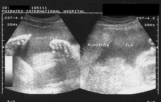
Polyhydramnios
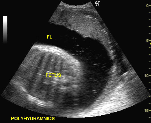
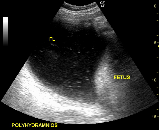
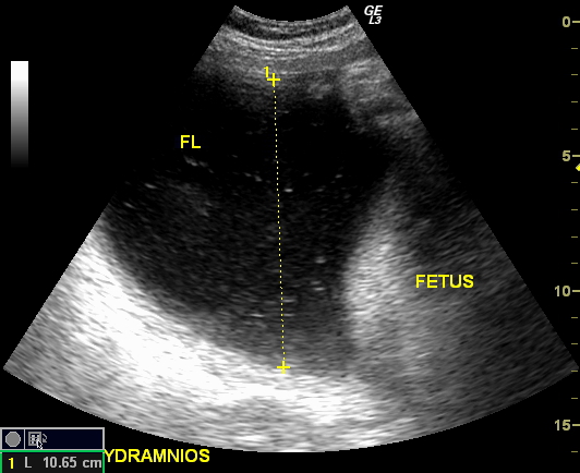
The above ultrasound images show excess amniotic fluid (the largest single pocket measuring 11 cms. approximately). This suggests mild to moderate polyhydramnios. Particulate matter seen in the fluid is due to fetal meconium. Sonography of polyhydramnios was done using a GE, Logiq-3 ultrasound machine.
Reference:
Hyperechoic amniotic fluid/ Liquor
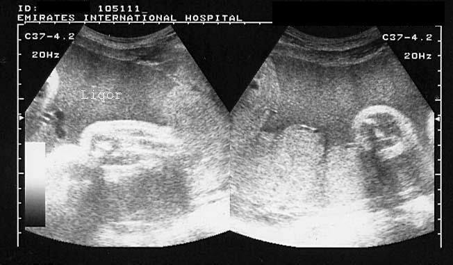

Increased echogenicity of Liqor amni These ultrasound images reveal markedly echogenic (hyperechoic) amniotic fluid. The fluid shows almost the same echogenicity as the placenta. Such appearances of the liquor amni on sonography are seen due presence of vernix caseosa (commonest cause), blood or meconium in the amniotic fluid. Images courtesy of Dr. Ravi Kadasne, UAE.
Reference:
1) http://www.thefetus.net/page.php?id=1763 (free article)
2)http://www.jultrasoundmed.org/cgi/content/abstract/13/2/95
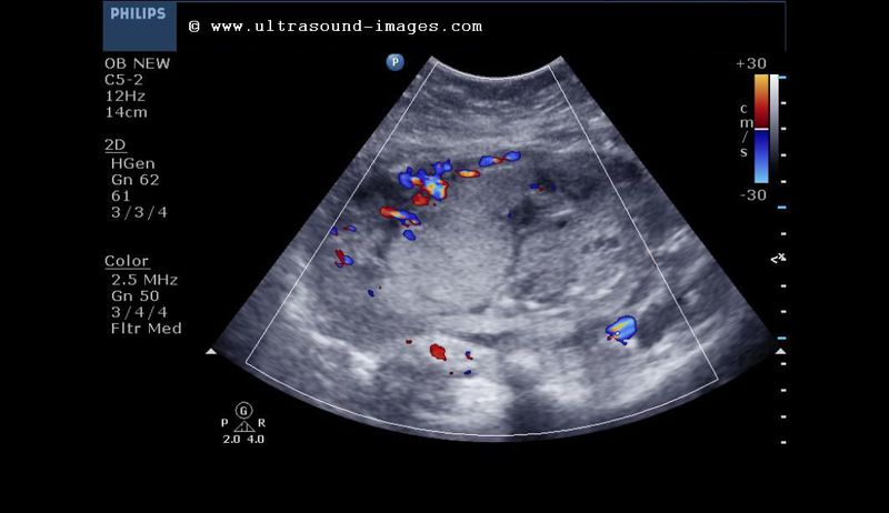
Free floating particles in Liqor amnii
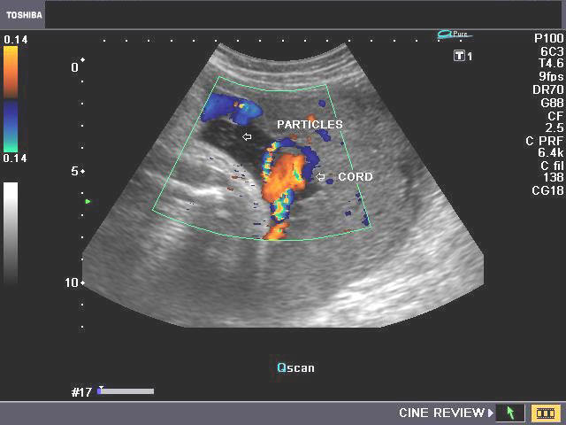

This was a 38 week old pregnancy showing multiple echogenic particles in the amniotic fluid (liqor amnii) on ultrasound/ Color Doppler imaging. Such freely mobile particles in the amniotic fluid are called free floating particles and are commonly seen in the 3rd trimester. Sometimes, particulate debris, when abundant, can be confused with normal umbilical cord floating in the amniotic fluid, on grey scale B-mode ultrasound. However, Color Doppler easily distinguishes between the two (see Doppler images above). Studies show some direct correlation between the size and number of free floating particles and levels of Maternal serum alpha feto-protein (MSAFP). These particles are presumed to be the result of vernix caseosa shed from the fetal skin. However, free floating particles may be visualized on ultrasound imaging even during the 2nd trimester (when vernix is not present). Have a look at the short ultrasound video clips of free floating particles in a 3rd trimester pregnancy at: http://cochinblogs.blogspot.com/2010/01/free-floating-particles-in-amniotic.html More ominously, such debris in the amniotic fluid may be caused due to hemorrhage into the fluid.
Reference:
http://www.jultrasoundmed.org/cgi/content/abstract/2/3/107 (abstract)
http://content.karger.com/ProdukteDB/produkte.asp?Doi=263858 (abstract)
Severe oligohydramnios in late 2nd trimester

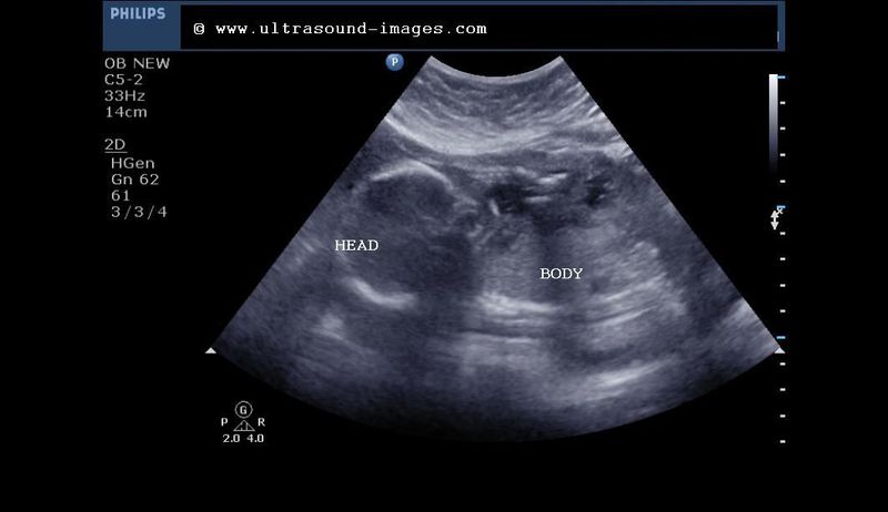
This is the case of a 19 week fetus with severe oligohydramnios. Fetus lies in breech presentation. The ultrasound images below show lack of any fluid pocket or very little amniotic fluid in any of the pockets.this patient had bleeding PV with a possible premature rupture of the amniotic membrane
Lack of fluid ( liqor amni) even around the Fetal abdomen or the Fetal limbs
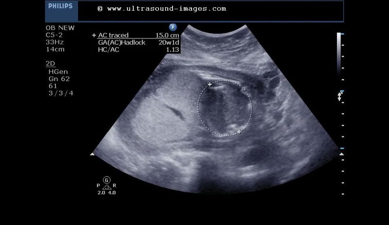
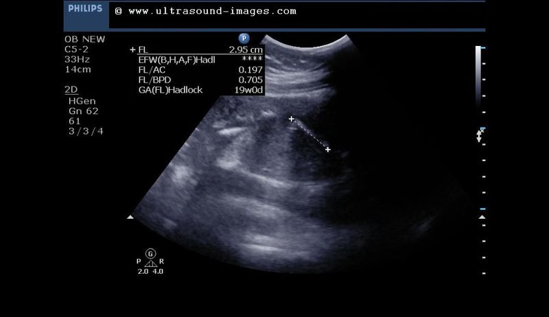

Among the Causes of oligohydramnios are- premature rupture of membranes, Fetal hypoxaemia, renal agenesis or dysgenesis, intake of certain drugs, Fetal demise and also Fetal IUGR. oligohydramnios is diagnosed when the four quadrant AFI or amniotic fluid index is less than five or the maximum vertical fluid pocket is less than 2 cm. Among the complications of such severe oligohydramnios are Fetal limb contractures and Fetal lung hypoplasia.
References:http://emedicine.medscape.com/article/405914-overview#a7
© 2026, Joe Antony, MD All images and content in this site are copyrighted. www.ultrasound-images.com When visiting this site, we may use cookies to improve the performance of this site. Use of this site means you accept the use of cookies. Read our privacy policy at this page: https://www.ultrasound-images.com/privacy-policy/