
Color Doppler ultrasound imaging of arteries and veins- page2:
Contents of this page
- Thrombosis of anterior Tibial vein:
- severe stenosis of femoral artery
- normal venous doppler of lower limb
- mild diabetic arteriopathy of lower limb
- severe diabetic arteriopathy of the lower limb
- thrombosis of the right anterior tibial and peroneal veins
- thrombosis of the lower half right popliteal vein
- a case of popliteal vein thrombosis
- colour doppler duplex study of hyperemia of lower limb
- aortic thrombosis- color and spectral Doppler imaging
- popliteal-vein-thrombosis-case-3
- Calcified atheromas of femoral artery- (calcific atherosclerosis)
- Takayasu Arteritis or Aortoarteritis or Pulseless disease
- Severe-stenosis-of-Rt-SFA-case-2
- Heel-and-toe-method
- pulsatile-venous-flow
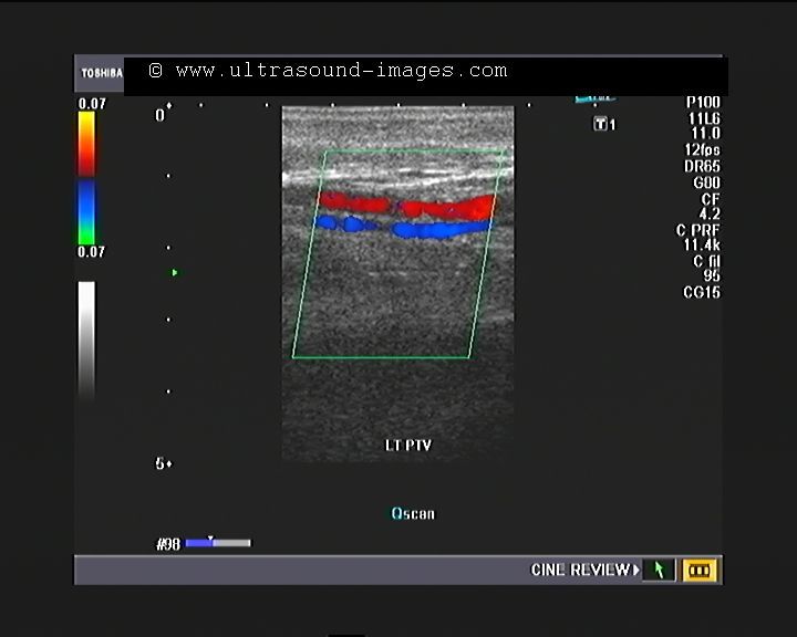
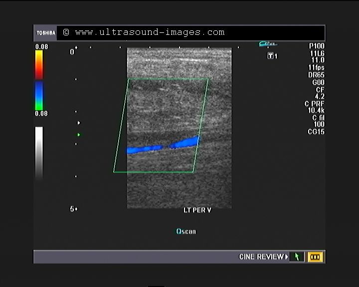
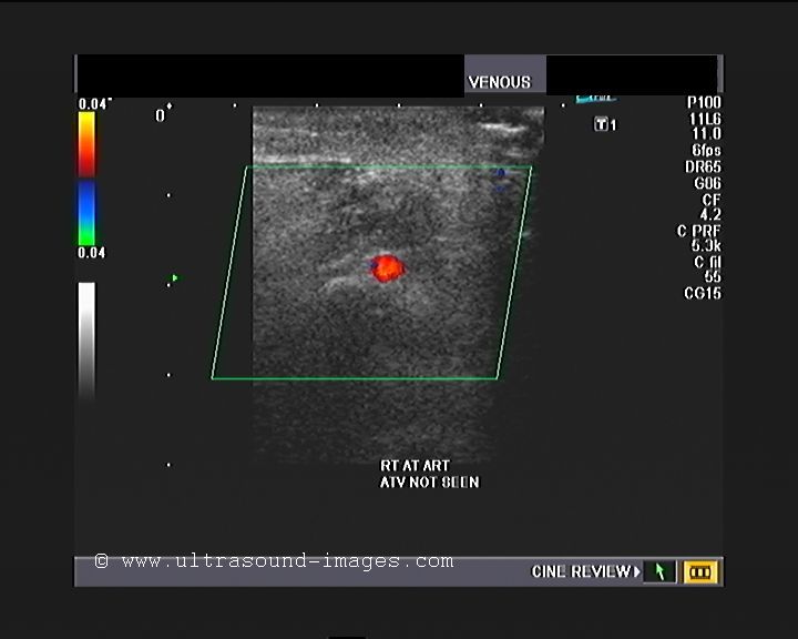
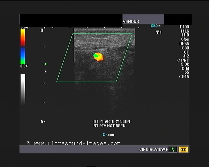
Thrombosis of anterior Tibial vein:
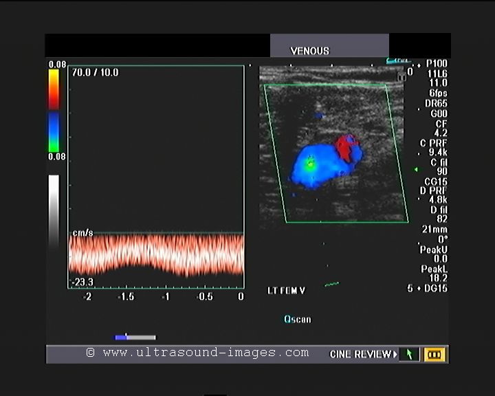
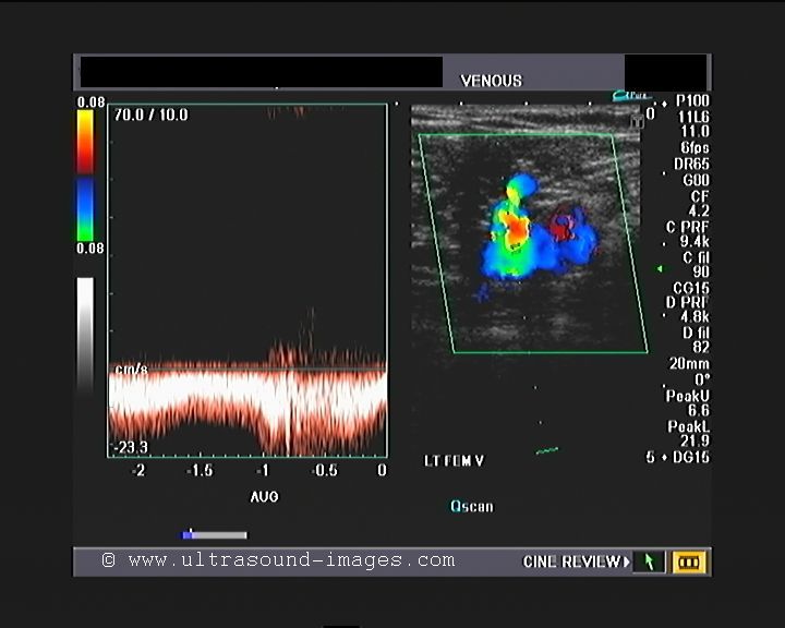
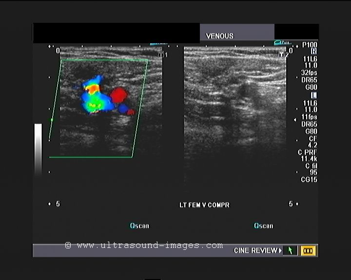
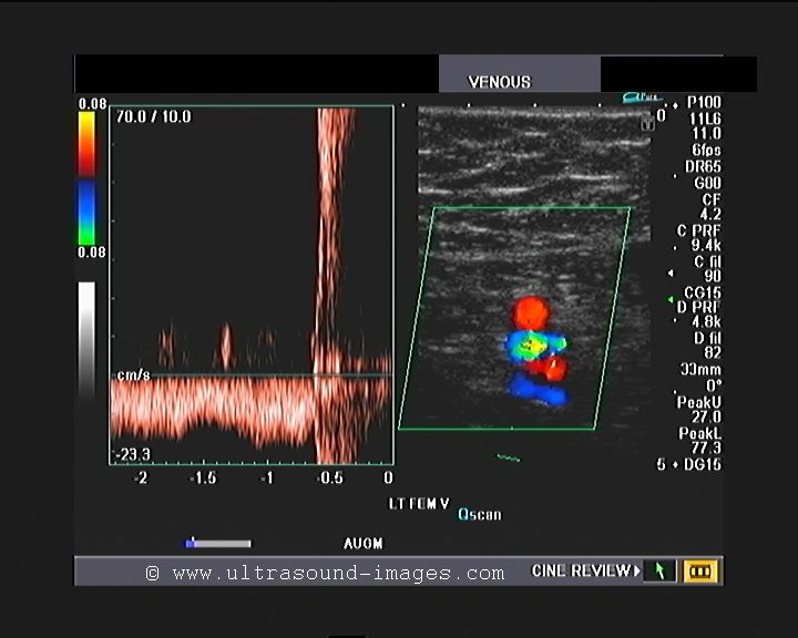


Two days later, she developed pain along the anterior aspect of the left leg which was severe in nature. there was also localised tenderness along the anterior aspect of the left leg with associated oedema.
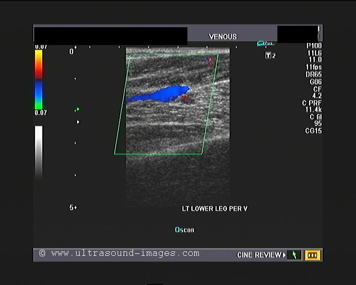
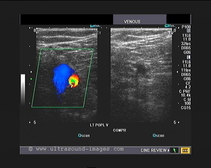
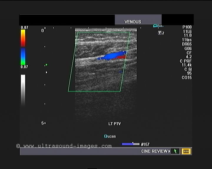
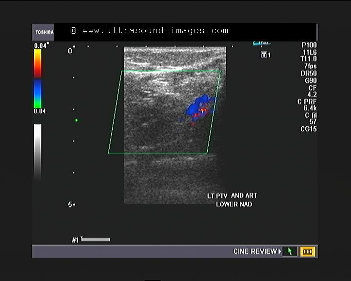
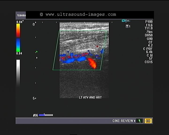
Colour Doppler imaging of the left lower limb show normal flow and colour Doppler appearances of the left femoral vein, the left popliteal vein, as well as the left peroneal vein and the posterior tibial vein.
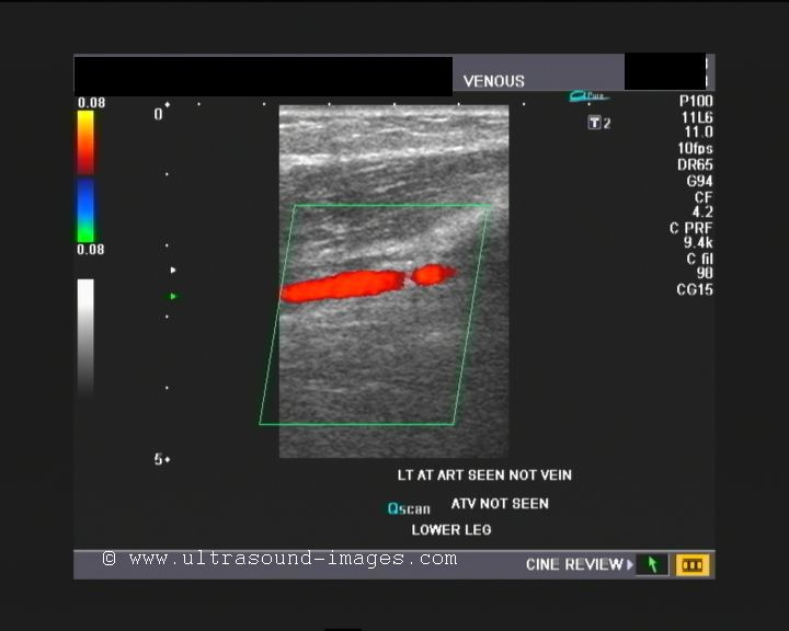
However, the left anterior tibial vein was not visualised alongside the left anterior tibial artery. There was no flow observed in the anterior tibial vein in its normal location near the anterior tibial artery.
These colour Doppler ultrasound images of the venous system of the left lower limb show clear evidence of thrombosis of the left anterior tibial vein. This explains the oedema and tenderness in this region.
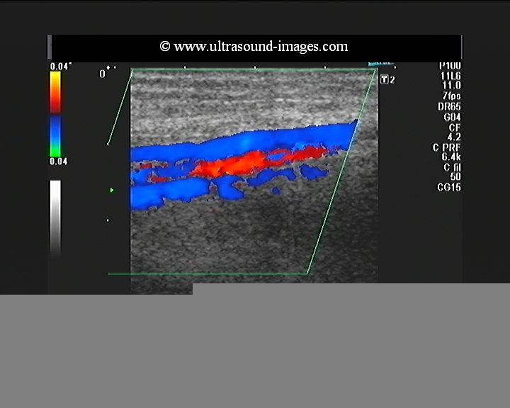
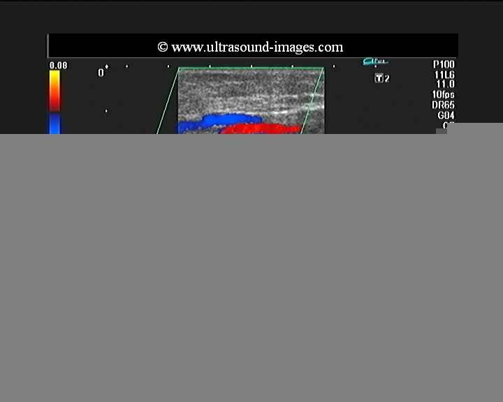
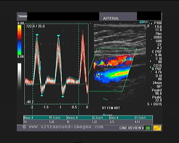
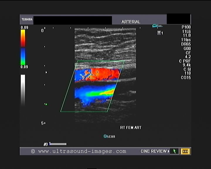
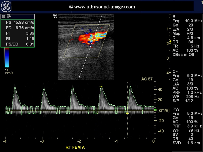
severe stenosis of femoral artery
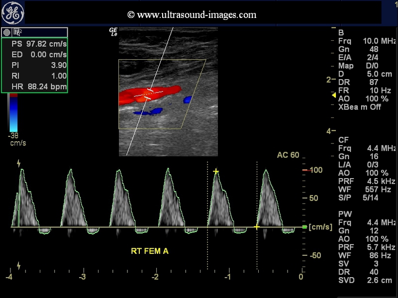
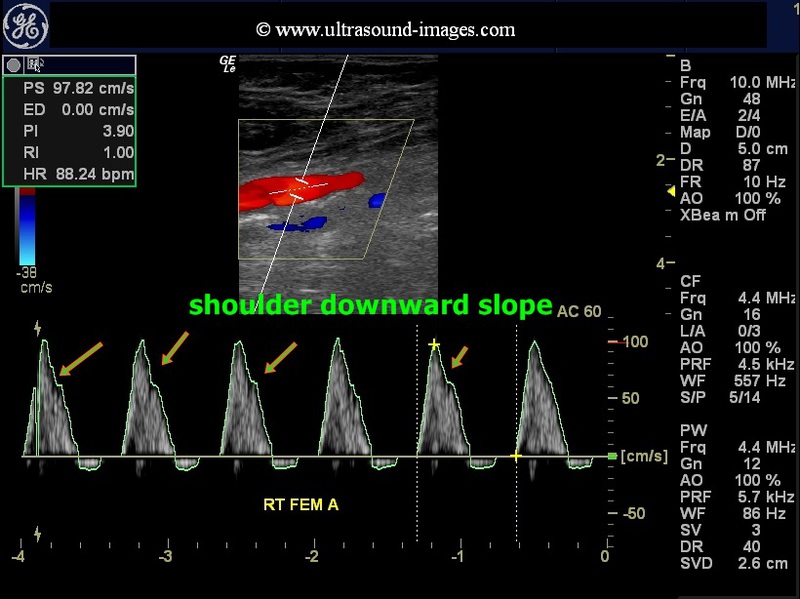
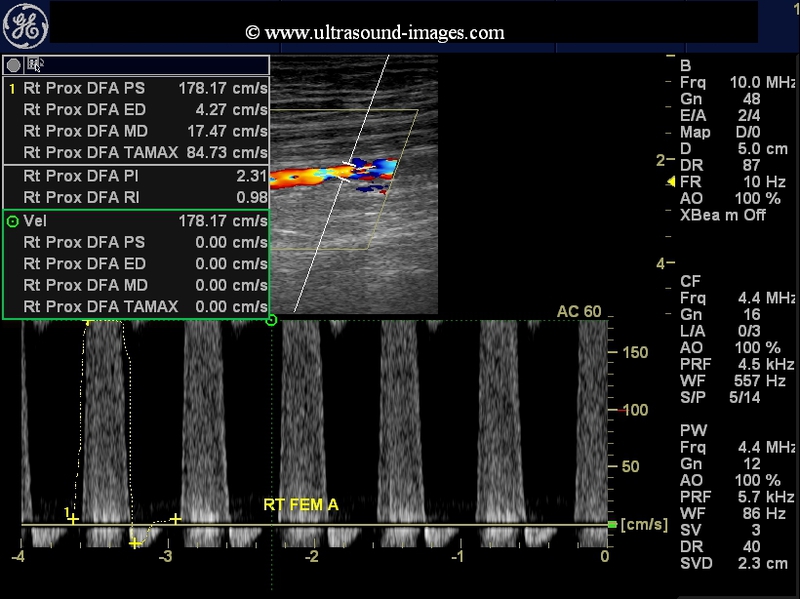
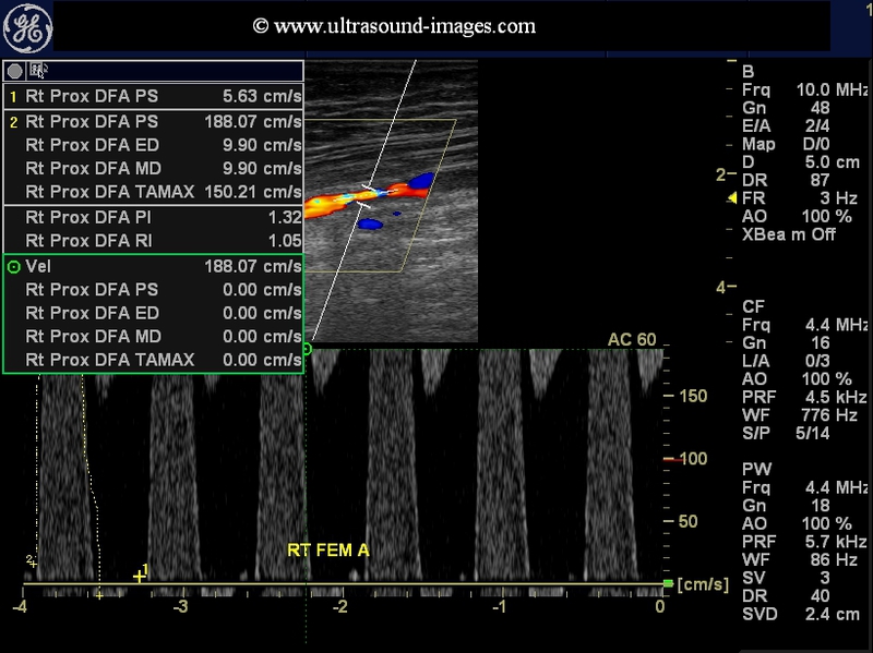

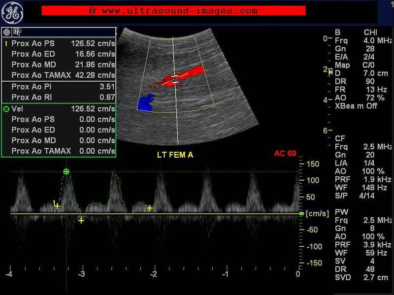
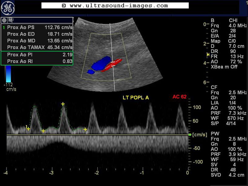
This elderly lady with a history of diabetes has evidence of ischemic changes of the right leg, with onset of gangrene of the toes. Colour Doppler study of the lower limb shows a severe stenosis of the right femoral artery, in its upper third. The diameter of the stenotic segment measured only 1.8 mm.
The prestenotic segment (Rt SFA/ femoral A) shows typical changes- steep upward slope of systole and shouldering of downward slope of systole (see green arrows in femoral A image).
Colour Doppler and spectral Doppler ultrasound examination of the stenotic segment of the right femoral artery shows a peak systolic velocity more than 190 cm/s (Vs). The peak systolic velocity in the pre-stenotic segment was 97 cm/s (Vp). The ratio Vs/Vp is greater than 2 suggesting a moderate to severe stenosis of the affected segment of the femoral artery.
The arterial tree below the femoral including the right popliteal, the right peroneal, posterior tibial and anterior tibial arteries all show moderate dampening off the blood flow in these arteries.
Reference: Doppler waveform changes in arterial stenosis
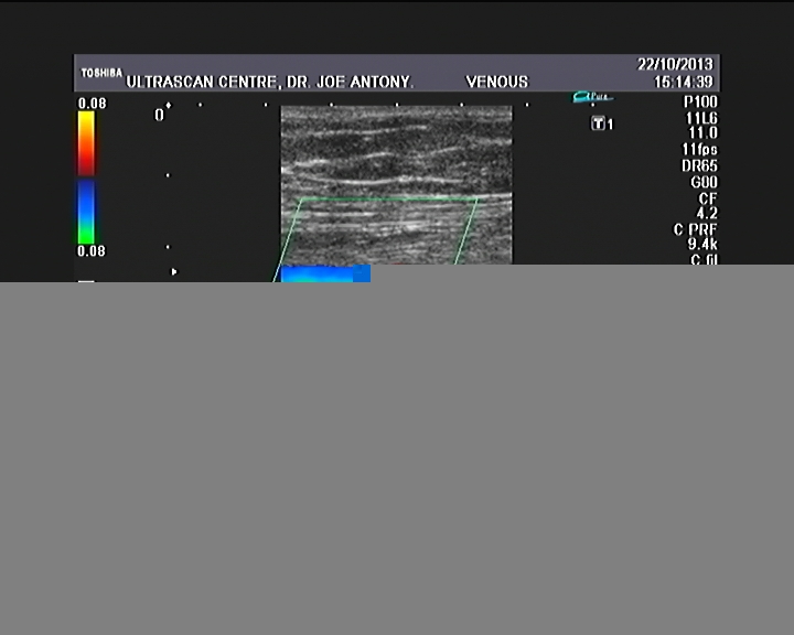
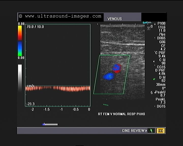
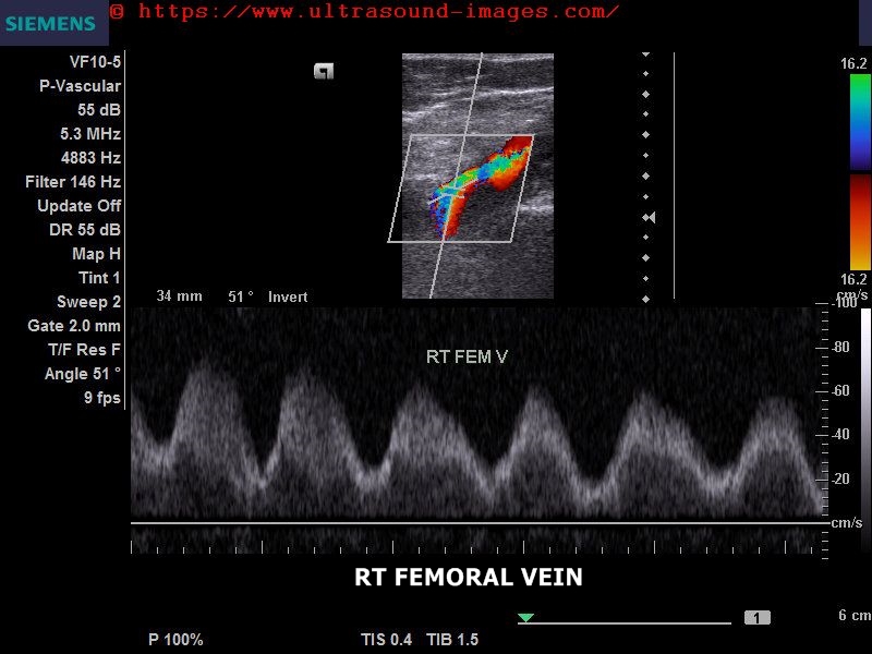
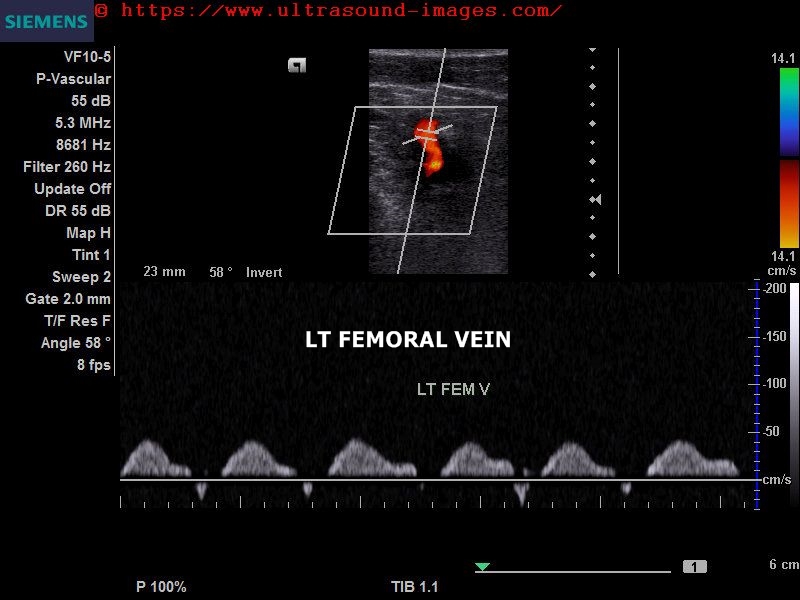
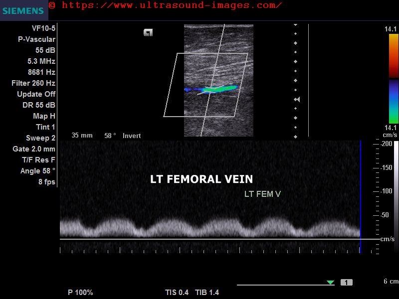
normal venous doppler of lower limb
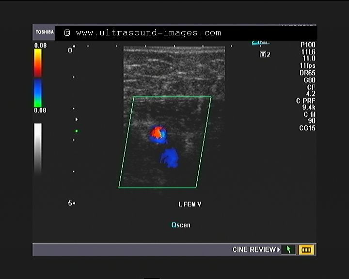


This patient with pain in the Left lower limb was evalulated with color and spectral Doppler ultrasound of the venous system of the left lower limb. The images reveal a normal color Doppler study of the veins, including the left femoral, popliteal, anterior and posterior tibial and peroneal veins. Often, the veins of the leg cannot be visualized in their entirety. It is often more practical to image the upper third and distal third of the anterior, posterior tibial and peroneal veins.
normal venous doppler of lower limb- case-2
this is yet another example of normal venous colour Doppler study of the left lower limb. The patient had in all probability, a mild form of deep vein thrombosis with oedema of the left lower limb, which almost completely resolved. On colour Doppler ultrasound study of the left lower limb, the veins both superficial and deep, appear totally normal. this study shows normal response to compression, augmentation and Valsalva manoeuvre in all the deep veins. Visualisation of the deep veins was excellent up to the ankle. Observe that the anterior and posterior tibial veins are paired, with two anterior tibial veins on either side of the anterior tibial artery and the same appearance in the case of the posterior tibial veins.
mild diabetic arteriopathy of lower limb

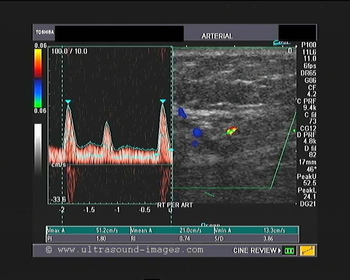
This elderly lady has pain in the right lower limb, below the knee. Ultrasound and colour Doppler imaging of the right lower limb shows early changes of diabetic arteriopathy or what is called as diabetic vasculopathy. This is characterised by the features shown in these colour Doppler ultrasound images;
1. Spectral broadening of the arterial waveform from the popliteal artery downwards
2. Mild decrease in peak systolic velocity below the popliteal artery
3. Early changes of loss of the tri-phasic spectral waveform is seen in peroneal artery
all these above-mentioned changes are typically seen in early diabetic arteriopathy suggesting mild stenosis in a diffuse fashion below the popliteal arter.
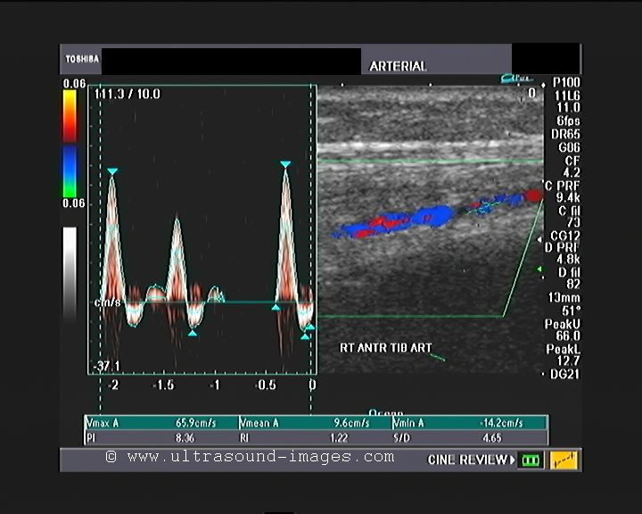
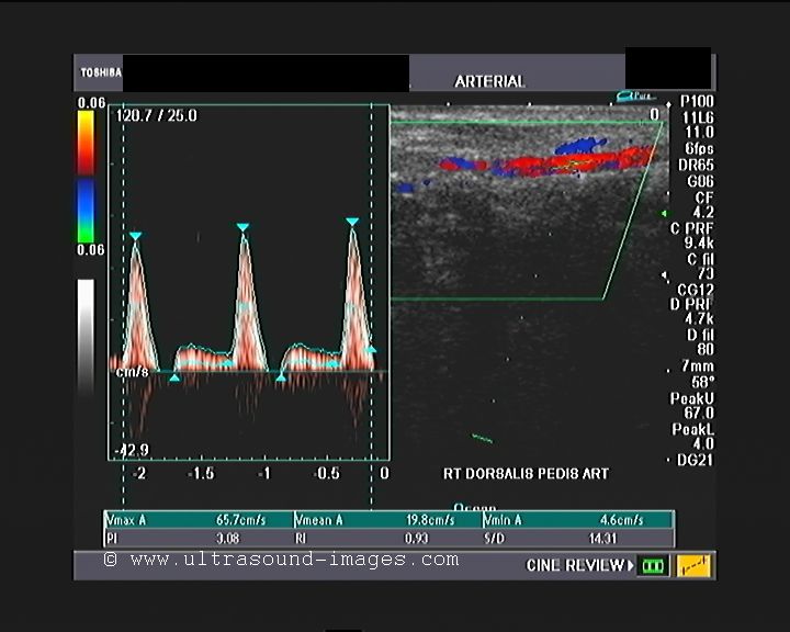
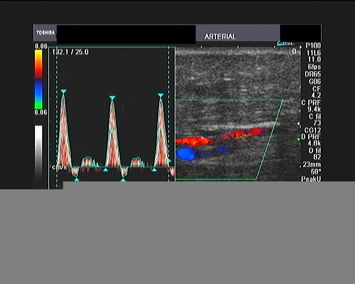
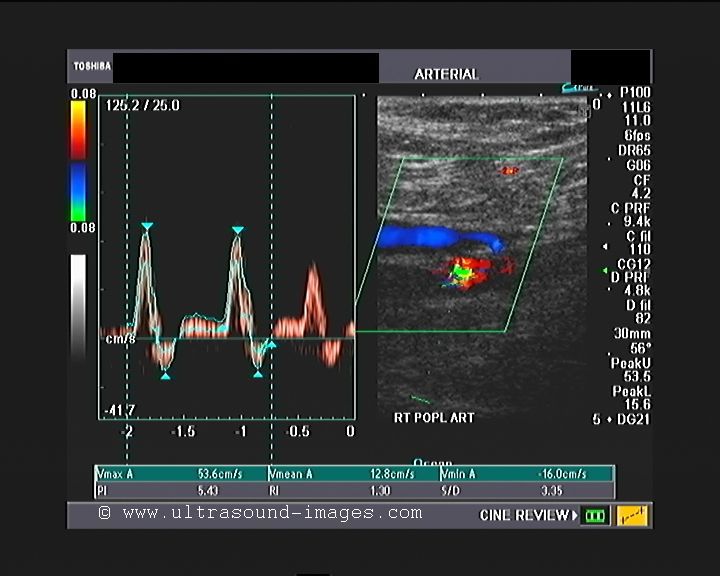
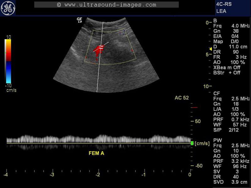
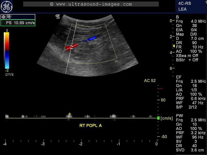
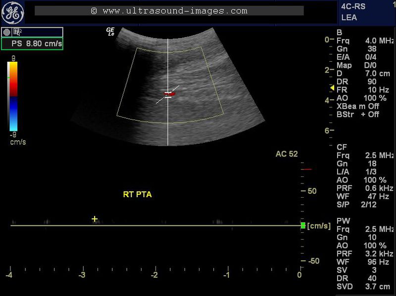
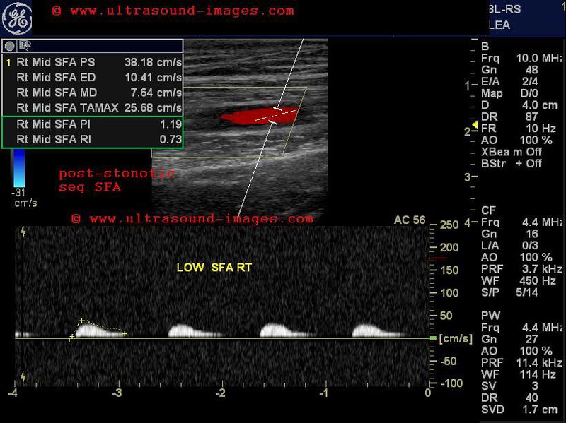
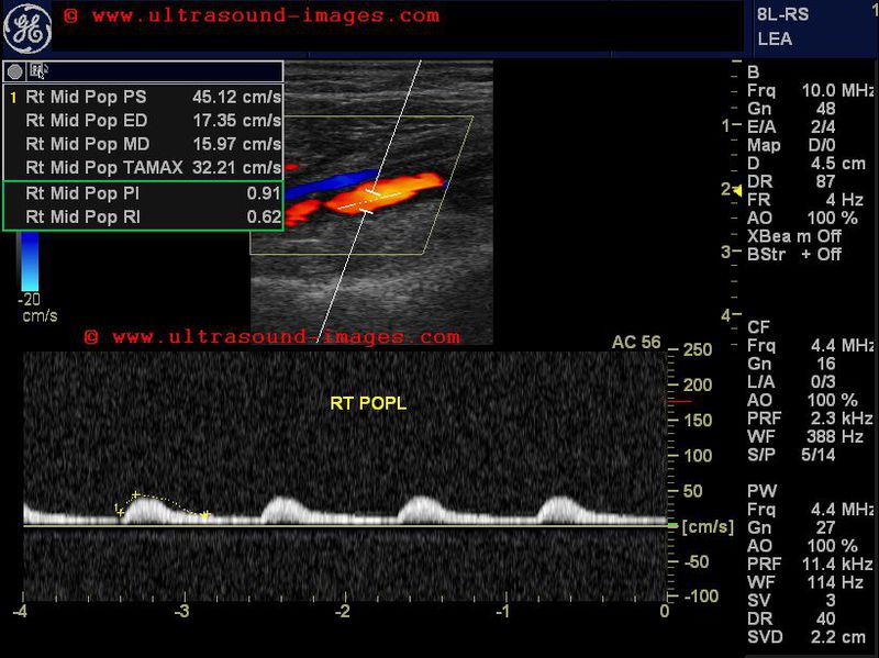
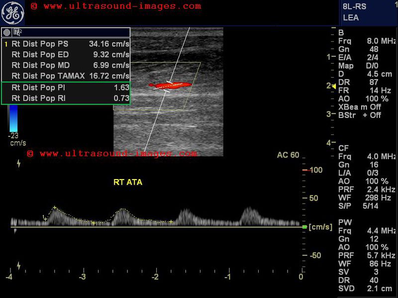
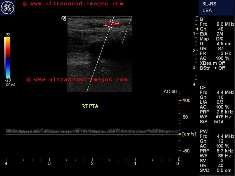
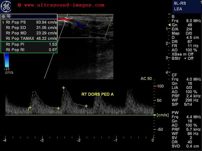
severe diabetic arteriopathy of the lower limb
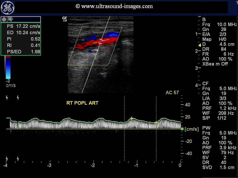
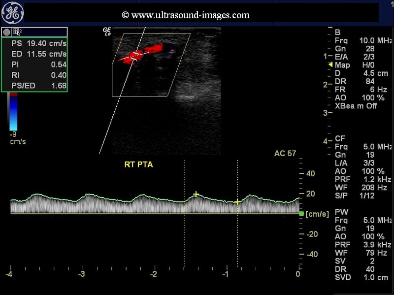
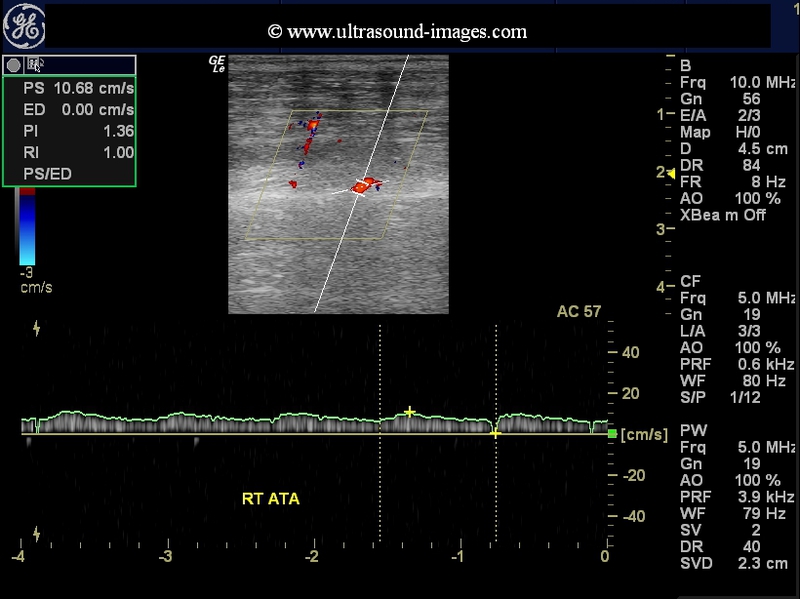
This elderly male diabetic patient had severe pain in the right leg. Colour Doppler ultrasound and spectral Doppler tracing reveal severe diffuse stenotic disease of the arterial system starting from the right popliteal artery to the dorsalis pedis artery. This kind of a picture is seen in long-standing diabetes mellitus resulting in severe arterial disease or arteriopathy. The waveform of the arteries affected including the popliteal artery, the anterior and posterior tibial arteries show loss of tri-phasic spectral waveform which is replaced with spectral broadening and an almost venous like flow pattern. This is known as severe dampening of the waveform and this also known as tardus parvus flow.
See this illustration of tardus parvus flow
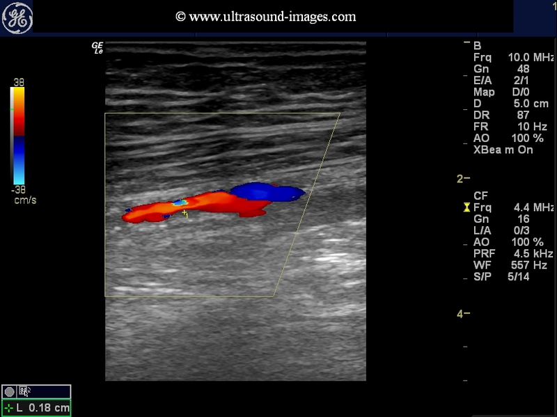
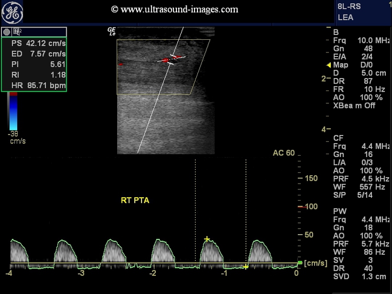
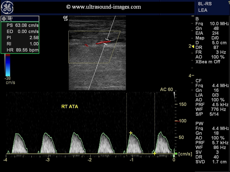
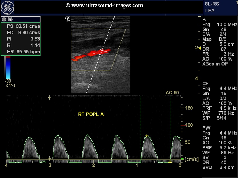
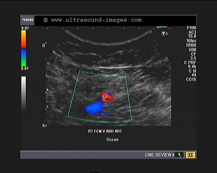
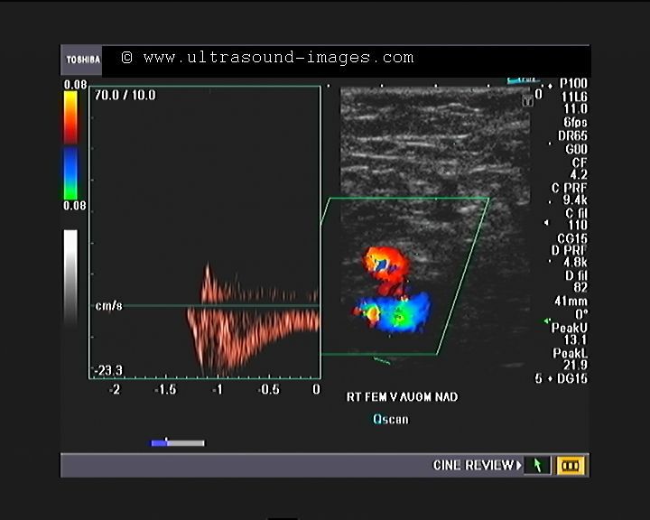
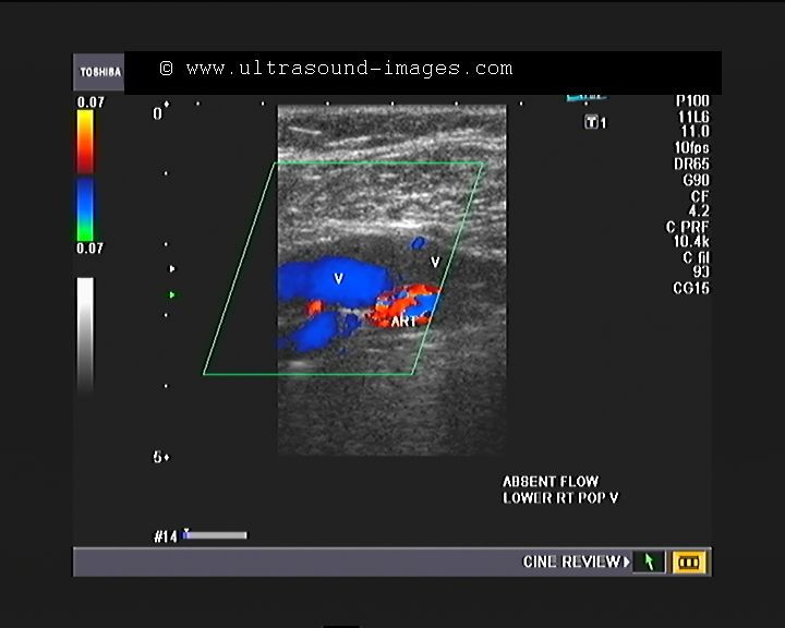
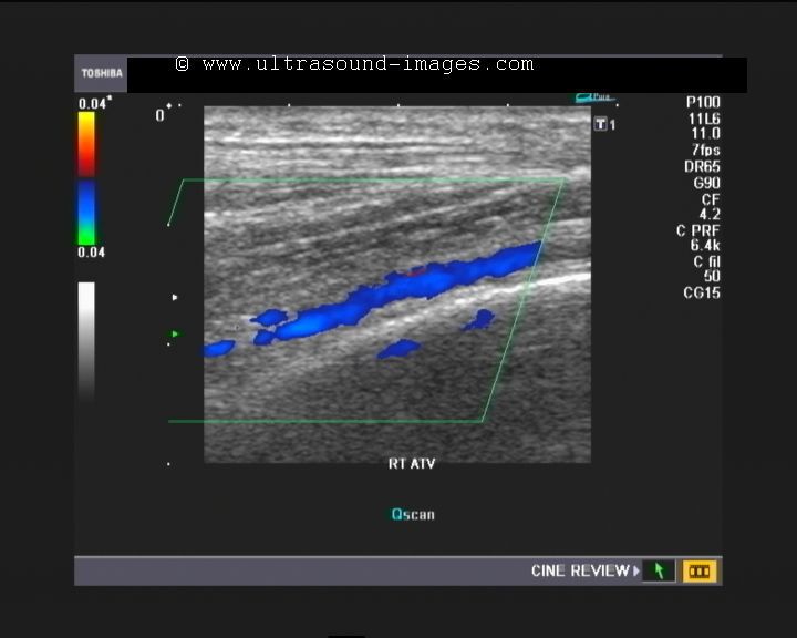
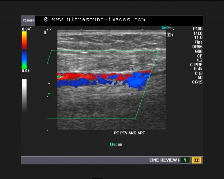
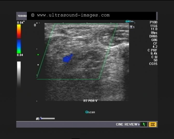
thrombosis of the right anterior tibial and peroneal veins

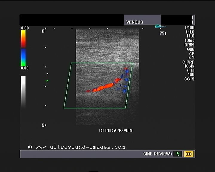


This his elderly lady had pain and edema of the right leg and foot. Colour Doppler ultrasound showed normal flow, compressibility and augmentation of the right femoral and popliteal veins. however ultrasound and Doppler study showed absent flow in the right anterior tibial and peroneal veins. Despite very low PRF settings, colour Doppler study failed to show any flow in these two veins. Initially, the right posterior tibial vein also did not show flow. However, with the patient in the sitting posture, normal flow was detected in the posterior tibial vein.
It must be noted, that most authors believe that colour Doppler study of the deep veins of the calf, namely the anterior and posterior tibial and peroneal veins, is not conclusive to diagnose a case of deep vein thrombosis involving the veins of the calf. However, the presence of normal flow on colour Doppler in these calf veins, helps to rule out deep-vein thrombosis involving the veins of the calf.
Most authors believe that deep vein thrombosis involving the calf veins is not a major pathology and can resolve spontaneously over a short period of time- that is a few weeks.
The following references are very useful in this study of and understanding of Doppler study of the veins of the lower limb:
![]()
http://pubs.rsna.org/doi/full/
(very good)... calf vein ultrasound below knee not needed for DVT study - conclusion
http://pubs.rsna.org/doi/full/
.. DVT study.. good images
http://gecommunity.
http://www.ncbi.nlm.nih.gov/
(small abstract)..incidence of calf and below knee vein DVT study doppler
http://www.ncbi.nlm.nih.gov/
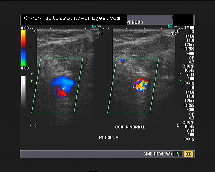
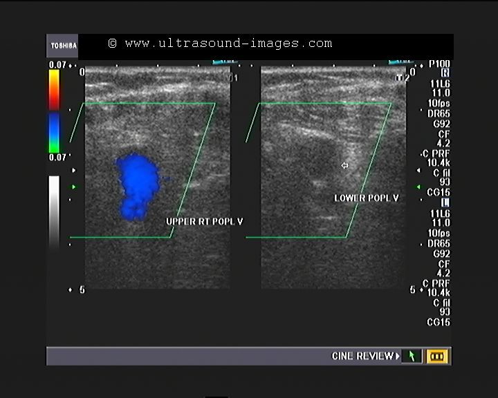
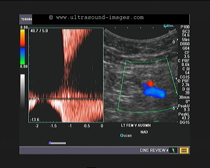
thrombosis of the lower half right popliteal vein
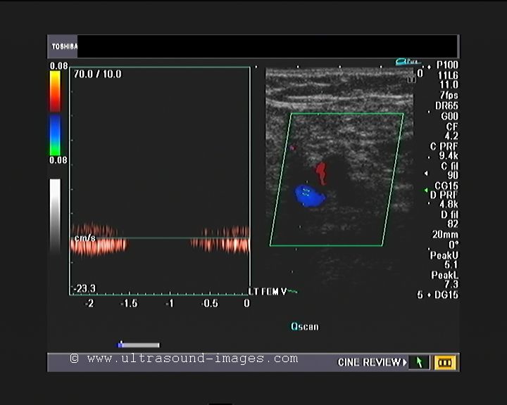
This elderly female patient had pain in the right popliteal region and upper right leg. Colour Doppler and spectral Doppler ultrasound revealed normal flow in the femoral vein and even in the upper right popliteal vein. However, no flow was observed in the lower half of the right popliteal vein, suggesting of thrombosis of the lower half of the right popliteal vein. Spectral Doppler ultrasound further confirmed this diagnosis by showing an absence of any waveform in this region.
normal flow in the right femoral vein
Normal flow is seen in the right femoral vein. However, augmentation of flow in the femoral vein was very weak despite sharp compression of the calf. This can indicate some kind of partial obstruction lower down.
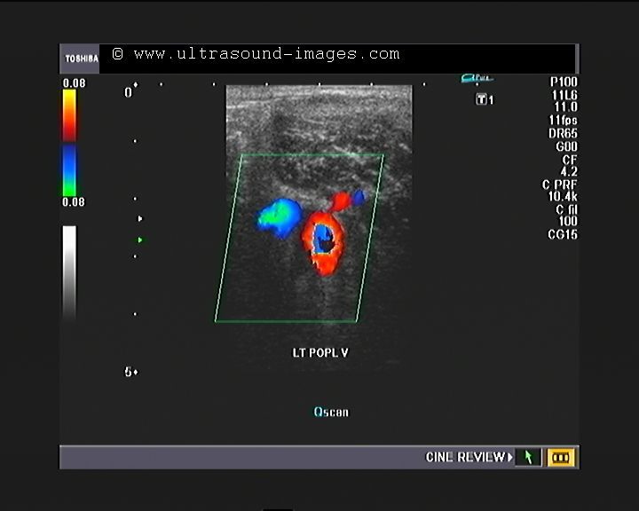
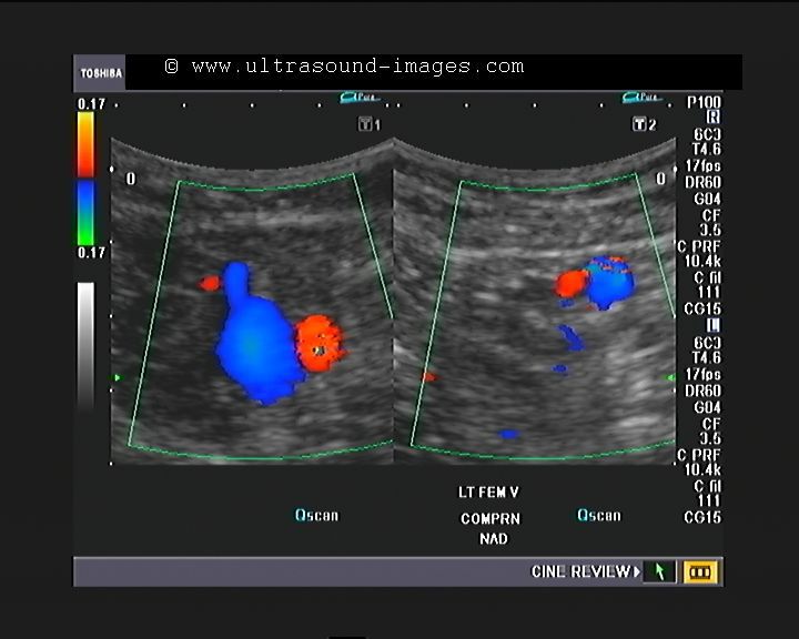
occlusion of right popliteal vein in lower half
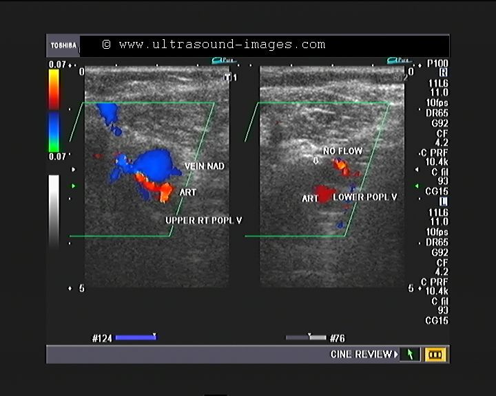
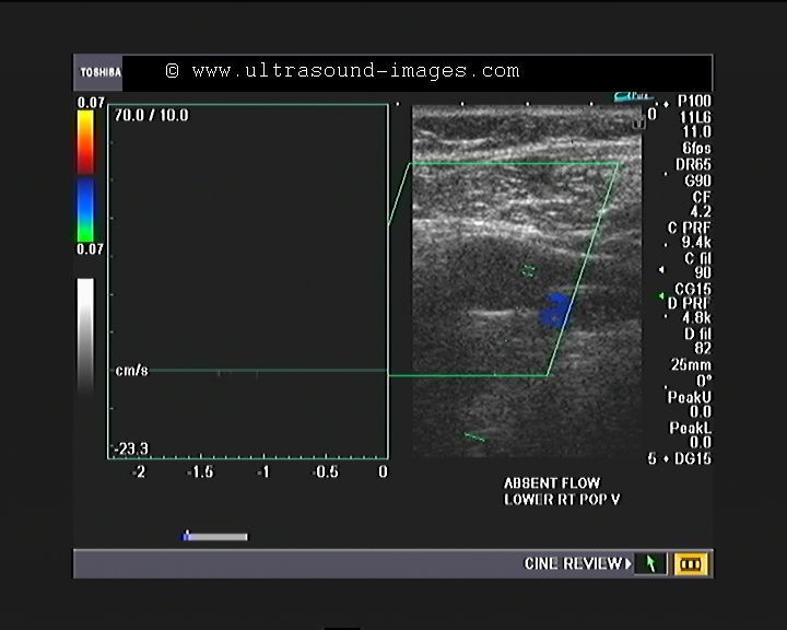
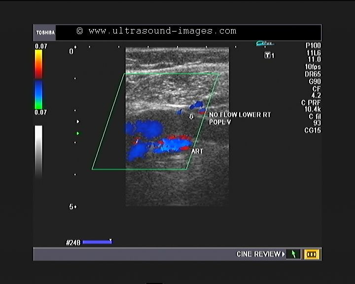
normal flow noted up to the upper half of the right popliteal vein. Both power Doppler ultrasound and colour Doppler ultrasound revealed lack of flow in the lower half of the right popliteal vein. This is diagnostic of occlusion by thrombosis of the distal right popliteal vein.
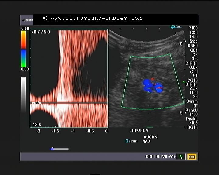
normal visualisation of the calf veins- contd
the same patient, ( continued)- the calf veins, namely the anterior tibial vein the posterior tibial vein and the peroneal vein appear normal. Normal flow or near normal flow was seen in these veins of the calf, possibly due to the development of colateral veins bypassing the occluded right popliteal vein.
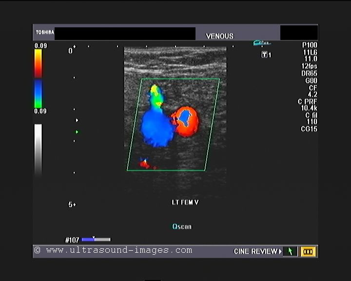
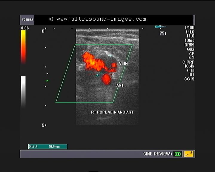
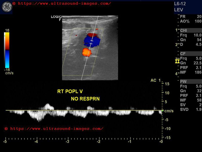
a case of popliteal vein thrombosis
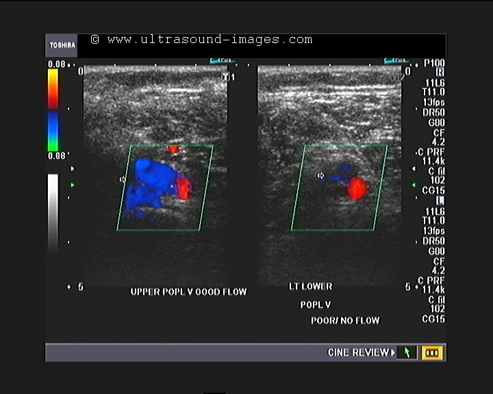


This middle-aged female patient presented with left calf pain and tenderness of a few days duration. She underwent medical treatment for a few days but has persistent mild calf pain. Colour Doppler and spectral Doppler ultrasound revealed normal flow augmentation and compressibility of the left femoral vein. However, the left popliteal vein, shows lack of flow in the lower half , suggesting left popliteal vein thrombosis. spectral Doppler ultrasound showed absent waveform and this was confirmed on colour Doppler ultrasound showing lack of flow signal in the lower half of the left popliteal vein. Thus, this colour Doppler ultrasound study confirms left popliteal vein thrombosis.
normal flow is observed in the deep veins of lower left leg, namely the anterior tibial, posterior tibial and peroneal veins.
Surprisingly, some flow signal was seen in the left popliteal vein on probe pressure and release; this was also seen on eliciting augmentation by pressure below the left calf. This suggests, that the left popliteal vein thrombosis maybe only partial, with some degree of recanalisation following medical treatment ( meaning thrombolysis).
colour doppler duplex study of hyperemia of lower limb
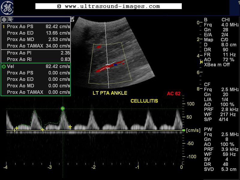

This male patient, aged 41 years had severe cellulitis of the left lower limb, mainly involving the calf and ankle. Colour Doppler and spectral Doppler duplex ultrasound study show the effects of hyperemia of the lower limb due to the cellulitis.
the chief effects of the hyperaemia of the lower limb are seen as:
1. Increased diastolic and end diastolic flow in the spectral Doppler waveforms of popliteal, anterior and posterior tibial arteries.
2. Reduce pulsatility of the waveforms of the popliteal, and the anterior and posterior tibial arteries.
3. Loss of triphasic waveform of the above-mentioned arteries, which is replaced by a monophasic waveform.
Thus, hyperaemia due to cellulitis and inflammation can produce dramatic changes in spectral Doppler waveform which may mimic those seen due to exercise and development of collateral vessels.
aortic thrombosis- color and spectral Doppler imaging


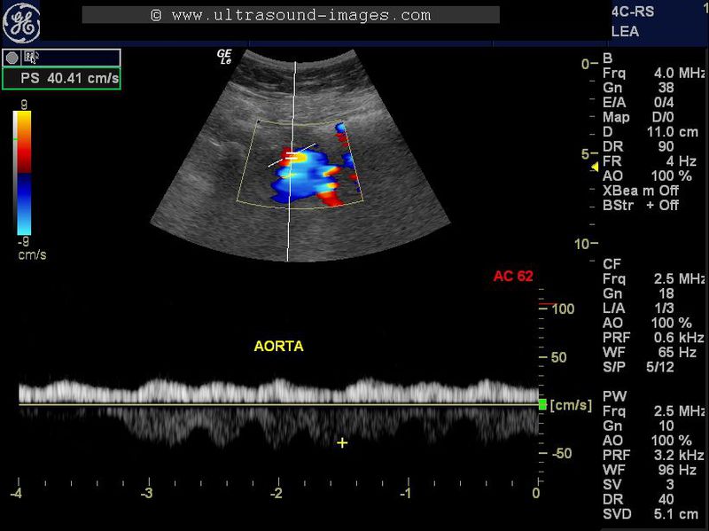
This elderly lady had pain in the right lower limb of acute onset. In addition, she had a history of atrial fibrillation the day before. Colour Doppler and spectral Doppler ultrasound imaging of both lower limbs and the aorta were done. This revealed marked dampening of flow in the arteries of the right lower limb as well as the left lower limb. The arteries we observed in this study included the femoral, popliteal and posterior tibial arteries. Evaluation of the lower abdominal aorta also showed a similar picture on spectral Doppler ultrasound. The colour Doppler and spectral Doppler ultrasound images shown above reveal a tardus-parvus flow pattern similar to that seen in veins. The PSV or peak systolic velocity was also extremely low throughout the right lower limb arteries as well as the left lower limb and the abdominal aorta. All these colour Doppler ultrasound findings point to an obstruction to the flow of blood somewhere along the abdominal aorta. This was confirmed on CT scan and MR imaging. The final diagnosis in this case was abdominal aortic thrombosis.
References:
colour and spectral Doppler ultrasound imaging in aortic thrombosis (free article and images)
popliteal-vein-thrombosis-case-3
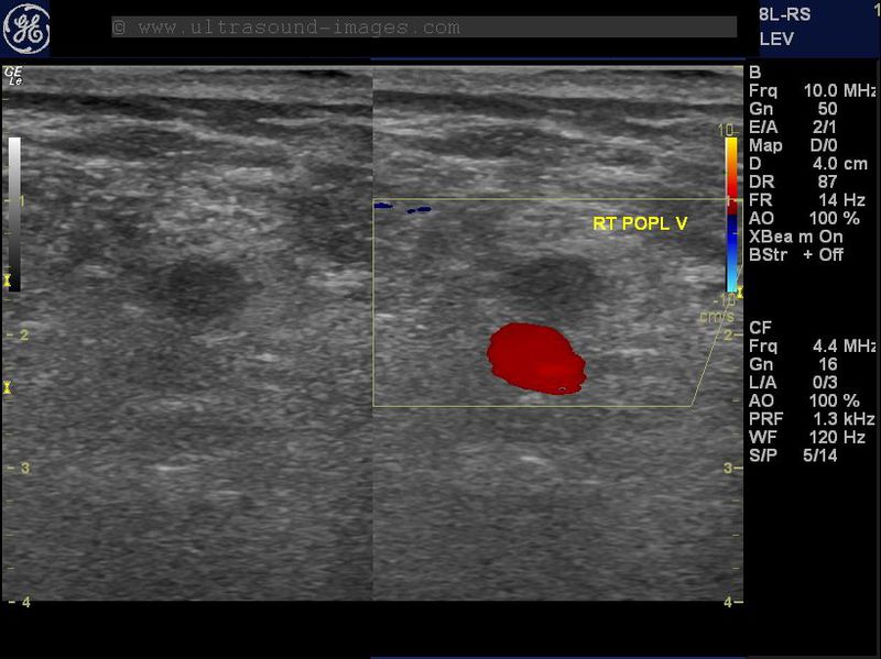
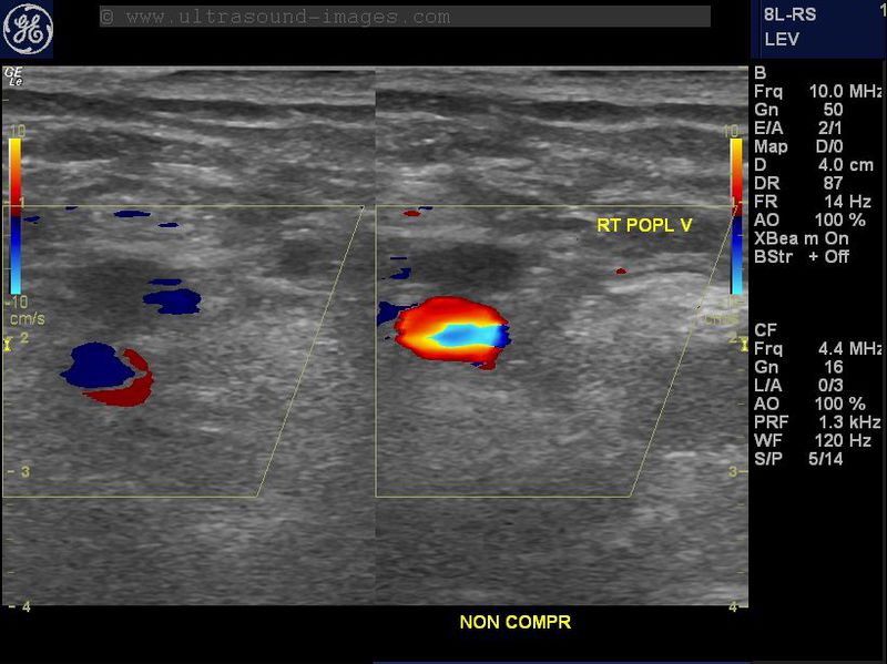
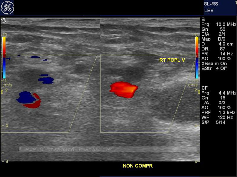
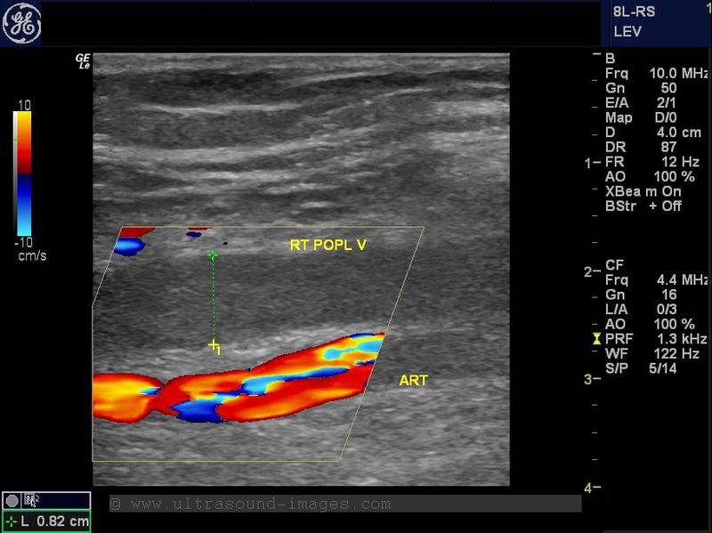
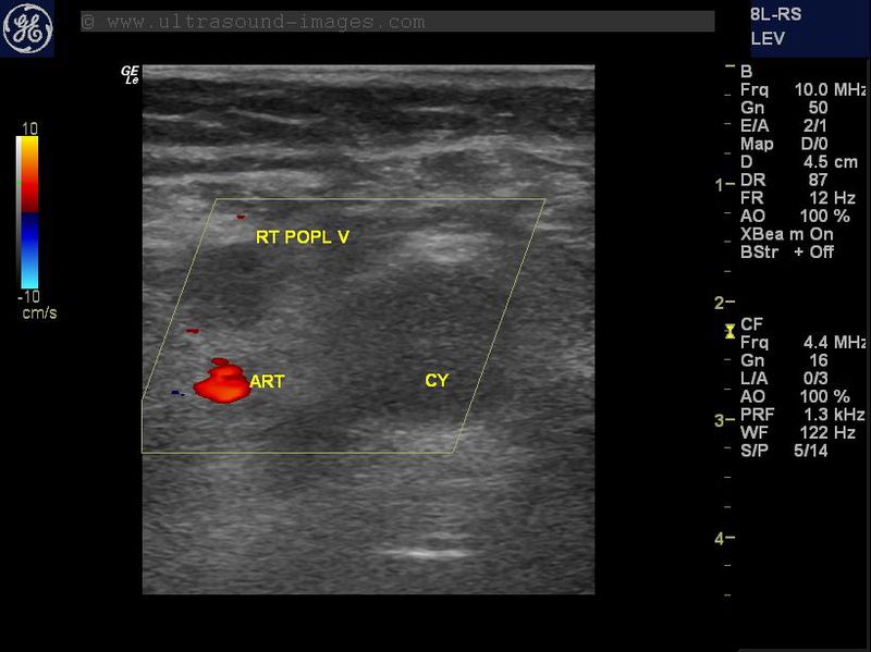
This is yet another case of right popliteal vein thrombosis in a lady complaining of pedal and leg edema. Again, the right popliteal vein shows absence of compressibility (non-compressible) despite pressure sufficient to cause deformity of the adjacent popliteal artery. In addition no flow is observed in the popliteal vein on color Doppler imaging. Further, a small cystic structure is seen medially, a Baker's cyst which is distinguished from the vessels nearby due to its oval shape and lack of Doppler signals. In addition, the right popliteal vein is dilatated at 8 mm. diameter with echogenic matter within it further confirming the presence of thrombus within it.
Calcified atheromas of femoral artery- (calcific atherosclerosis)
B mode image (left) and color Doppler image on right
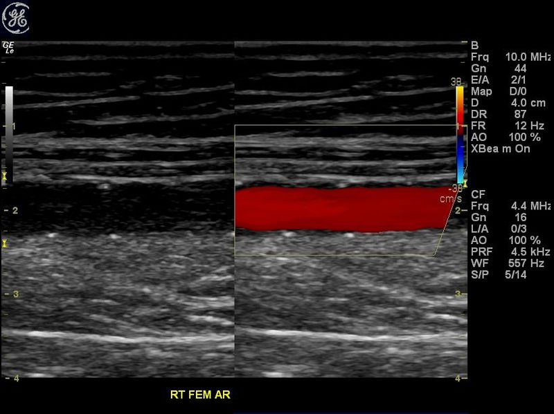
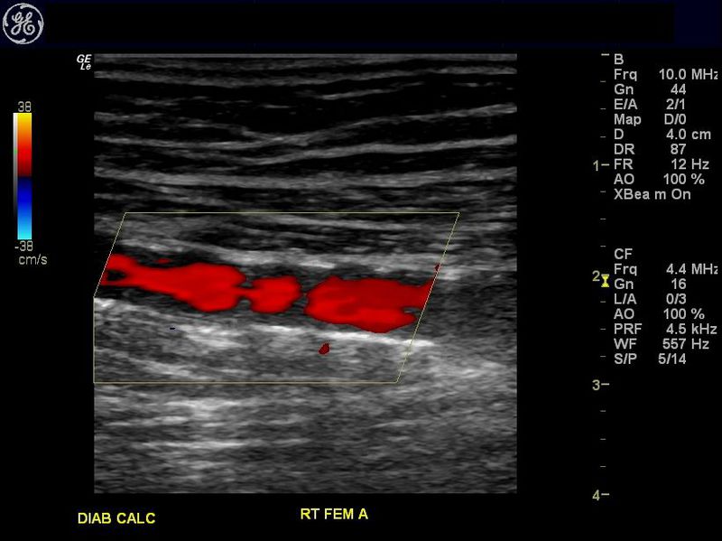

Most long term diabetic patients have extensive atherosclerosis of the arteries of the lower limb. In addition, there are calcific atheromas in these arteries. The ultrasound and color Doppler images above show a typical case of calcific atheromas of the right femoral artery, seen on the B-mode image as hyperechoic foci along the walls of the femoral artery. These calcific atheromas produce acoustic shadowing and areas of color "drop out" on color Doppler images. The areas of attenuation or color "drop out" are seen as breaks in the color flow signal within the artery (as shown above).
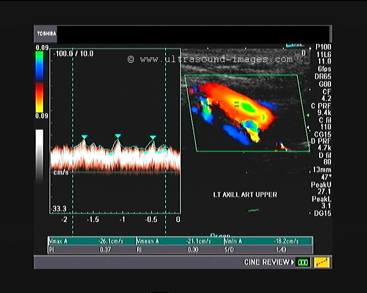
Takayasu Arteritis or Aortoarteritis or Pulseless disease
Colour Doppler ultrasound and spectral Doppler trace of the left axillary artery

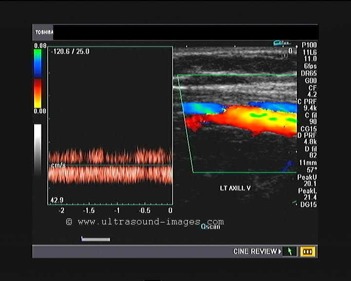
Colour Doppler ultrasound image of the left brachial artery and vein and spectral Doppler trace of the left brachial artery
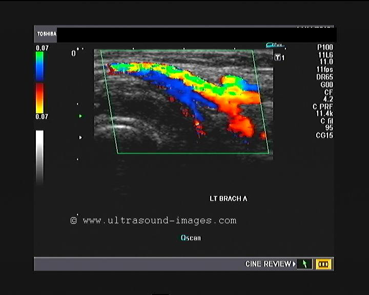
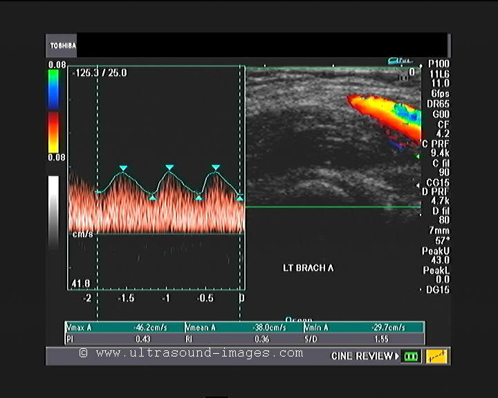
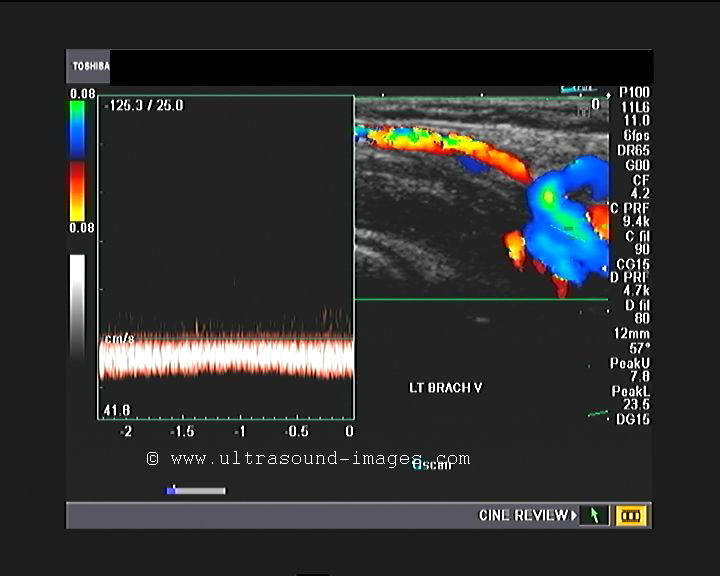
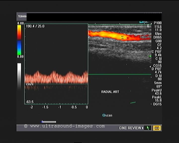

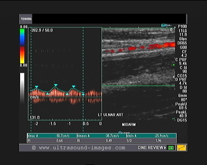
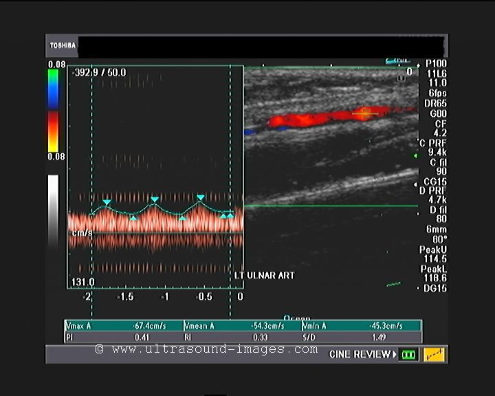
This young woman of 28 years age presented with pain and numbness of both upper limbs. colour Doppler ultrasound study of the left upper limb was done to evaluate the arterial system of this limb. All the arteries, starting from the left axillary artery, the left brachial artery, the left ulnar and radial arteries all show a tardus parvus pattern suggesting a high-level severe stenosis, possibly at the level of the left subclavian artery. all the arteries shown here show very low pulsatility and an almost venous flow pattern. Similar features can be expected in the arterial system of the right upper limb also. Such findings are typical of a condition known as Takayasu arteritis all a aorto-arteritis or pulse less disease. In Takayasu arteritis the common carotid artery as well as other major vessels may show extensive narrowing and stenosis due to wall thickening of a uniform nature. this ultrasound appearance is known as macaroni sign. Unlike atherosclerotic narrowing of the arteries, in Takayasu arteritis, there is uniform rather than segmental thickening of the arterial wall. Among the earliest and commonest sites of involvement, is the left subclavian artery and later the aorta, common carotid arteries as well as other major arteries.
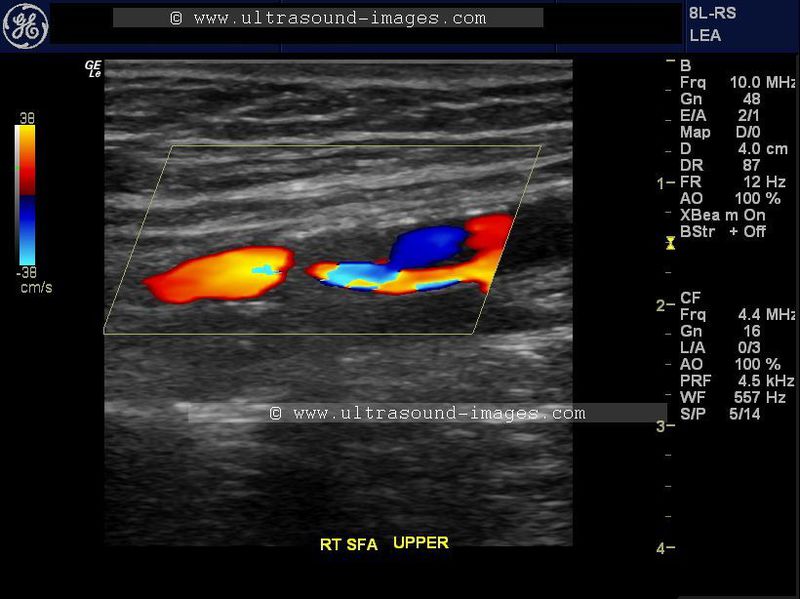
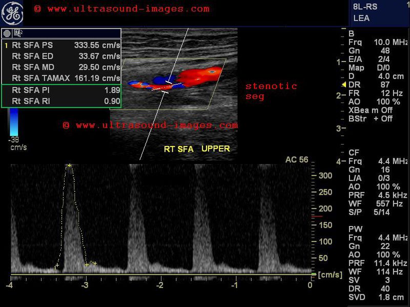
Severe-stenosis-of-Rt-SFA-case-2
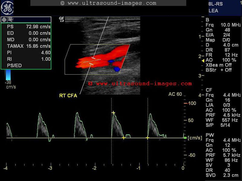
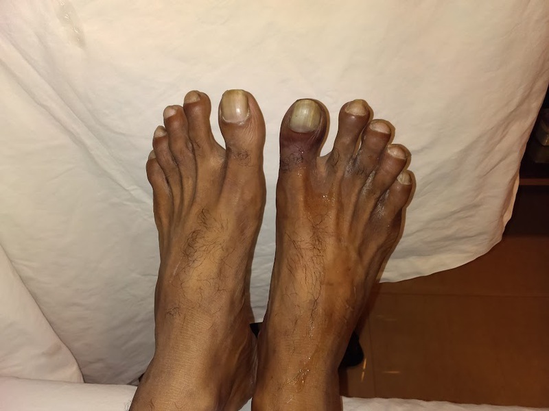

This diabetic male patient shows evidence of darkening of the right toes (see snap above), s/o ischemic changes and onset of pre-gangrene.
Doppler and spectral Doppler waveforms show a severe stenosis of the right SFA with Vs/ Vp ratio in this case = 330/72 almost 5 suggesting a severe stenosis in the right SFA. The prestenotic segement of CFA shows typical shouldering of the descending slope of systolic wave and poor diastolic flow suggesting distal obstruction. A likeness of characteristic "thumping" can be seen in this flow (prestenotic segment).
The distal SFA shows marked dampening of flow. The popliteal Artery and arteries of the leg show marked reduction of flow velocities with tardus parvus flow to venous like flow patterns. The dorsalis pedis A shows flow due to collateral supply.
Heel-and-toe-method
Heel and toe method of arterial Doppler ultrasound:
Very often, the sonologist / radiologist is able to align the spectral Doppler bar with the blood flow in the artery, but then the angle of insonation cannot be fixed in the 30 to 60 degree range.
Or, conversely, the angle of insonation is correct (30 to 60 degrees), but then the spectral Doppler bar is no longer parallel to the arterial flow.
In such situations the heel and toe method is useful (see color Doppler ultrasound video above).
Here the toe (cranial end) or toe (caudal end) of the probe is gently rocked to obtain the optimum angle yet remaining parallel to the flow.
pulsatile-venous-flow
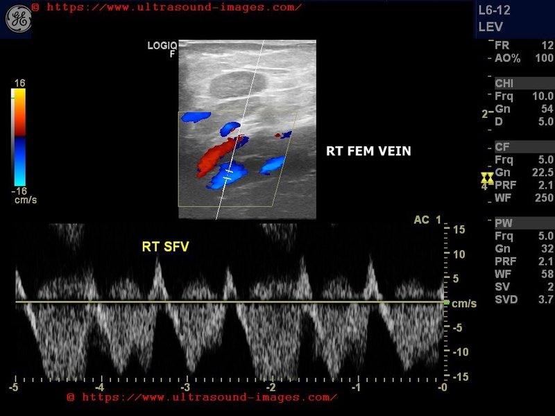
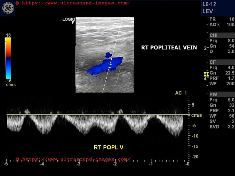
case-1:

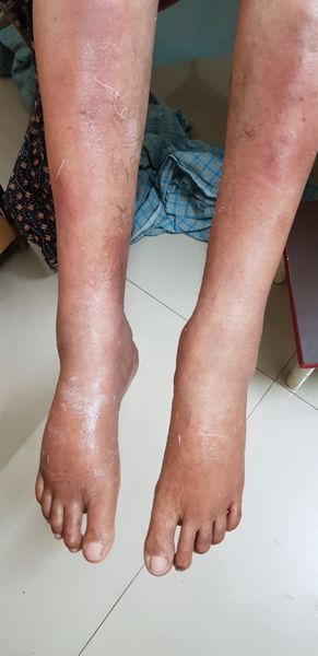
=Bilateral lower limb edema below knees
= bilateral markedly pulsatile flow in spectral Doppler ultrasound study of femoral and popliteal veins
= normal deep veins of the lower limb show gentle variation with respiration
= In this case the deep veins of the lower limbs show severe pulsatile flow (almost resembling arterial flow)
= almost always the result of increased Rt. atrial pressure or Rt. heart failure.
Reference: Pubmed article on Rt. heart failure and pulsatile flow in veins
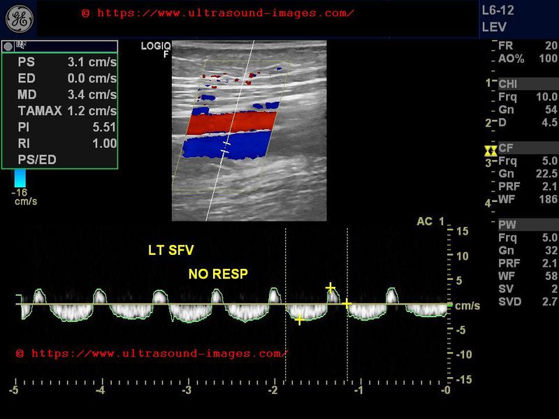
case-2: pulsatile venous flow in lower limbs
pulsatile flow- case-3:
© 2026, Joe Antony, MD All images and content in this site are copyrighted. www.ultrasound-images.com When visiting this site, we may use cookies to improve the performance of this site. Use of this site means you accept the use of cookies. Read our privacy policy at this page: https://www.ultrasound-images.com/privacy-policy/