
Ultrasound images of diseases of fetal urogenital system
Contents of this page
- Fetal hydronephrosis
- Fetal polycystic kidneys (autosomal recessive polycystic kidney disease- ARPKD)
- Sonography of another case of infantile polycystic kidney disease (autosomal recessive polycystic kidney disease) or ARPKD
- Fetal simple renal cyst
- Posterior urethral valve
- Fetal ureterocele
- Fetal bilateral multicystic kidney disease
- Fetal cross fused ectopia
- solitary fetal kidney
- fetal-pelvic-kidney
- fetal-urogenital-sinus-persistent-cloaca
- fetal-megacystis
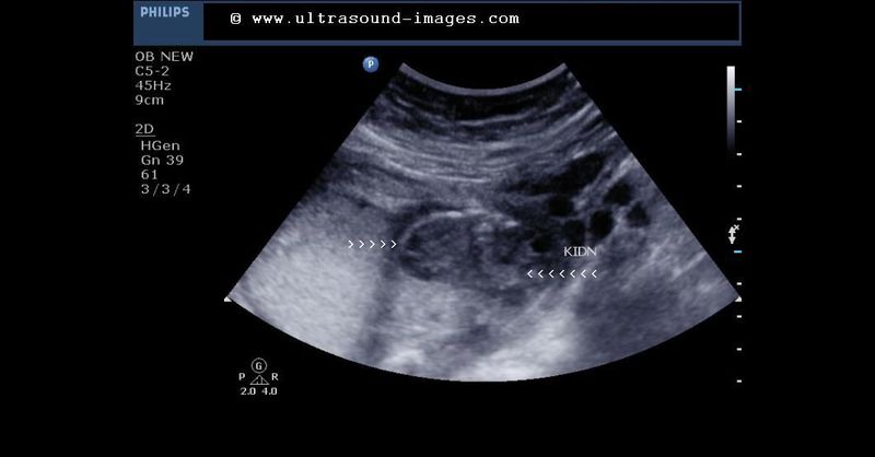
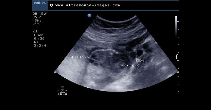
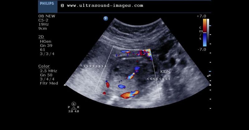
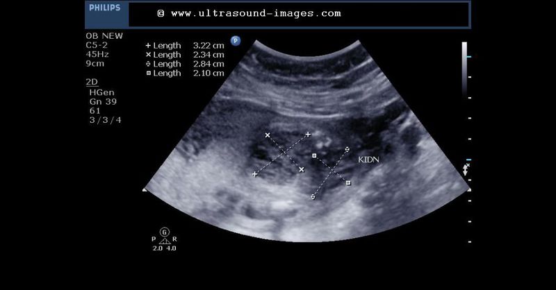
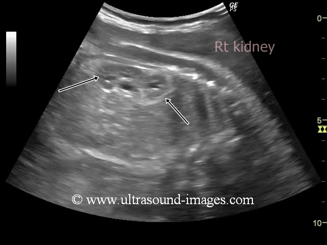
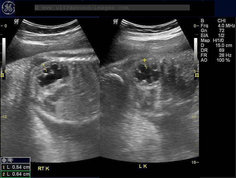
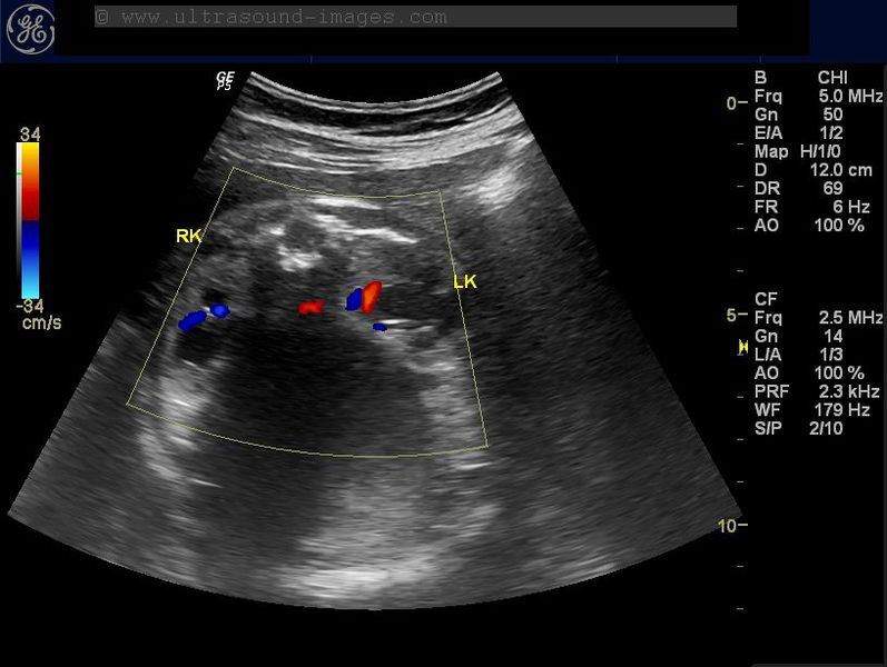
Fetal hydronephrosis
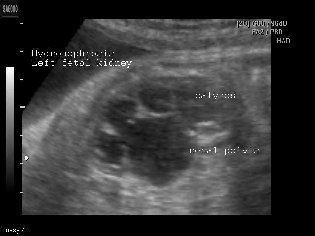
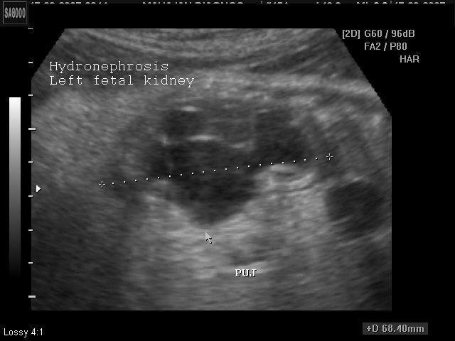
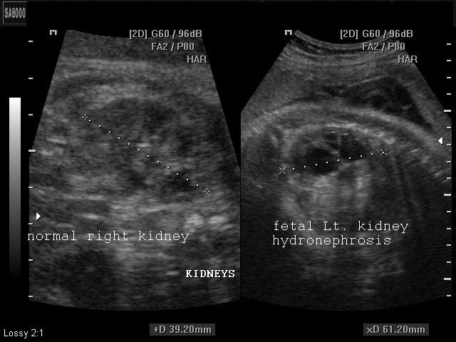
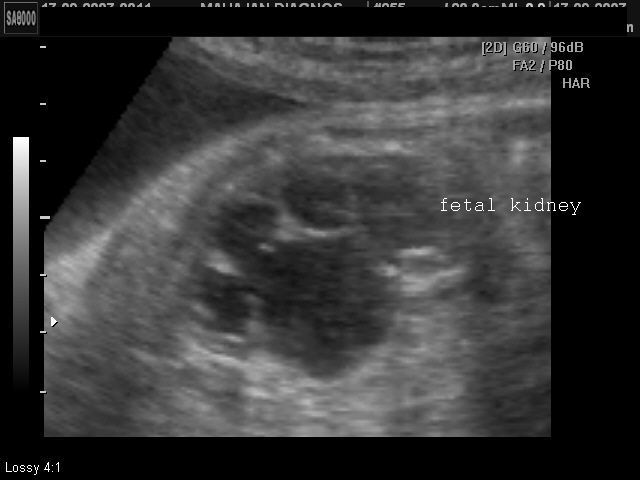
These ultrasound images reveal unilateral hydronephrosis of the left fetal kidney. The renal pelvis and calyces appear dilated with thinning of the renal parenchyma in the affected kidney. The commonest cause of such hydronephrosis is PUJ (Pelvi-ureteral junctional) obstruction, usually functional. The left ureter does not appear to be dilated. For cases of dilatation of the renal pelvis, it is labelled as abnormal if the RPD (renal pelvic A-P diameter) is more than 10mm (after 20 weeks gestational age). If less than 20 weeks, more than 4mm. is abnormal. These ultrasound images are courtesy of Dr. Arun Mahajan, Delhi, India.
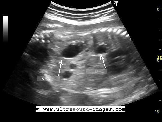
Fetal polycystic kidneys (autosomal recessive polycystic kidney disease- ARPKD)
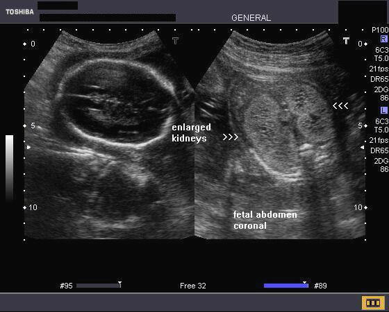
Case 1:
Sonography of the fetal abdomen in this 2nd trimester fetus reveals- 1) bilateral grossly enlarged fetal kidneys, almost filling the entire abdomen 2) markedly hyperechoic fetal kidneys with minute anechoic areas within them. 3) oligohydramnios 4) poorly distended urinary bladder. This ultrasound image is diagnostic of fetal autosomal recessive polycystic kidney disease (ARPKD). This image was taken by Dr. Durr-e-Sabih, Pakistan, using a Toshiba Nemio-30 color doppler machine.
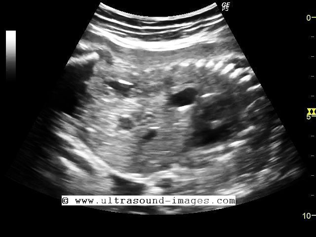
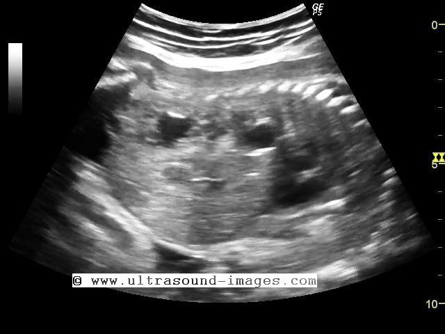
Sonography of another case of infantile polycystic kidney disease (autosomal recessive polycystic kidney disease) or ARPKD
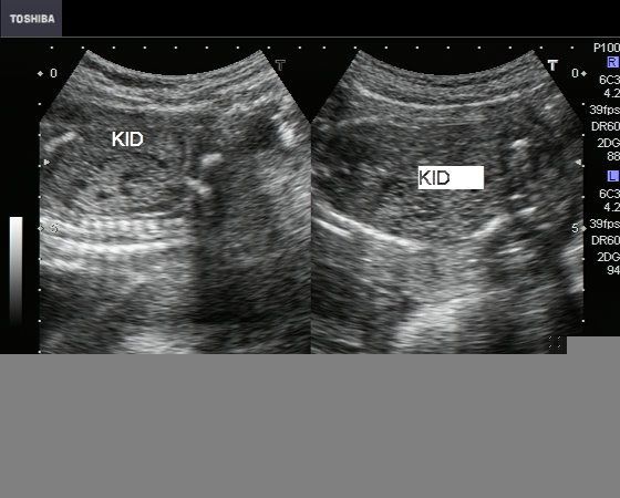

Case 2:
These ultrasound images (another case) also show grossly enlarged, echogenic fetal kidneys with minute cystic lesions (1 to 2 mm.) within them. The cystic lesions in this case, as well as the previous case show a typical rosette like pattern that is typical of autosomal recessive polycystic kidney disease. These ultrasound images are courtesy of Dr. Jaydeep Gandhi, Mumbai India. The machine used is a Nemio 30 from Toshiba.
Reference: E-medicine article on ARPKD
http://www.jultrasoundmed.org/cgi/reprint/22/1/105.pdf (free article and images).
Fetal simple renal cyst
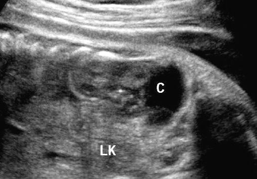
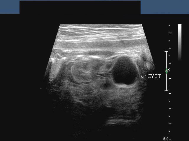

Solitary cyst of fetal kidney. This fetus of 30 weeks gestational age underwent sonography of the fetal abdomen. Ultrasound images show a small anechoic rounded lesion of the lower pole. The walls of the lesion are smooth and the contents appear to be clear fluid with no septae or tissue. There is also evidence of posterior acoustic enhancement. These ultrasound findings (images) are diagnostic of simple renal cyst (left kidney). Solitary renal cysts in fetus are a rare occurrence and may be of little clinical importance, but need to followed by serial scans for changes in characteristics or size. The lower right shows a 3-D ultrasound image of the fetal renal cyst. All images are courtesy of Ravi Kadasne, MD, UAE.
Reference http://www.sonoworld.com/fetus/page.aspx?id=547 (sonoworld article on sonography of case of fetal renal cyst )
case-2-simple-renal-cyst
Yet another case of fetal simple renal cyst- this time in the left fetal kidney. This cyst is large at 8 cms. and the left kidney is compressed by the huge renal cyst (see ultrasound and Power Doppler images above). This case is courtesy of Jorge Hernandez, MD.
Posterior urethral valve
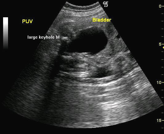

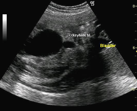
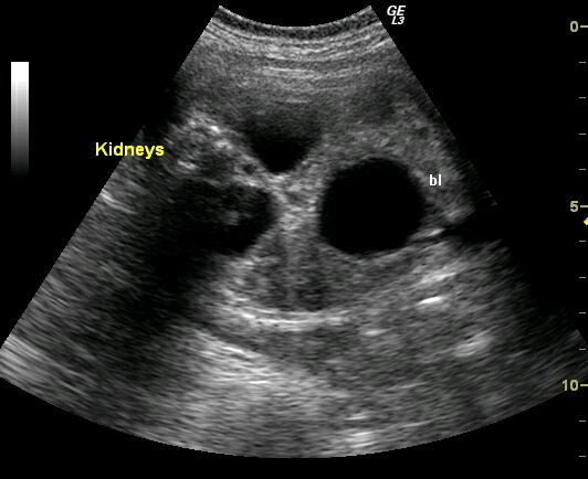
Sonography of this 3rd trimester fetus shows 1) keyhole urinary bladder (an overdistended bladder with the dilated prostatic urethra forming a keyhole like appearance). 2) hydronephrotic kidneys (bilateral) with thinning of the renal cortex. These ultrasound images suggest fetal lower urinary tract obstruction, probably a posterior urethral valve. Some degree of pelvi-ureteric junction obstruction may also be present. Images by Joe Antony, MD, India. Images taken using a GE- Logic 3 ultrasound system.
Fetal ureterocele

A rare case of ureterocele detected sonographically in 3rd trimester fetus Sonography of the fetal urinary bladder, shows a sac like structure (arrow) in the region of the distal end of the fetal left ureter. This ultrasound image suggests fetal ureterocele. Ureterocele is often seen in adults, but very rarely in a fetus. It is caused by defect during in embryogenesis of the fetal ureter. Ultrasound image is courtesy of Dr. Latha Natarajan, India.
Reference:http://www.thefetus.net/page.php?id=561(free article and images)
Fetal bilateral multicystic kidney disease


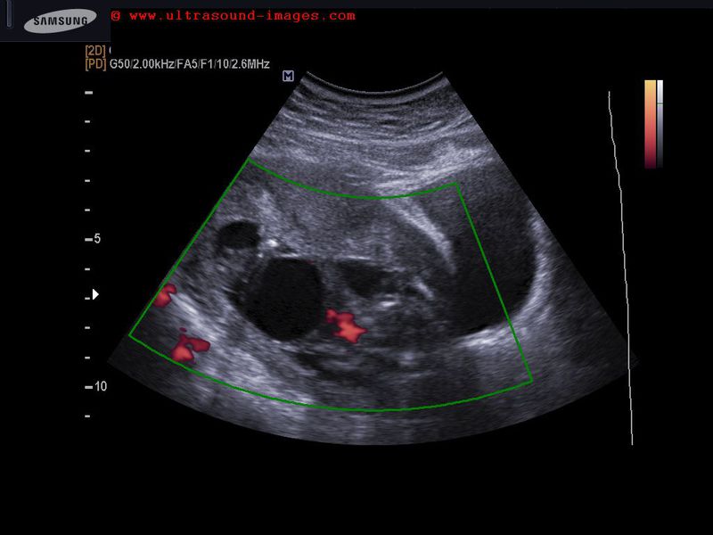
This 20 Week Old Fetus shows enlarged kidneys on both sides with severe oligohydramnios. In addition, the kidneys appear echogenic with multiple cysts, of varying sizes, within the Fetal renal tissue. Colour Doppler Study shows the renal arteries on both sides but little or no vascularity within the Fetal renal tissue. These Findings are consistent with a diagnosis of multicystic kidney disease, in this case, bilateral in nature. Multicystic dysplastic kidney disease is usually unilateral, however in this case it is bilateral. Bilateral multicystic dysplastic kidneys are more common in female foetuses. Multicystic Disease of the Kidneys is usually the result of chronic obstruction in the early stages of Fetal development. Bilateral multicystic dysplastic kidneys in foetuses is invariably fatal. The Main Differential Diagnosis in multicystic dysplastic kidneys is autosomal recessive polycystic kidney disease of the fetus. However, in multicystic kidneys, as in this case, the Fetal kidneys appear poorly defined in outline. In autosomal recessive polycystic kidney disease of the fetus, the kidneys are markedly enlarged with well defined margins. Besides, in autosomal recessive polycystic kidney disease, the cysts are small in size and are of uniform size and arranged in a rosette pattern. In multicystic dysplastic kidney, as in this case, the cysts are of larger size and of various sizes.
References:http://www.sonoworld.com/fetus/page.aspx?id=553
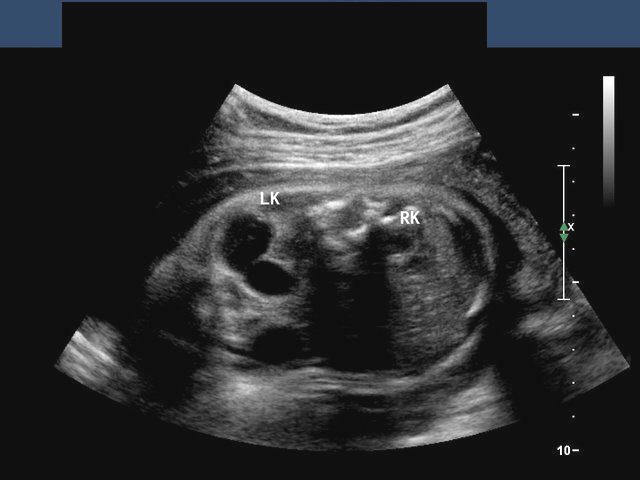
Fetal cross fused ectopia
This late second trimester fetus shows both kidneys located to the right of the fetal spine. No kidney was seen to the left of fetal spine. Both kidneys appear to be partially fused- with mild hydronephrosis of both upper and lower moieties of the fetal kidneys. This Sonographlc appearance ( . See the ultrasound images above), suggests Cross fused ectopia of the fetal kidneys. These images are courtesy of Dr Sunil Yadav, MD. Cross fused ectopia is a comparatively rare anomaly of the fetal kidneys and is often associated with obstructed changes in one of both moieties. If possible, one must try to identify the fetal ureters to look for dilation of these structures. Complete or partial duplication of the fetal ureters maybe present.
References: 1) http://www.jultrasoundmed.org/content/30/4/578.full
2) http://www.ultrasound-images.com/kidneys.htm#Normal%20variants
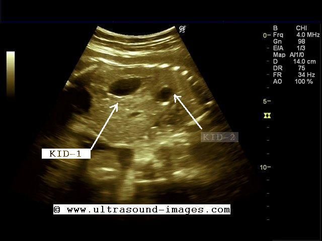
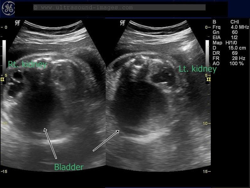
solitary fetal kidney
Absence of one of the fetal kidneys or solitary fetal kidney is a comparatively rare anomaly. Detection of unilateral agenesis (or absence of one of the fetal kidneys) during prenatal sonography is possible with most ultrasound machines. In this case, ultrasound images show absence of the left fetal kidney or solitary right kidney. Every effort must be made to detect the other kidney to rule out ectopic kidney (lower lumbar or pelvic kidney). The absence of the left renal artery also confirms the diagnosis of unilateral agenesis of the left kidney. Ultrasound images of unilateral renal agenesis are courtesy of Ravi Kadasne, MD, UAE.
References:http://radiopaedia.org/articles/renal-agenesis
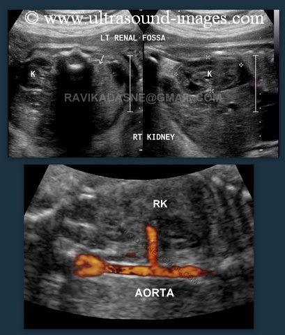
unilateral-agenesis-fetal-kidney-case-2
This fetus also shows a similar condition with unilateral absence of one of the fetal kidneys, in this case agenesis of the left kidney. Note the presence of the normal right kidney in the right renal fossa. The left kidney is not imaged in the left renal fossa. Also note again, the absence of the left renal artery, while the right renal artery is well visualised. These images were captured using the logic P5 ultrasound system. Both images show axial sections of the fetal abdomen.
(Key: RT.K= RT. KIDNEY of fetus; NON VIS= non visualized left fetal kidney)
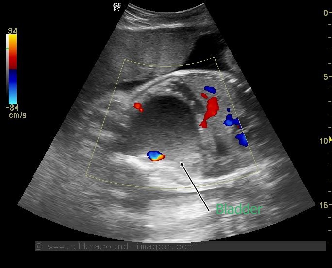
fetal-pelvic-kidney
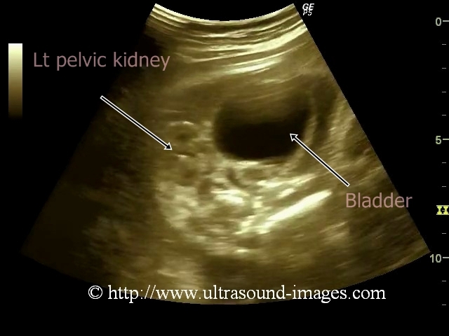
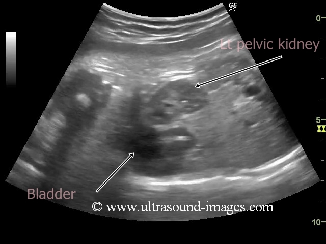
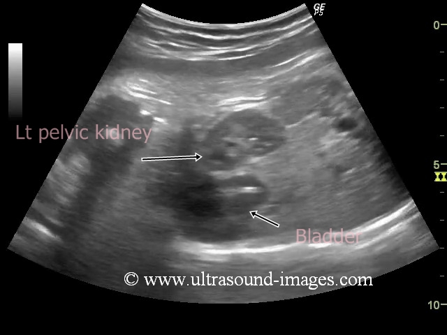
This second trimester pregnancy shows the normal right kidney located in the right renal fossa. The Fetal left kidney was not visualised in the left renal fossa. However, a scan below the left lumbar region of the fetus, and into the pelvis reveals a hyper distended urinary bladder with the left kidney located just to the left of the urinary bladder. The ultrasound images above show one of the commonest anomalies involving renal ectopia, namely, pelvic kidney. A pelvic kidney though one of the commonest renal anomalies, must however, trigger a detailed examination of the fetus for other congenital anomalies involving the renal, genital, cardiac and skeletal systems. Pelvic kidneys may be bilateral and are often associated with undescended testes and hypospadias. these images of pelvic kidney are courtesy of Sunil Yadav, MD.
References:sonographic imaging of fetal pelvic kidney
fetal-urogenital-sinus-persistent-cloaca

This 34 week fetus shows a spherical cystic structure in the lower part of the fetal abdomen, besides the severe bilateral hydronephrosis. The cystic "mass" shows particulate fluid matter, which is mildly echogenic on sonography (see ultrasound images above). This cystic "mass" represents the markedly distended fetal urinary bladder. The two umbilical arteries are seen on either side of the urinary bladder confirming its identity. The differential diagnosis in such a case include bilateral PUJ (or UPJ- pelviureteric junction) obstruction, posterior urethral valve (if male), and ovarian cyst in case of female fetuses and urogenital sinus or persistent cloaca (female). Post natal ultrasound imaging confirmed the presence of a persistent cloaca anomaly or urogenital sinus defect in this fetus. There was a single orifice exiting from a common chamber into which the rectum, vagina and uterus and urinary bladder seemed to empty (this common chamber is the cloaca which is normally present in the first trimester of pregnancy). The presence of echogenic material within the bladder is the result of mixing of fetal meconium and fecal particles from the fetal rectum with the urine. Very often, persistent fetal cloacal defect is associated with fetal ascites and distended fetal bowel as well as calcifications in the urinary bladder.
References:
http://www.jneonatalsurg.com/ojs/index.php/jns/article/view/13/28
fetal-megacystis

This 14 weeks old fetus shows -
-large cystic lesion in the abdomen extending till diaphragm from the fetal pelvis
- size of cyst is 3 cms.
-oligohydramnios
-aseptate cystic lesion
These are all typical findings of megacystis or hyperdistended urinary bladder usually caused by bladder outlet obstruction, which can be caused by posterior urethral valves.
D/d: ovarian cyst - however, in fetal ovarian cyst, there may not be evidence of oligohydramnios
see more at: sonography of fetal megacystis
© 2019, Joe Antony, MD All images and content in this site are copyrighted. www.ultrasound-images.com When visiting this site, we may use cookies to improve the performance of this site. Use of this site means you accept the use of cookies. Read our privacy policy at this page: https://www.ultrasound-images.com/privacy-policy/