
Ultrasound images of diseases of the prostate
Contents of this page
- Benign prostatic hyperplasia
- Color doppler imaging of prostatitis
- Some more images of acute prostatitis (another patient)
- Carcinoma prostate- Ultrasound and Color Doppler imaging
- Prostate calcification
- Seminal vesicle calculi (seminal vesicle calcification)
- Large cyst of seminal vesicle
- Prostatic utricle cyst
- Calcified cyst of the prostatic utricle
- Imaging of the normal seminal vesicles, and vas deferens
- 3D Ultrasound image of prostate calculus
- 3D ultrasound images of large prostate calculus- case-2
- 3D-ultrasound-prostatic-urethra
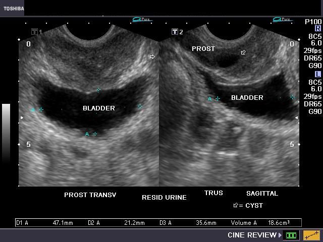
Benign prostatic hyperplasia
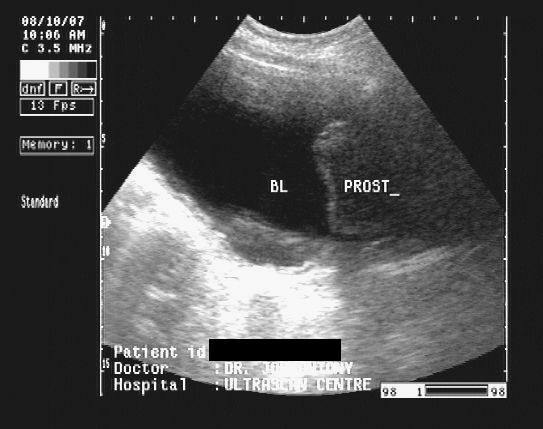
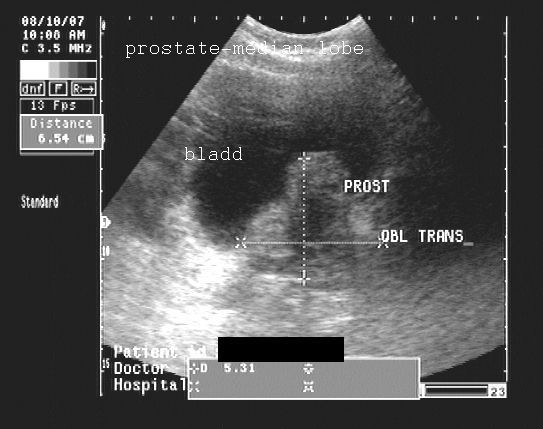
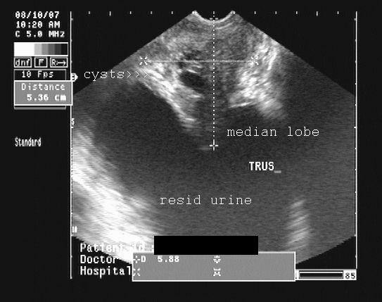
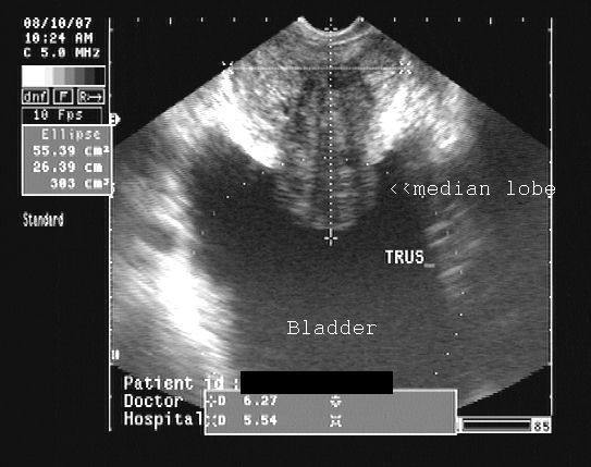
This 80 yr. old male patient presented with lower urinary tract symptoms (LUTS). Transabdominal ultrasound scan images reveal obvious intravesical enlargement of the enlarged median lobeof the prostate. Post-voiding trans-rectal ultrasound scan (TRUS) images reveal- 1) large volume of residual urine (303 cc) (more than 40 cc. is abnormal). 2) gross enlargement of the prostate mainly involving the transition zone. 3) intra-vesical enlargement of median lobe. 4) few small cysts in inner gland 3) there is also evidence of corpora amylacea and nodularity in the transition zone. 5) the peripheral zone is compressed by the enlarged transition zone. Diagnosis: these ultrasound images are diagnostic of benign hyperplasia of prostate.
Reference:
1) http://www.guideline.gov/summary/summary.aspx?ss=15&doc_id=3740&nbr=2966(free article)
2)http://www.pubmedcentral.nih.gov/articlerender.fcgi?artid=1502354 (free article)
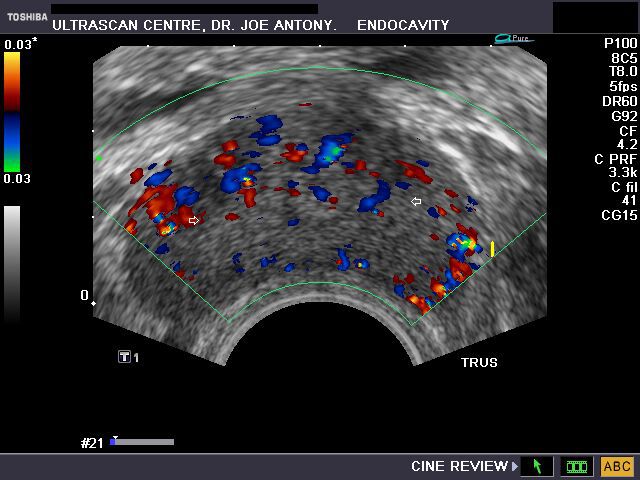
Color doppler imaging of prostatitis
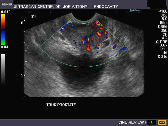
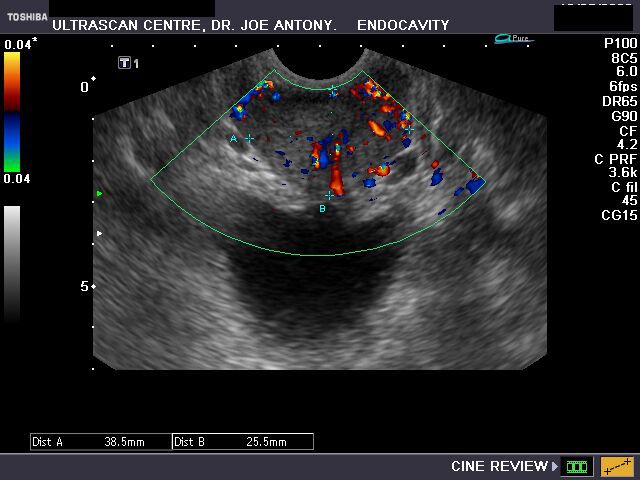

The above TRUS ultrasound and color doppler images in a young male patient show a) hypoechoic prostate b) gross augmentation of vascularity in the prostate tissue. These ultrasound findings suggest presence of acute prostatitis. Ultrasound images taken using Nemio XG color doppler machine (Toshiba).
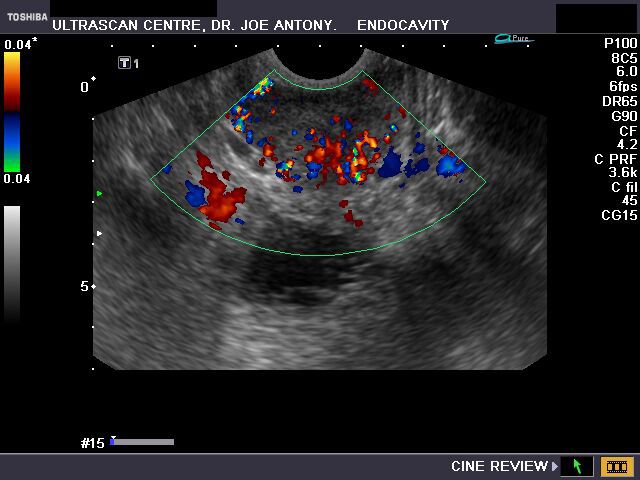
Some more images of acute prostatitis (another patient)
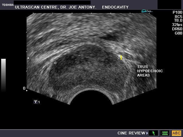
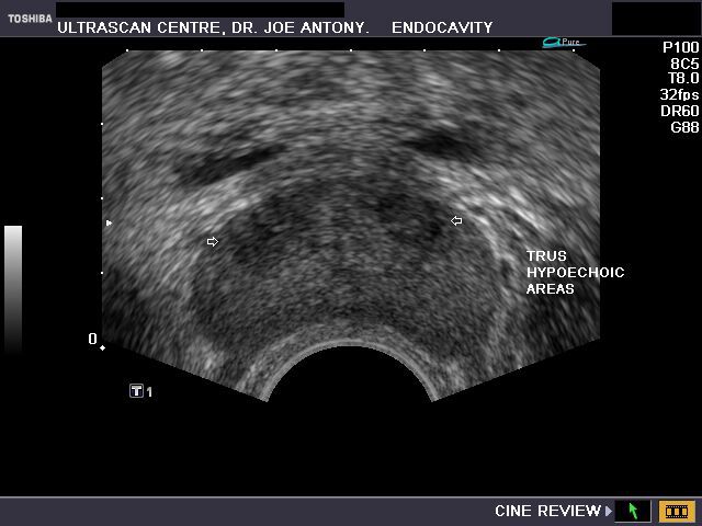
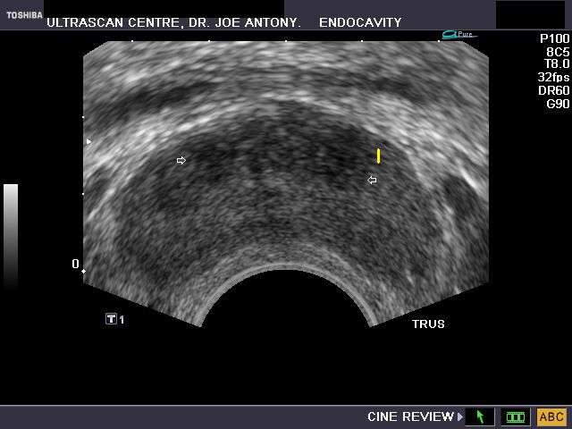
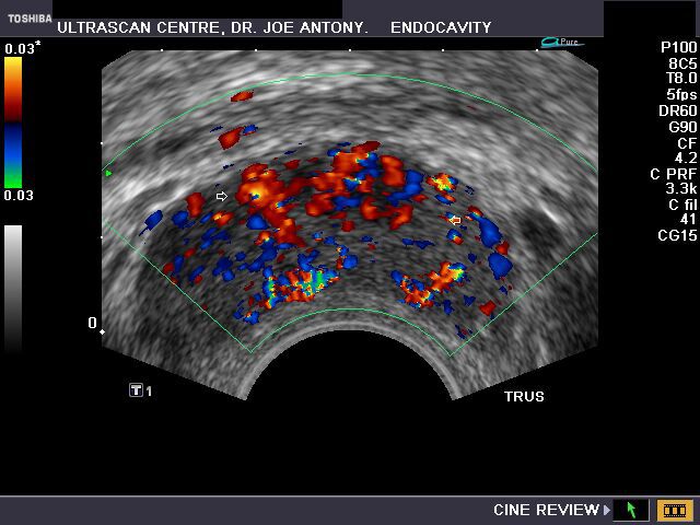
TRUS images of prostatitis.
Note the markedly hypoechoic patches in the inner zone of the prostate (arrowed), which appear overtly vascular on color doppler imaging
Carcinoma prostate- Ultrasound and Color Doppler imaging
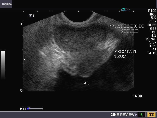

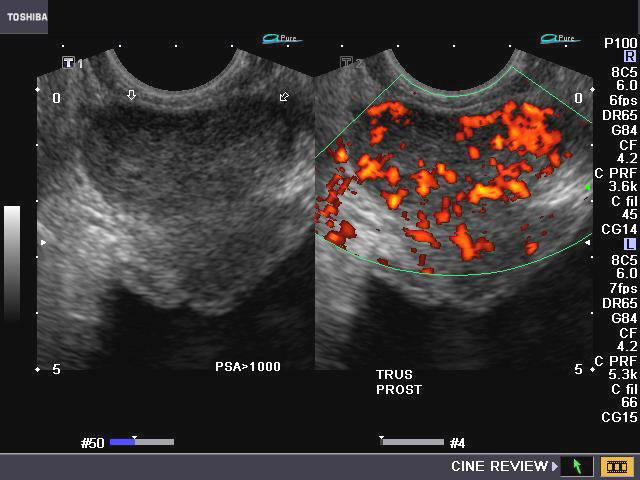

Sonography of the prostate using TRUS (Transrectal ultrasound) was done in this elderly male patient with hard nodule palpable on DRE (digital rectal examination) of the prostate. The hard nodule was felt in the left half of the prostate. PSA study showed very high values (> 1000 ng/ml) (normal < 4 ng/ml). Ultrasound images (TRUS) reveal a hypoechoic lesion involving much of the left peripheral zone. Color and Power Doppler images (TRUS) reveal marked vascularity in the region of the nodule (left peripheral zone). These ultrasound image findings are typical of carcinoma of prostate. There is also evidence of benign prostatic hypertrophy. All ultrasound images by Joe Antony, MD, India, using a Nemio- XG machine.
Reference:
1)http://drjoea.googlepages.com/home2
2)http://emedicine.medscape.com/article/457757-overview
Prostate calcification
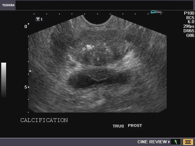
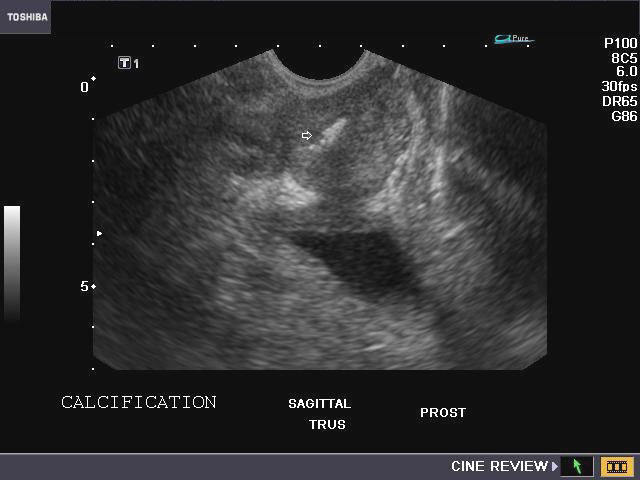
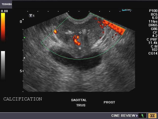
Calculi or calcific foci in prostate.
This middle aged patient underwent TRUS imaging (transrectal ultrasound) of the prostate for prostatism (symptoms related to the prostate). TRUS images show multiple hyperechoic foci (arrows), each of 4 to 7 mm. in the inner gland of the prostate and also along the prostatic urethra. Power Doppler image (bottom) shows normal flow in the prostate. These ultrasound images suggest prostatic calcification or calculi. Calcific foci in prostate are associated with normal aging process in the male and may be the result of formation of corpora amylacea. These are formed by calcification of secretions of the gland. It is also seen in chronic inflammation of the prostate (chronic prostatitis).
Seminal vesicle calculi (seminal vesicle calcification)
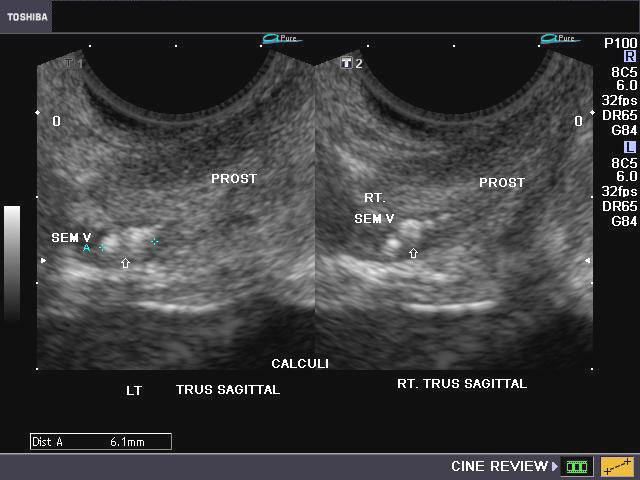
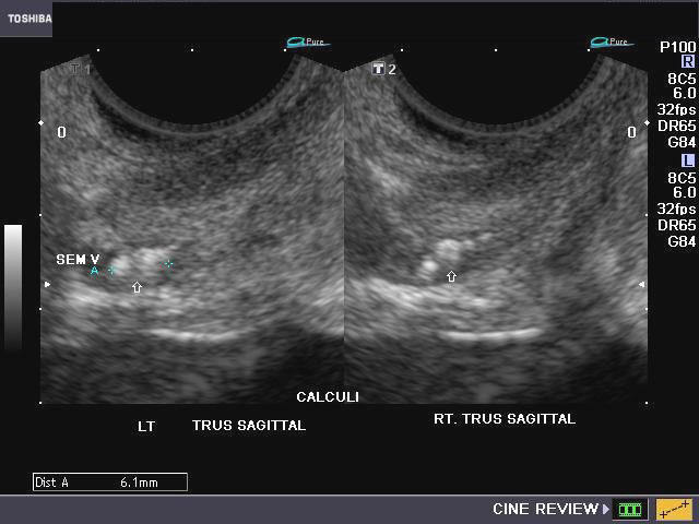

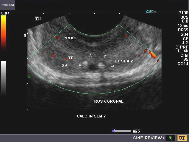
Multiple bilateral stones/ calculi in seminal vesicles (seminal vesicular lithiasis)
This middle aged male patient presented with a history of hemospermia (passage of blood in semen) with mild pain during ejaculation. Sonography of the abdomen was normal. Transrectal ultrasound (TRUS) of the prostate and seminal vesicles showed multiple echogenic foci/ lesions in the terminal (proximal) part of the seminal vesicles, bilaterally. The ultrasound images show multiple seminal vesical calculi bilaterally, each measuring 2 to 4 mm. in size. Studies suggest that such stones are related to inflammation, obstruction or diabetes mellitus. The ultrasound image on bottom right shows Power Doppler study of the prostate; no abnormal flow was found. Calculi in this case can cause poor flow of semen during ejaculation, hemospermia and painful ejaculation.
Reference:http://www.bioline.org.br/pdf?is06009 (free article and images)
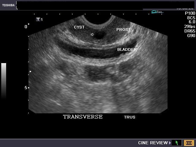
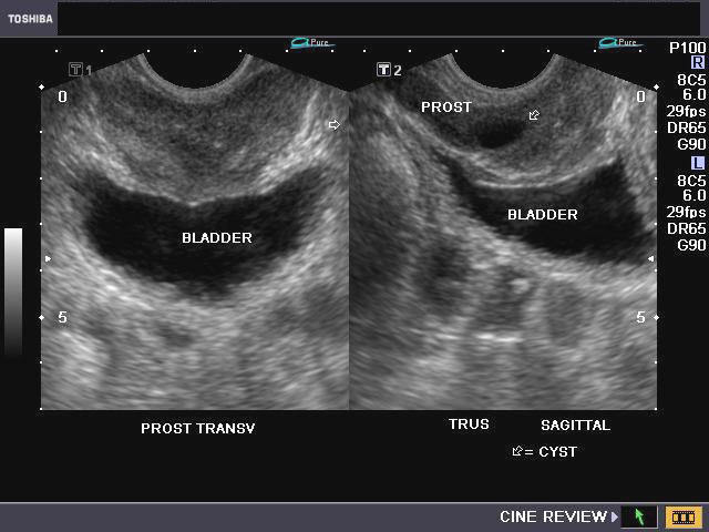
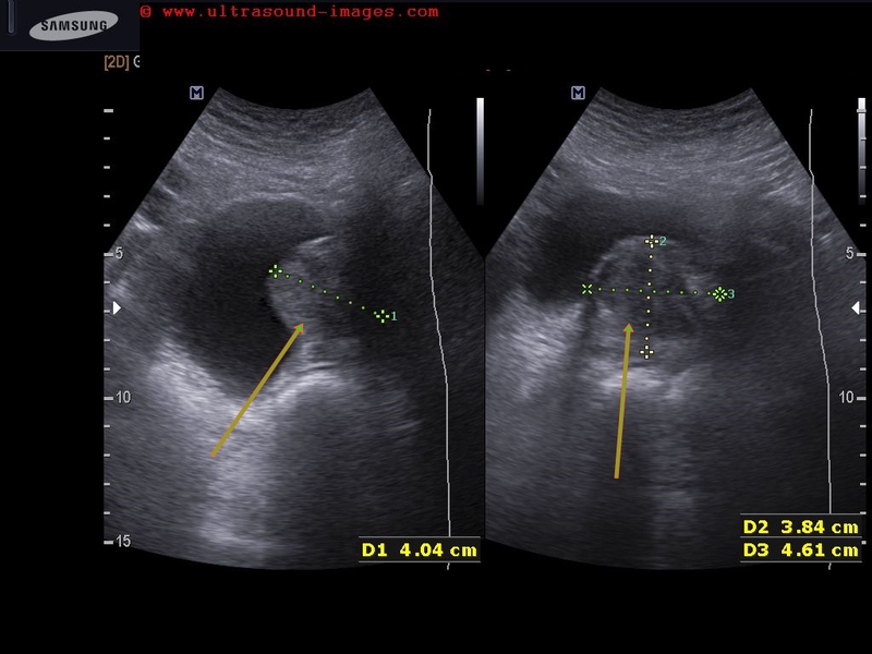
Large cyst of seminal vesicle
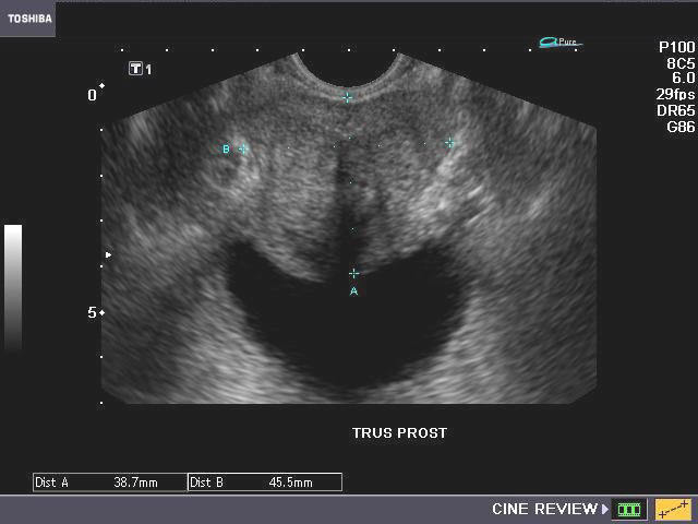
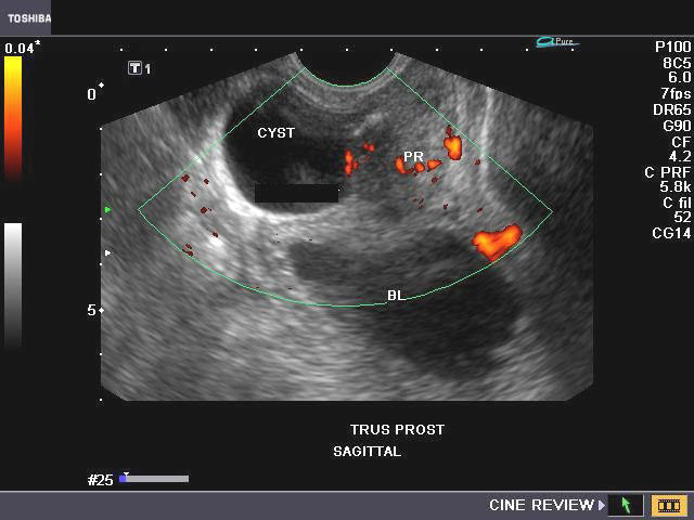


This elderly male patient had history of dysuria and urgency and other symptoms of prostatism. TRUS sonography (Transrectal ultrasound) showed a large cyst just above and to the left of the prostate. The cyst measured 3.6 x 2.8 cms. and showed clear fluid contents. Color and Power Doppler showed no significant findings. The cyst contained no septae or solid nodules and was non calcific. These findings suggest a large cyst of the left seminal vesicle. Both kidneys appeared normal. (BL= BLADDER; PROS= PROSTATE). The TRUS image of prostate (transverse)- in topmost row shows evidence of co-existing early benign prostatic hypertrophy. The main differential diagnoses in this case include: ejaculatory duct cysts; these are usually in the midline and have a tapering appearance; the others include prostatic utricle cysts which are present within the midline of the prostate. Seminal vesicle cysts may be congenital or acquired.
Reference: http://www.ajronline.org/cgi/reprint/175/1/177(free article and images).
Prostatic utricle cyst
The above ultrasound images (TRUS) show a typical prostatic utricle cyst. The cyst is located in upper half of the prostate in the midline. The image in lower row shows small amount of post voiding residual urine, one of the results of such cystic lesions in the prostate. The close proximity of the utricle cyst to the prostatic urethra, produces dysuria and some degree of retention of urine. Conservative treatment is usually sufficient to remedy this condition, though cyst enucleating may be the ultimate solution. See case 8- the cyst in this case is well within the prostate, and rules out the possibility of seminal vesicle cyst (case 8 above). However, it can be difficult to distinguish utricle cyst from ejaculatory duct cysts (both of these lesions can also cause infertility in the male).
Reference: http://emedicine.medscape.com/article/457757-overview
Calcified cyst of the prostatic utricle
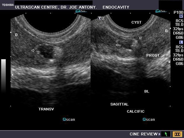

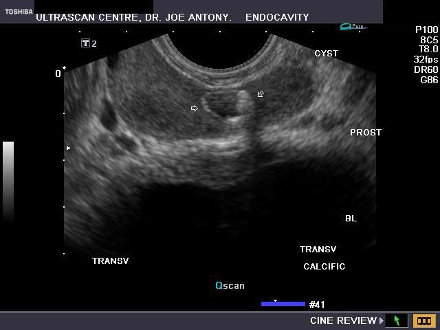
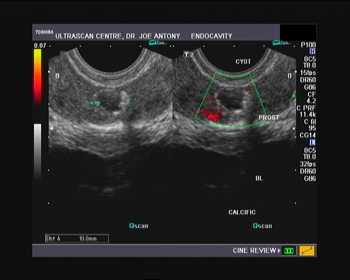
This late middle aged male patient presented with lower urinary tract symptoms. TRUS ultrasound shows a 9 mm. midline cyst of the prostate; what is interesting is the markedly hyperechoic rim of the prostate cyst suggesting calcification of the cyst walls. This is an unusual appearance for what is obviously a cyst of the prostatic utricle with almost no literature available. Power Doppler TRUS image (on right above), shows no significant changes in vascularity of the prostate and suggesting absence of prostatitis at present. The calcification of the walls of this midline utricle cyst of the prostate may be the result of dystrophic changes.
References: http://radiographics.rsna.org/content/10/4/635.full.pdf+html
Imaging of the normal seminal vesicles, and vas deferens
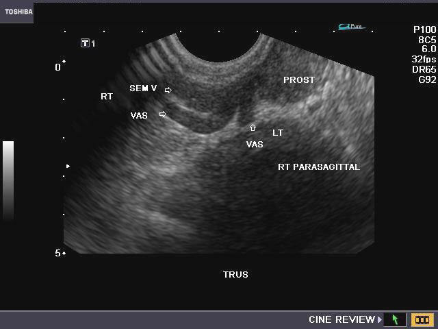
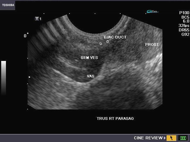
The vas deferens is seen joining the seminal vesicle and entering the prostate to form the ejaculatory duct in these TRUS ultrasound images. There is no defect in any of these structures in these images.
3D Ultrasound image of prostate calculus
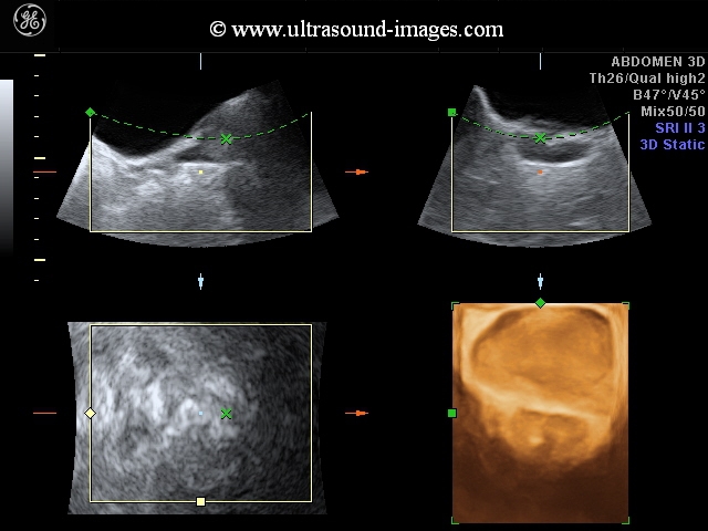

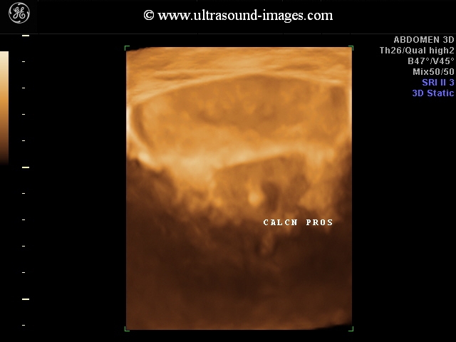
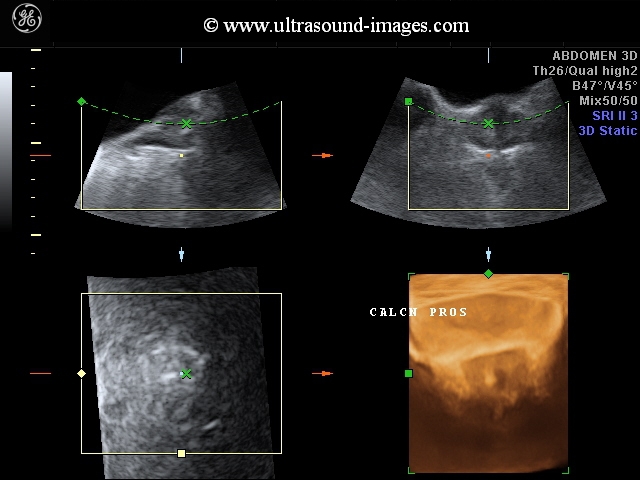
These 3-D ultrasound images of prostate and adjoining part of the bladder show a relatively small calculus of 5 mm lodged in the prostatic urethra. The 3-D surface rendering shows the internal surface of the urinary bladder in fine detail. Such prostatic calculi may cause pelvic or perineal pain and dysuria and a certain degree of urinary obstruction.
3D ultrasound images of large prostate calculus- case-2

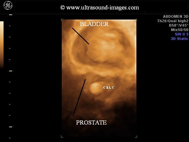

in this patient we see a large prostate calculus measuring almost 12 mm. The 3-D ultrasound images show the prostatic calculus lodged within the posterior urethra. Surgical intervention may be required to remove such large calculi in the prostate.
3D-ultrasound-prostatic-urethra
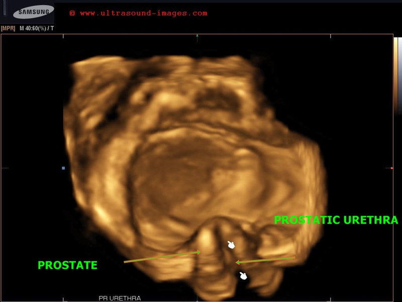
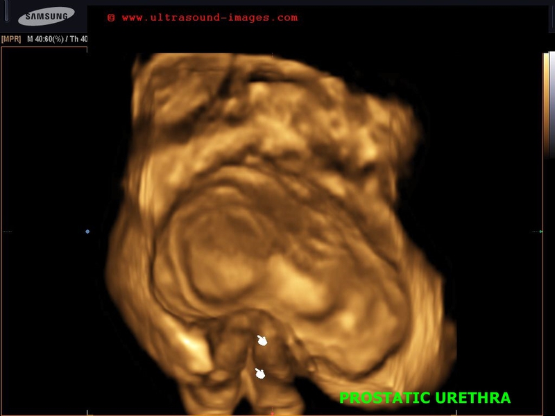
The above images show 3D ultrasound images of prostate in a patient of benign hypertrophy of the prostate or prostatomegaly. The 3D images show the full extent of the prostatic urethra (arrows) much alike the coronal reformat images of the uterus showing the endometrium. This kind of imaging can show the degree and kind of obstruction in lower urinary tract outlet. (Images captured using a Samsung Accuvix XG ultrasound system).
© 2019, Joe Antony, MD All images and content in this site are copyrighted. www.ultrasound-images.com When visiting this site, we may use cookies to improve the performance of this site. Use of this site means you accept the use of cookies. Read our privacy policy at this page: https://www.ultrasound-images.com/privacy-policy/